Abdominal Wall Defect
Description
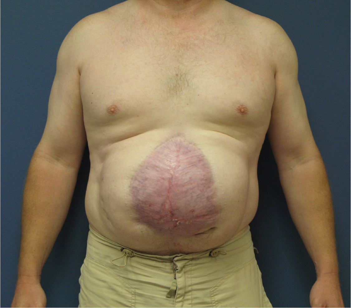
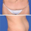
Large midline abdominal wall hernia consisting of myofascial defect with overlying skin.
The rectus abdominis muscles have migrated laterally.
There is no evidence of visceral incarceration.
Work-up
History
Etiology of defect: Congenital, previous surgery, trauma, resection.
Duration of defect, management thus far.
Nutritional status.
History of smoking.
Steroids or immunosuppressive medications.
Physical examination
Body mass index (BMI): weight (kg)/[height (m)]2.
Abdominal wall defect description
Location
Midline or lateral.
Upper, middle, or lower abdomen.
Tissue defect
Skin and subcutaneous tissue.
Myofascial.
Full thickness.
Size of defect.
Condition of surrounding tissues.
Preexisting incisions.
Pertinent comorbid conditions (diabetes, autoimmune diseases, coronary artery disease, etc).
Pertinent imaging or diagnostic studies
Computed tomography of the abdomen with contrast may be helpful to delineate the extent of the defect, the related anatomy, and other issues (e.g., bowel adhesions, abscesses).
Pulmonary function testing should be performed if there is preexisting respiratory compromise or suspicion for loss of domain from a large hernia.
Related posts:

Stay updated, free articles. Join our Telegram channel

Full access? Get Clinical Tree


