Atrophic Scar Management


Atrophic Scar Management


Joanna G. Bolton

Lisa A. Zaleski-Larsen

Mitchel P. Goldman


Enhanced awareness of scar pathophysiology and advances in technology and applications including laser, light and energy devices, injectable, surgical and topical techniques (both alone and in combination) provide new hope to millions of patients affected by scars worldwide. The term “cosmetic” belies the enormous potential psychosocial impact of a scar on a particular individual (see Chapters 4 and 24). The primary focus of this chapter is on the aesthetic treatment of atrophic and flat scars, with particular attention to the significantly distressing conditions of acne scarring and striae. Other chapters have detailed the pathophysiology and treatment of hypertrophic and keloid scarring, and these will be discussed briefly within the context of acne scarring.

Treatment of scars to improve their cosmetic appearance has progressed significantly over the past decade.1 The complexity of the wound-healing process dictates that a multifaceted approach is used in the minimization of scarring as well as in improving the aesthetic appearance of existing scars. With this in mind, these various facets will be discussed: (1) medical intervention—use of topical and intralesional agents that may influence the wound-healing cascade, decrease inflammation, or help remove the outer layers of skin; (2) laser, light, and energy-based interventions—use of various devices to improve the color, texture, and contour of scars and influence the wound-healing cascade; (3) soft tissue augmentation and novel approaches for transfer of material to help lift and blend scars; and (4) surgical intervention—for purposes of this chapter, defined as minor procedures that attempt to revise scars, usually in combination with other modalities (i.e., punch excision and/or grafting, subcision, dermabrasion, needling). The importance of “watchful waiting” and the “tincture of time” for optimal cosmesis cannot be overemphasized with any intervention.

The visibility of a scar depends on its width, texture, color, contrast, and flatness. Perceptions of how a scar appears is individualistic, with scars considered nearly imperceptible or normal by some being considered psychologically devastating by others. As pointed out by Tsao et al.,2 there are basically three types of scars: (1) atrophic scars (most commonly seen in acne and postvaricella scarring), (2) exophytic scars (hypertrophic and keloid scars), and (3) flat scars, which are considered normal scars, and are frequently dyspigmented after surgery or trauma to the skin, and which gradually diminish over time with or without intervention. Although the majority of flat, hyperpigmented scars are considered “normal,” they are frequently a concern of patients who seek advice and treatment for optimal cosmetic outcomes.

The authors of this chapter have chosen to include a discussion of striae, as the literature for this clinically challenging and commonly distressing condition has grown
in recent years. Striae can be considered a form of atrophic scarring, resulting from various mechanisms of skin stretching. Millions of people are affected and bothered by this common condition and may seek evaluation and a discussion of treatment options.


Scar History

Assessment of scar etiology, duration, and developmental history is important. Scars less than 1 year old are normally more erythematous than are older scars, and although they may be amenable to pulsed-dye laser (PDL) and/or intense-pulsed light (IPL) treatment, the patient should be educated that a degree of spontaneous improvement is expected over the first 12 to 15 months (see Chapters 8 and 9). Generally, scars that are greater than 1 year in duration and continue to worsen or are aesthetically concerning to the patient should be considered for intervention to prevent further abnormal scar growth and to expedite improvement.

Information on prior scar treatment should be solicited at the initial evaluation. Atrophic scars that have been treated with prior dermabrasion with resultant dermal thickening may not be vaporized as readily with carbon dioxide (CO2) laser resurfacing, possibly reducing the final clinical response.3 On the contrary, hypertrophic and keloid scars that have only received intralesional corticosteroid injections may elicit a more robust response to subsequent laser treatment.

Systemic medication use is an important consideration. Historically, there have been concerns that patients currently taking, or with a recent history of completing, a course of oral isotretinoin are at risk for delayed reepithelialization and resultant atypical scarring from acne scar treatment. The concern stems from the medication’s mechanism of action on adnexal structures, reducing or otherwise affecting the number and size of pilosebaceous glands that are essential for optimal wound healing. Historical clinical practice recommendations suggest that patients wait 6 to 12 months following discontinuation of isotretinoin therapy before undergoing resurfacing or surgical scar revision. However, these non-scientifically proven recommendations have been contradicted by recent studies. In a retrospective study (n = 110), patients taking oral isotretinoin (0.5 mg/kg/d) for acne or hirsutism were compared with those receiving only topical acne medications.4 Both groups underwent invasive treatment for acne scars and/or laser hair removal.5 Wound healing did not appear delayed nor was it associated with any adverse effects or atypical scarring in the patients taking isotretinoin. In another study, 80% of patients exhibited better than “fair” improvement and no aggravated acne scars, hypertrophic scars, or keloids following treatment with a 1,550-nm erbium-doped fiber laser to reduce acne scars while taking low-dose isotretinoin (10 mg per day) for at least 1 month.6

A history of immunosuppressive medications, such as chronic systemic steroids or biologic therapy, or presence of certain medical conditions, such as uncontrolled diabetes mellitus, should also give pause to major scar revision because of concerns for adverse outcomes from poor wound healing.


Acne Scars


Scar Pathogenesis

Scarring occurs as a result of damage to the skin during the healing of active acne. However, the pathogenesis remains incompletely understood. Acne can occur in any region where there is an abundance of pilosebaceous glands, especially the face, shoulders, back, and chest.7 The risk factors for developing permanent scarring are multifactorial, including genetic predisposition, a delay or nonadherence in acne treatment, and behaviors by patients that can induce excessive inflammation and mechanical trauma to the skin, impeding the healing process and worsening outcomes (i.e., acne excoriae). Not every patient with acne develops scars, and clinically it can be difficult to predict who will. Severe disease, such as nodulocystic acne, is most likely to result in scarring, but even patients with mild acne can develop scars.8

The most accepted hypothesis for the pathogenesis of atrophic acne scars points to dysregulated inflammation that affects fibroblastic function, culminating in a relative collagen deficiency and tissue atrophy.9 It has been demonstrated that acne patients prone to scarring have a particular cellular milieu different than that in those patients who do not scar.10 A cascade of pro-inflammatory cytokines and mediators, including activator protein (AP)-1, matrix metalloproteinase (MMP)-1 (collagenase-1), and other MMPs, are overexpressed in inflammatory acne lesions, leading to prolonged inflammation, follicular rupture, and perifollicular abscess formation. The well-recognized role of MMPs in collagen matrix degradation explains the tissue atrophy characteristic of depressed scars11 (Fig. 17-1). A study by Holland et al.12 demonstrated that patients with a propensity to scar had a greater nonspecific inflammatory response lasting for a longer duration when compared with those patients who did not scar. This could be due to an ineffective immune response to damaged tissue and abnormal wound healing. An overzealous healing response coupled with limited collagen lysis during the remodeling phase can create a raised, firm nodule of fibrotic tissue, whereas depressions or pits may result from inadequate replacement of enzymatically degraded collagen fibers and subcutaneous fat.13 Consequently, inflammatory acne lesions can result in permanent scars, and the longer treatment is delayed, the worse the scarring that results.

Community-based studies report acne prevalence is 90% in adolescent patients and persists into adulthood in approximately 12% to 14% of cases.14 Acne scarring is estimated to occur in up to 95% of acne patients, and 30% may develop significant scarring.15 Other studies have reported much lower incidences of scarring, ranging from 0.17% to 14%.16,17,18 Any degree of active acne and acne scarring may have a profoundly negative impact on daily activities, social life, self-esteem, and relationships, all of which make them both common conditions for which treatment is sought or would be of benefit. It has even been noted that the ability to acquire employment in adulthood may be limited in those with acne scarring.19 Until recently, very few studies had explored the negative impact caused by postacne scarring. Fried et al.20 confirmed that acne scars
have a substantial negative impact on the overall social and functional well-being of affected individuals. In their study, an overwhelming 85.4% of subjects revealed they were unhappy looking at themselves in the mirror, with 84.4% feeling less attractive owing to their acne scars.


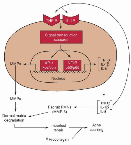
FIGURE 17-1 Hypothetical model of the pathophysiology of inflammatory acne and dermal damage. In inflammatory acne lesions, NF-κB signaling is activated. As a consequence, NF-κB-driven inflammatory cytokine genes (e.g., TNF-α and IL-1β) are induced. These primary cytokines will propagate the inflammatory response by acting on endothelial cells to elaborate adhesion molecules (eg, ICAM-1) to facilitate recruitment of inflammatory cells into the skin. TNF-α and IL-1β will also stimulate the production of secondary cytokines, such as IL-8, which can aid in chemotaxis of inflammatory cells. By working through their cell surface receptors, TNF-α and IL-1β not only amplify the NF-κB signaling cascade, but also activate MAP kinases to stimulate AP-1-mediated gene transcription. As a consequence of AP-1 activation (cJun induction), AP-1-driven MMPs are elaborated by resident skin cells. Along with MMP-8 and neutrophil elastase brought in by PMNs, they degrade the matrix. This is followed by matrix synthesis and repair, which is imperfect. Most of the imperfections would leave clinically undetectable deficits in the organization or composition, or both, of the extracellular matrix. However, when they occur to a significant extent throughout time, accompanied by sustained procollagen synthesis, acne scarring becomes clinically visible. (Used with permission from Kang S, Cho S, Chung JH, et al. Inflammation and extracellular matrix degradation mediated by activated transcription factors nuclear factor-kappaB and activator protein-1 in inflammatory acne lesions in vivo. Am J Pathol. 2005;166:1691-1699.)

Although it appears that acne severity is directly proportional to the severity of the acne scarring,15 it is important to note that levels of psychosocial distress may not be accurately predicted by one’s degree of disfigurement.21 With this in mind, it must be emphasized to patients that the most effective means of minimizing or preventing acne scarring and its associated devastating psychosocial difficulties is to treat acne early and aggressively.


Scar Classification

The nomenclature for categorizing types of acne scars has not been entirely settled in the literature.4 Acne scars are primarily classified according to whether there is a net loss or gain of collagen in the lesion. For the purposes of practicality and ease in treatment selection, acne scars can be categorized as either “atrophic” or “hypertrophic,” with “hypertrophic” including both hypertrophic and keloidal scars10 (Table 17-1). Eighty to 90% of people with acne scars have atrophic scars compared with a minority who show hypertrophic scars and keloids.14 Proper scar classification is important because differences in clinical subtype help guide appropriate therapeutic options on the basis of surface, depth, and three-dimensional architecture22 (Table 17-2).
In other words, treatment of acne scars must be individually directed for each patient depending on the size, type, and severity of scars present.23 The anatomic location of the scar and the patient’s skin type may also be important considerations. Clinicians may encounter scars with more than one physical characteristic, such as pigmentation or erythema, in addition to being atrophic or hypertrophic. These may be termed “hybrid” scars.4 Various modalities, single or combined, have been used to treat acne scars. Limited efficacy and problematic side effects have made the gold standard or “home-run” treatments challenging to determine.


Table 17-1 Classification of Acne Scars


Scar Classification


Clinical Features


Atrophic


Loss of dermal collagen (primarily) Most common form of acne scarring


Ice pick (60%-70%)


Deep and narrow pitting (<2 mm)


V-shaped, sharply demarcated epithelial tracts that extend into the dermis or subcutaneous tissue


Boxcar (20%-30%)


Round or oval depressions


Shallow (<0.5 mm) or deep (>0.5 mm) 1.5-4 mm diameter


Sharply demarcated vertical edges with a wide base; do not taper to a point at base and are clinically wider at the surface than ice pick scars


Rolling (15%-25%)


Wide and shallow undulations (>4-5 mm)


Superficial skin tethered by fibrous anchors to dermis or subcutaneous tissue, leading to shadowing and gently sloped edges that merge with normal-appearing skin


Hypertrophic


Excess collagen deposition


Hypertrophic


Pink, raised, firm papule Confined to the area of previous damage


Keloidal


Red to purple, firm papules and nodules Extend beyond original wound borders


Adapted from Gozali et al.,23 Levy and Zeichner,8 and Fabbrocini et al.14


Table 17-2 Most Common Treatment Modalities Based on Scar Type


Atrophic Scars


  • Laser (ablative and nonablative; fractionated and nonfractionated)


  • Light therapy (i.e., IPL)


  • Energy therapy (i.e., RF)


  • Injectable dermal fillers


  • Autologous fat transfer


  • Chemical peels


  • Subcision


  • Punch techniques


  • Dermabrasion/microdermabrasion


  • Needling


  • Topical retinoids


Hypertrophic Scars


  • Silicone gel sheeting


  • Intralesional corticosteroids


  • Cytotoxic agents: bleomycin, 5-fluorouracil


  • Cryosurgery


  • Radiotherapy


  • Laser (CO2, erbium, pulsed dye)


IPL, intense-pulsed light; RF, radiofrequency; CO2, carbon dioxide. Adapted from Gozali et al.,23 Levy and Zeichner,8 and Fabbrocini et al.14


Atrophic Scars

Atrophic scars are dermal depressions commonly caused by collagen destruction during the course of an inflammatory skin disease, such as cystic acne or varicella,3 though there are many causes and risk factors (Table 17-3). Histologically, atrophic scars show loss of not only collagen in the dermis but also cells in the epidermis and subcutaneous fat, all contributing to the clinical appearance of a depression of the skin.7 Atrophic scars cause significant patient morbidity and are reported to worsen with age because of the natural lipoatrophy, which further accentuates the scars.24


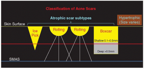
FIGURE 17-2 Classification of acne scars. Yellow line represents skin surface. Yellow/gray-shaded areas within scar subtypes represent loss of tissue. Brown-shaded area represents gain of tissue. Red line roughly denotes depth of ablation and resurfacing capability of the CO2 laser. Blue line represents SMAS to which fibrous bands (white lines) adhere, creating rolling scars. (Adapted by Dr. Joanna G. Bolton from Jacob CI, Dover JS, Kaminer MS. Acne scarring: a classification system and review of treatment options. J Am Acad Dermatol. 2001;45:109-117.)


Table 17-3 Main Causes and Risk Factors for Developing Atrophic Scars


Cause/Risk Factor


Inflammatory


Acne


Cyst


Discoid lupus erythematosus


Infective


Postvaricella


Trauma


Injury


Burn


Iatrogenic (surgery)


Skin stretching/striae


Pregnancy


Obesity


Rapid weight loss/gain


Puberty/growth spurt


Medications (i.e., steroids, ACTH)


Patient factors


Genetics (tendency toward atrophic scarring)


Previous atrophic scars


Ehlers-Danlos syndrome


Primary anetoderma


Adapted from Patel L, McGrouther D, Chakrabarty K. Evaluating evidence for atrophic scarring treatment modalities. J R Soc Med. 2014;5(9):1-13.


In 2001, Jacob et al.22 unified the previously vague terminology used to describe atrophic acne scars by proposing a classification based on width, depth, and a three-dimensional architecture of acne scars: ice pick, rolling, and boxcar scars (Fig. 17-2). Influenced extensively by Goodman,25,26,27 this new classification system married scar anatomy with the available effective treatment options, facilitating precise identification of scar type with enhanced, more reproducible treatment outcomes. Ice pick scars are reported as the most prevalent subcategory at 60% to 70%, boxcar scars are second at 20% to 30%, and, finally, rolling scars at 15% to 25%.

Ice pick scars are narrow (<2 mm), deep, sharply marginated epithelial tracts that extend vertically to the deep dermis or subcutaneous tissue. With this type of scar, the opening is typically wider than the deeper infundibulum (forming a “V” shape) (Fig. 17-3). But they may also be wider at a variety of levels in the dermis so that laser ablation may actually produce a wider scar/depression. Boxcar scars are round to oval depressions with sharply demarcated vertical edges, usually wider at the surface than ice pick scars (˜1.5 to 4 mm) and do not taper (“U” shape). They may be
shallow (0.1 to 0.5 mm) or deep (>0.5 mm). Shallow boxcar scars are within reach of most resurfacing treatments, but deeper boxcar scars and the tip of the infundibulum of ice pick scars are resistant to improvement in the absence of full-thickness treatment of the scar.


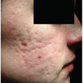
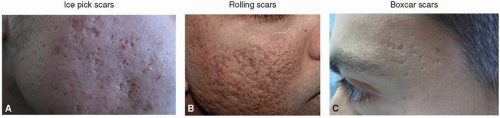
FIGURE 17-3 Atrophic acne scars. A: Ice pick, B: rolling and boxcar, C: boxcar. All three subcategories can be deep and coexist, making a singular clinical identification difficult. (Photo permission granted for (B) by Dr. Mitchel P. Goldman, MD; (A, C) Copyright © 2010 Gabriella Fabbrocini et al.)

Rolling scars occur from dermal tethering of otherwise normal-appearing skin, usually measuring >4 mm in diameter. Abnormal fibrous bands attached to the superficial musculoaponeurotic system (SMAS) create a shadowed rolling or undulating appearance to the overlying skin (“M” shape). Although they tend to be shallow, correction of the subdermal anchoring component is required for treatment success. In general, boxcar and rolling scars are thought of as wider and more superficial than ice pick scars; however, all can be deep and intermixed on the same patient.


Hypertrophic Scars

Hypertrophic and/or keloidal scars are raised scars that present a different challenge for aesthetic treatment. Hypertrophic scars are elevated, firm erythematous scars formed at the sites of healed acneiform lesions. The result is thick, hyalinized collagen bundles consisting of fibroblasts and fibrocytes intermixed with various densities of blood vessels. Hypertrophic scars generally remain within the confines of the original integument injury and may regress without intervention over months to years. In contrast, keloids are typically reddish-purple, raised, dense and rubbery nodular scars that extend beyond the borders of the inciting wound and do not normally regress spontaneously over time. Although they can be seen in all skin types, hypertrophic scars and keloids most frequently arise at sites of moderate-to-severe acne in patients with darker skin types (Fitzpatrick III to VI) because of inherited metabolic alterations in collagen. The scars are especially likely on the chest, shoulders, and back. Controlling inflammation associated with acne is paramount in skin of color, given the higher risk for long-lasting sequelae such as distressing postinflammatory hyperpigmentation (PIH) and, in severe cases, keloids.28 Unfortunately, undertreatment or delay in treatment of acne is common and increases the risk of hypertrophic and keloid scarring.


Table 17-4 Goodman and Baron Qualitative Grading Scale of Postacne Scarring


Grade


Level of disease


Characteristics


1


Macular


  • Erythematous, hyper- or hypopigmented flat marks


  • Represent a problem of color as opposed to problems with contour and texture


2


Mild


  • Mild atrophic or hypertrophic scarring that may not be obvious at social distances of 50 cma or greater (i.e., mild rolling, small soft papular scars)


  • May be covered adequately by makeup or the normal shadow of shaved beard hair, or body hair if extrafacial, in men


3


Moderate


  • Moderate atrophic or hypertrophic scarring that is obvious at social distances of 50 cm or greater (i.e. more significant rolling, shallow boxcar, mild-to-moderate hypertrophic or papular scars)


  • Not easily covered by makeup, or facial/body hair in men


  • Still able to be flattened by manual stretching of the skin (if atrophic)


4


Severe


  • Severe atrophic or hypertrophic scarring that is evident at social distances greater than 50 cm (i.e. punched out deep boxcar and ice pick, gross atrophy, significant dystrophic, hypertrophic, or keloid scars)


  • Not easily covered by makeup, or facial/body hair in men


  • Not able to be flattened by manual stretching of the skin


a 50 cm = 1 feet, 6 inches.


Adapted from Goodman GJ, Baron JA. Post acne scarring: a qualitative global scarring grading system. Dermatol Surg. 2006;32(12):1458-1466; Fabbrocini G, Annunziata MC, D’Arco V, et al. Acne scars: pathogenesis, classification and treatment. Dermatol Res Pract. 2010;2010:893080.


Further complicating matters, hypertrophic and keloid scars and the three different subtypes of atrophic scars may coexist on the same patient, making clinical differentiation between individual scars difficult and optimal treatment plans more complex. For this reason, Goodman and Baron29,30 proposed qualitative and quantitative grading systems to help classify the level of scarring and to differentiate the clinical features. The qualitative scale, summarized in Table 17-4, is simple and universally applicable. It incorporates four
different grades to identify the level of postacne scarring. In those affected with mild acne, the pattern and grading is often easy to assess. However, in severe cases, different patterns are simultaneously present and may be difficult to differentiate. Their quantitative global postacne scarring assessment tool30 is a more complex grading pattern that assigns scores on the basis of the type of scar and the number of scars present.

In summary, treatment plans for postacne scarring must be individually determined for each patient depending on the types of scars and level of disease present. The various classification systems provide tools for more effective clinical assessments and allow treatments to be tailored to the specific type of scarring noted.


Treatment Options for Acne Scars

Many treatment options are now available to help improve the cosmetic appearance of atrophic acne scarring. These include, but are not limited to, chemical peeling; laser, light, and energy-based treatments; injectable dermal fillers; autologous fat and fibroblast transplantation; subcision; punch excision techniques; dermabrasion; needling; and a variety of combination therapies. There are also promising procedures on the horizon, such as therapies with stem cells, epidermal growth factor, hair transplantation, laser-assisted delivery, and autologous platelet-rich plasma.

At the time Jacob et al.22 published their landmark article in 2001, their four treatments of choice for acne scars were reported as punch excision, punch elevation, subcision, and laser resurfacing, with the best improvement noted when laser resurfacing followed one or all of the minor surgical interventions. Although these methods still hold significant ground in the treatment of acne scarring, over time the armamentarium of treatment techniques has expanded.23,31,32,33 The pros and cons of the most common modalities are highlighted in Table 17-5.


Table 17-5 Pros and Cons of the Most Common Treatment Modalities for Atrophic Scars


Treatment


Pros


Cons


Ablative laser therapy


Quick


Can also improve wrinkling


Multiple treatments, expensive, less well tolerated, technically more involved, side effects, long downtime


Nonablative laser therapy


Quick


Minimal side effects


Minimal to no downtime


Can also improve wrinkling


Expensive, multiple treatments (more than ablative)


Chemical peels


Quick


Easier to administer


More affordable


Multiple treatments, higher-strength concentrations less well tolerated but needed for better results, side effects, long downtime


Dermabrasion


Quick


Minimal side effects (if administered correctly)


More affordable


Multiple treatments, dubious long-term maintenance of results, posttreatment scarring and complications


Injectable dermal fillers


Quick


Easy to administer


Initial results in literature appear promising


Expensive, long-term efficacy unclear, repeated treatments may be needed


Autologous fat transfer


Use of patient’s own fat


Few side effects


Good for forehead scars


Expensive, technically involved, dubious long-term maintenance of results


Subcision


Well-known and published technique


More affordable Easy to utilize


Multiple treatments, significant side effects and downtime, discomfort during treatment, delay in seeing results


Tretinoin-iontophoresis


Good initial results


Technically involved procedure with special equipment, side effects, dubious long-term treatment maintenance


Adapted from Patel L, McGrouther D, Chakrabarty K. Evaluating evidence for atrophic scarring treatment modalities. J R Soc Med. 2014;5(9):1-13.


The less common hypertrophic acne scars have a variety of treatment options, mostly similar to treatment of hypertrophic or keloidal scars resulting from any cutaneous mechanical trauma and discussed more in depth elsewhere in the text (see Chapters 10, 13, 14, and 16). Treatment options include, but are not limited to, lasers, dermabrasion, excision, cryosurgery, radiotherapy, compression with silicone sheeting, and injection of medications such as corticosteroids, bleomycin, and 5-fluorouracil. Hypertrophic scars are generally less responsive to ablative epidermal treatments like chemical peels.

It is very difficult to give clear guidelines as to which therapy is best, because the choice of treatment(s) will depend on individual patient characteristics such as skin type, type of scar, scar location, previous attempted treatments, presence of active acne, associated downtime, patient expectations with treatment, and the willingness to trial combination therapies. From the cosmetic perspective, the ultimate goal of any intervention is for improvement, not for a total cure or perfection.31 Of utmost importance when performing aesthetic treatments is educating the patient on realistic expectations with the various therapies.
Stressing postprocedure aftercare regimens, expected healing times, unpredictability with any intervention, and emphasizing the need for multiple treatment sessions spaced over many months to achieve desired appearance is essential. “Under promise, over deliver” results is a good rule of thumb.


Table 17-6 Hierarchy of Therapy for Atrophic Acne Scars, in Descending Order of Efficacy


CO2 ablative laser


1,450-nm diode laser/Nd:YAG laser


Long-pulsed/combined 585/1,064-nm laser


Glycolic acid/biweekly peels


Percutaneous collagen induction/TCA peels


Subcision


Autologous fat transfer


Injectables


Dermabrasion


Topical retinoids


Based on Clinical Evidence GRADE scores assigned to categorize all interventions according to their likely effectiveness based on type of study, quality, dose response, consistency of results, and significance of results. Treatments listed on the same lines were both equally effective. Adapted from Patel L, McGrouther D, Chakrabarty K. Evaluating evidence for atrophic scarring treatment modalities. J R Soc Med. 2014;5(9):1-13.


Another consideration is out-of-pocket expense for patients. Acne scarring is the result of an inflammatory skin disease wrought with psychological impact20; however, treatment of these scars is often deemed “not medically necessary” and, thus, may not be covered by health insurance plans (see Chapters 4 and 25). The patient’s ability to afford suggested recommendations must be considered when treatment options are offered. Because of finances, inexpensive or incomplete regimens with less demonstrated efficacy may be attempted in futility, leaving patients and physicians frustrated. Patel et al.7 examined 41 published studies reporting treatment modalities for atrophic acne scarring and found evidence of a hierarchy favoring CO2 ablative therapy and nonablative laser therapy as the most efficacious treatment modalities (Table 17-6), both of which come with significant expense in the hundreds to thousands of dollars.

A general review of the most common acne scar treatment modalities, including some therapies on the horizon, is provided below.


Topical Retinoids

Retinoids are vitamin A derivatives that are an important part of any aesthetic regimen with the goal to improve the appearance of facial skin. They are typically thought of as a treatment for acne vulgaris by regulating follicular hyperkeratinization and reducing inflammation. In addition, topical retinoids stimulate collagen formation, improve elastic fibers, and have been shown to increase dermal collagen synthesis. This broad range of effects explains their application in both photoaging and scars.34,35 In fact, monotherapy with topical retinoic acid can improve acne scarring because of these mechanisms.8 Application of tretinoin 0.05% for 4 months improved the appearance of atrophic ice pick acne scars in one study.36 Iontophoresis has been used with tretinoin to provide increased tissue concentrations of the medication. It is a noninvasive method able to enhance transdermal drug delivery using a small electrical current applied by an iontophoretic chamber containing a similarly charged active agent and its vehicle.37 Three-times weekly iontophoresis with tretinoin 0.025% gel was efficacious in improving acne scarring in 93% of study subjects.38 A follow-up study to this showed the procedure was associated with a statistically significant decrease in scar depth in 94% of patients.39 Tretinoin-iontophoresis has minimal side effects aside from erythema and stinging that are sometimes reported.40


Chemical Peels and CROSS Treatment

The German dermatologist P. G. Unna is credited with first discussing trichloroacetic acid (TCA) as a skin peeling agent in 1882.14 The treatment of acne scars with chemical agents dates back to the first reported use of phenol in the 1950s.41 There are various chemicals used to peel skin today. Peels continue to be preferred by patients because they are relatively inexpensive, noninvasive, easily obtainable, and have the added potential to simultaneously improve skin pigmentary and textural problems. The function of a chemical peel, in part, is to accelerate the normal process of exfoliation by destroying the outer damaged layers. Different agents have different depths of penetration, and therefore chemical peels can be divided into four different groups on the basis of the histologic level of necrosis that they cause.23 The general classification of peeling agents is listed in Table 17-7.

A worrisome disadvantage of chemical peels is penetration that is often not uniform, yielding unpredictable and potentially uneven results. Furthermore, there are significant risks of PIH, milia, secondary infection, and additional scarring, especially if time of application is too long or too concentrated in a localized area. As one would expect, these risks are higher with stronger, deeper peels, such as TCA (>35%) and phenol.42 Chemical peels should be used with caution in patients with darker skin tones (Fitzpatrick skin types IV to VI), given their inherent propensity to induce hyperpigmentation (see Chapter 18). It is recommended that superficial peeling agents be used in these patients, such as glycolic acid or Jessner solution (resorcinol, salicylic acid, and lactic acid).43

Of the various products, glycolic acid (in concentrations ranging from 5% to 70%) is the most commonly used peeling agent.44,45,46 Since most glycolic peels are intended to be superficial, they are well tolerated with few complications and very mild postprocedure erythema and desquamation. It is an α-hydroxy acid sold over the counter in low concentrations in daily skin care products. Concentrations of 30% to 70% are generally used in chemical peels to achieve the depth needed for thinning of the stratum corneum, epidermolysis, and dispersion of basal layer melanin. The latter mechanism is the reason these peels are often employed in the treatment of melasma as well. Glycolic acid increases dermal hyaluronic acid (HA) and collagen gene expression by increasing secretion of IL-6.47 Five sequential sessions of 70% glycolic acid every 2 weeks is suggested for best results with acne scars.14 The superficial nature of these peels limits efficacy, and neutralization is
mandatory. Additionally, superficial peeling agents include Jessner solution and low-concentration (10% to 30%) TCA. Similar to light glycolic peels, they affect only the epidermis when applied correctly and are best utilized to treat only the most superficial acne scars and PIH.


Table 17-7 Classification of Peeling Agents


Depth of Penetration


Histologic Level


Peeling Agents


Very superficial


Destruction of the stratum corneum without creating a wound below the stratum granulosum


  • Glycolic acid, 30%-50%, brief application time (1-2 min)


  • Jessner’s, applied in 1-3 coats


  • TCA 10%, applied in 1 coat


Superficial


Destruction of part or all of the epidermis, anywhere from the stratum granulosum to the basal cell layer


  • Glycolic acid, 50%-70%, variable application time (2-20 min)


  • Jessner’s, applied in 4-10 coats


  • TCA 10%-30%


Medium depth


Destruction of the epidermis and part or all of the papillary dermis


  • Glycolic acid, 70%, variable application time (3-30 min)


  • TCA 35%-50%


  • Augmented TCA (i.e., CO2 + TCA 35%; Jessner’s + TCA 35%; glycolic acid 70% + TCA 35%)


Deep


Destruction of the epidermis and papillary dermis, extending into the reticular dermis


  • Phenol 88%


  • Baker-Gordon phenol formula


Jessner solution preparation is made from resorcinol (14 g); salicylic acid (14 g); lactic acid (85%, 14 g); and ethanol (100 mL).


Adapted from Gozali MV, Zhou B, Luo D. Effective treatments of atrophic acne scars. J Clin Aesth Dermatol. 2015;8(5):33-40.


Higher concentrations of TCA (35% to 50%), alone or augmented with other acids or carbon dioxide (CO2) laser, and glycolic acid 70% applied for up to 30 minutes provide what are considered medium-depth peels, extending down to part or all of the papillary dermis. Deep chemical peels, such as TCA >50% and phenol-based peels, cause destruction extending into the reticular dermis. Medium-depth and deep peels are more effective than superficial chemical peeling agents for ice pick and deep boxcar atrophic scars, but are more commonly associated with the aforementioned higher risks.

The application of TCA to the skin causes epidermal cellular necrosis and necrosis of dermal collagen, resulting in protein denaturation (keratocoagulation) observed readily as “white frost”23,31 (Fig. 17-4). The degree of the white frosting correlates with the depth of solution penetration. The dead cells are sloughed and the skin undergoes reepithelialization.42 During this process, there is an increase in the production of collagen, elastin, and glycosaminoglycans.48 The fact that TCA penetration can be easily evaluated by the color of the frost allows for easier assessment of uniformity of chemical application. Although TCA is a generally low-cost peel and may thus be favored, the associated painful stinging and burning is poorly tolerated at concentrations >25% over large areas.


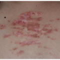
FIGURE 17-4 Patient after two coats of 30% trichloroacetic acid (TCA) with a dense white frost representing keratocoagulation. (Photo permission granted by Joseph Niamtu, DMD.)

Phenol peels are used infrequently because they traditionally require cardiopulmonary monitoring and intravenous hydration because of direct cardiotoxicity from phenol.8 Commercial preparations have experimented with lower concentrations of phenol, for which monitoring and hydration are not necessary. An example is a peel that combines low concentrations of phenol (approximately 2%) and unknown proprietary concentrations of TCA, salicylic acid, retinoic acid, glycolic acid, and vitamin C (Vi Peel, Vitality Institute Medical Products, Culver City, CA) (Fig. 17-5).

While full-face peels are commonly performed, the CROSS technique (chemical reconstruction of skin scars), or dot peeling, using a high-strength TCA (65% to 100%) has been found to be a useful solo or adjunctive treatment for ice pick and small boxcar scars24,31,49,50 (Fig. 17-6). The CROSS technique entails stretching the skin and using a fine wooden toothpick to apply TCA to the bottom of the ice pick or boxcar scar, which leads to destruction of the epithelial tract.23 TCA is applied for a few seconds until the scar displays the characteristic white frosting.14 Neocollagenesis ensues in the subsequent healing phase (2 to 6 weeks), filling in the depressed scar sites. Momentary mild-to-moderate burning pain is typically reported with application, but no local anesthesia or sedation is needed. On average, about 25% improvement of scars is noted with one CROSS session, with increasing efficacy reported up
to 70% or more after three to six treatments at intervals of 2 to 4 weeks.51,52 Patient satisfaction was rated higher with 100% TCA versus a 65% concentration, at 94% satisfaction versus 82%, respectively, in the study by Lee et al.52 However, Fabbrocini et al.49 have shown that a lower TCA concentration (50%) has similar results with fewer adverse reactions. The CROSS technique has also been successful in the treatment of atrophic postvaricella (chickenpox) scarring, with over 80% of patients demonstrating moderate to marked improvement following six treatments with 70% TCA.53


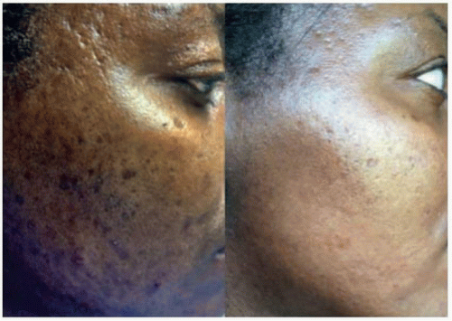
FIGURE 17-5 Acne scarring with PIH treated with a commercially available acid peel (before and after four Vi Peel treatments and a proprietary topical regimen including bleaching cream). (Photo permission granted by Melissa McGuire, Vi Aesthetics.)


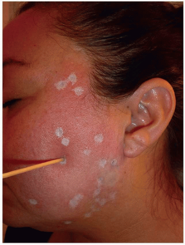
FIGURE 17-6 CROSS technique (chemical reconstruction of skin scars), or dot peeling, using a high-strength TCA (65% to 100%) has been found to be a useful solo or adjunctive treatment for ice pick and small boxcar scars. (Photo permission granted by Joanna G. Bolton, MD.)

The major advantage of the CROSS technique is that adjacent normal tissue and adnexal structures are spared, promoting more rapid healing with a lower complication rate.51,52,54 A number of studies have demonstrated this technique can avoid postpeel scarring and reduce the risk of hyper/hypopigmentation,55,56,57 making it particularly efficacious in darker skin types for which full-face, higher-strength peels are not normally recommended. However, one author (MPG) has found an unacceptable rate of hypopigmented scars in patients with Fitzpatrick skin type III to IV with this technique.


Dermabrasion/Microdermabrasion

Arguably one of the most effective but operator-dependent therapies for acne scarring is dermabrasion.31 Dermabrasion is considered the first major advance in the treatment of acne scars.33 Although it has largely fallen out of favor with the advent of resurfacing lasers, it remains commonly available outside dermatology offices and a basic understanding is appropriate. It is a facial resurfacing procedure that mechanically abrades damaged skin in order to promote reepithelialization and repigmentation by migration of cells to the healing surface from adjacent adnexal structures (hair follicles, sebaceous glands, and sweat ducts). Thus, the neck, chest, and back are not ideally suited for treatment because of the relative paucity of adnexal structures.58 The wound-healing process is accompanied by new collagen formation, remodeling of structural proteins, and a smoothened appearance of scarred skin.59 The technique completely removes the epidermis and penetrates to the level of the papillary or reticular dermis, allowing successful treatment of rolling and shallow boxcar scars. Deep boxcar and ice pick scars are not optimally treated.

Microdermabrasion, a superficial variant of dermabrasion, only removes the outer layer of the epidermis and essentially accelerates the natural process of exfoliation.60 Because of its less aggressive nature, microdermabrasion typically produces better textural improvement of fine wrinkling and PIH, although very superficial acne scars may benefit from deeper settings. There are variable results seen with either form of treatment, and multiple sessions are usually required.

Each procedure employs different instruments with a different technical execution. Dermabrasion is accomplished by use of a high-speed brush, diamond cylinder, fraise, or manual silicone carbide sandpaper.31 All microdermabraders include a pump that generates a stream of aluminum oxide or salt crystals with a hand piece and vacuum to remove the crystals and exfoliate the skin.61 Unlike dermabrasion that requires local and sometimes general anesthesia because of significant pain and bleeding, microdermabrasion can be repeated at short intervals, does not require anesthesia, and is associated with a lower rate and less severe complications.60

Dermabrasion has many potential complications, most of which are operator and technique dependent.8 These include prolonged erythema and healing time, eczema, milia, bacterial or viral infection, hypertrophic or keloidal scarring, unroofing of unapparent wide-based scars, telangiectases, photosensitivity (requiring strict postprocedure sun protection), treatment demarcation lines, and prolonged or permanent hyper-/hypopigmentation.62 The pigmentary concerns are greatest for dark skin types. Postprocedural
hypertrophic scarring has been reported to be a potential risk, first noted in patients undergoing dermabrasion following a recent course of oral isotretinoin therapy. This complication originally prompted the recommendation to wait 6 to 12 months for scar revision following isotretinoin use.33,63 Despite this recommendation, there are reports of patients undergoing dermabrasion with concurrent or recent isotretinoin therapy without hypertrophic scar formation and research is ongoing.64


Needling

Skin needling, also called collagen induction therapy or needle dermabrasion, is a more recently employed technique for acne scars. In its simplest form for small areas, a 26G to 30G needle may be introduced into the skin to a controlled depth of about 2 to 3 mm with repeated stabs.65 For larger areas, needling more commonly involves using a tattoo gun without pigment or a sterile roller composed of hundreds of fine, sharp needles to puncture the skin 1.5 to 2 mm to the level of the mid-dermis13,14,32 (Fig. 17-7). Following facial skin disinfection and a topical anesthetic in place for 60 to 90 minutes, the procedure is achieved by rolling the tool until bleeding and microbruising occurs, which initiates the complex cascade of growth factors that finally results in collagen production.23,54 The needling device is applied to the acne-scarred areas four to six times during a treatment session and should be rolled in four directions: horizontally, vertically, and diagonally left and right.40 Results are generally appreciated after 6 weeks, but the full effect can take 3 months or more. Skin texture will continue to improve over a 12-month period, typical of skin remodeling. For best results, most patients require three to four treatments approximately 4 weeks apart.14 The number of treatments required depends on the individual collagen response and on the desired results. Histology shows thickening of skin and a dramatic increase in new collagen and elastin fibers.


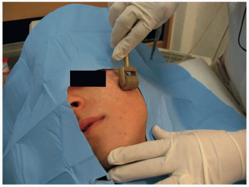
FIGURE 17-7 Needling procedure utilizing a sterile roller comprising hundreds of fine, sharp needles to puncture the skin 1.5 to 2 mm (level of mid-dermis). (Copyright © 2010 Gabriella Fabbrocini et al.)

Similar to dermabrasion, rolling and shallow boxcar acne scars are most optimally treated with needling. However, compared with dermabrasion and other resurfacing procedures such as chemical peels and laser, this technique has many advantages. Skin needling can be safely performed on all skin types with less risk of PIH, the procedure does not result in treatment demarcation lines, the recovery period is 2 to 3 days shorter than other resurfacing procedures, and needling is much less expensive for the individual patient and to incorporate into a dermatology practice.23,66 Significant contraindications are anticoagulant therapy, bleeding disorder, active skin infection, history of injectable filler in the previous 6 months, and personal or family history of hypertrophic and keloidal scars.40


Subcision/Microsubcision

In the subcutaneous incisionless (subcision) technique, a specialized needle, typically a closed lumen 18G, 11/2-inch with triangular cutting tip (Nokor Admix needle, Becton Dickinson & Co, Franklin Lakes, NJ), is inserted percutaneously and passed in multiple directions to release fibrotic bands in the dermis and subcutaneous tissue, similar to a “mini-scalpel”4 (Fig. 17-8A and B). First described by Orentreich in 1995,67 it is most useful for tethered rolling scars that have normal quality skin at the base of each scar. However, it can be helpful for any depressed scars on the face. This approach results in the scar being “released” and allows organization of blood and neocollagenesis to take place beneath the scar, helping to lift and smooth the contour.67 It has become a first-line treatment for many isolated, moderately bound-down, atrophic scars.68 Both Goodman69 and Jacob et al.22 provide excellent reviews of how to perform standard subcision following marking and administration of local anesthetic.

Microsubcision (MSUBx, Suneva Medical, Inc., San Diego, CA) is a newer technique that utilizes a standard hypodermic needle instead of a true subcision needle. Essentially, the needle is “passed back and forth” underneath a depressed scar to create a potential space, or pocket, which is then filled with a fibrin clot, or more commonly an injectable dermal filler. Both subcision and microsubcision can effectively be combined with dermal fillers, which occupy the space rather than relying solely on the created blood clot8 (see section on tissue augmenting agents).

Advantages to subcision include being easy to perform, being inexpensive, having modest downtime, being safe for various skin types, having low rate of complications, and having remarkable and persistent improvement.23 Complications associated with the procedure include pain, bleeding, bruising, infection, transient discoloration, possible acne exacerbation (requiring intralesional corticosteroid injection), additional, worsened, or hypertrophic scarring, and recurrence/persistence of the treated scar. It may be necessary to perform variable depths of sweeping, fanning, or lancing with the needle to disrupt the fibrous bands, and multiple treatment sessions or attempts may be required.31 As such, the procedure must be performed judiciously to avoid damage to adjacent structures such as nerves and large vessels. Attaching a 3-mL syringe to the needle can allow for easier needle handling and better leverage.


FIGURE 17-8 A: Schematic depicting subcision. A regular lumen, 18G, 11/2-inch hypodermic needle or a triangular, solid-tipped Nokor Admix needle (inset photo; Becton Dickinson and Co, Franklin Lakes, NJ) may be used to undermine and separate fibrous bands (white bands). Nokor needle photo is for illustration only – the level of treatment should remain above the SMAS. (Adapted by Dr. Joanna G. Bolton from Jacob CI, Dover JS, Kaminer MS. Acne scarring: a classification system and review of treatment options. J Am Acad Dermatol. 2001;45:109-117.) B: Subcision technique with Nokor needle inserted intradermally and being used to undermine a bound-down scar. Scars identified for treatment are marked preprocedure with blue ink. Piston-like motion used to release the fibrous bands; the skin is elevated to improve traction, facilitate needle motion, and avoid underlying structures. The needle may be placed on a 3-mL syringe for easier gripping and better leverage. (Photo permission granted by Joanna G. Bolton, MD.)

An interesting split-face study contrasted subcision on one side and a combination of subcision and nonablative laser on the other.70 The results of the combination treatment suggested a synergistic effect between these modalities. Another study compared the effect of the 100% TCA CROSS method against subcision in treating rolling acne scars.71 Twenty patients of skin types III and IV with bilateral rolling acnes scars received one to three sessions of the 100% TCA CROSS technique on the left side of the face and subcision for scars on the right side. The mean decrease in size and depth of scars was significantly greater for the subcision side compared with the 100% TCA CROSS. In addition, more side effects in the form of pigmentary alternation (25%) were observed with the CROSS method.


Punch Excision Techniques

Punch excision techniques are minimally invasive surgical treatments mainly indicated for ice pick or small boxcar scars. According to diameter, depth, and shape of the scar, a biopsy punch of appropriate size is used to excise the scar potentially followed by closure, elevation, or grafting.40

With punch excision and closure, the scar is excised and sutured (6-0 or smaller suture) after undermining, in a direction parallel to the relaxed skin tension lines. The goal is to trade a larger, deeper scar for a smaller, linear closure that will hopefully be less noticeable. If a depressed scar has a normal surface texture, punch incision to the subcutaneous tissue followed by elevation of the base and suturing to the level of surrounding skin may improve scar appearance. Retraction of the tissue occurs during the healing phase, resulting in a leveled surface.22 Finally, in punch excision with grafting, a scar is excised and replaced with either an autologous split-thickness or full-thickness punch graft or prepackaged dermal graft material. The pre- or postauricular region or the gluteal fold are the most used donor sites for autologous grafts.40 This is probably best for sharp-walled or deep ice pick scars, but is painstaking as often 20 or more replacement grafts are required in a single session.23 Laser skin resurfacing with the concurrent use of punch excision techniques further improves facial acne scarring.22,72


Laser Treatment

Acne scar treatment with lasers varies depending on the type of scar. Ice pick scars often do not respond well to laser treatments, and in the view of the authors are better
treated with the CROSS technique, punch techniques, and radiofrequency treatments. Shallow boxcar scars have been successfully treated with ablative and nonablative fractionated lasers in addition to subcision, dermal fillers, skin needling, radiofrequency, and/or other surgical corrections such as excision and closure, rhytidectomy, and punch-grafting techniques. Deeper boxcar scars are better treated with punch-grafting or excisional techniques and/or radiofrequency devices. Rolling acne scars have been treated with ablative and nonablative fractionated lasers in addition to dermal fillers, dermabrasion, subcision, skin needling, and/or radiofrequency devices. Hypertrophic and keloidal scars are better treated with the 585-to-595-nm PDL, 515-to-1,100-nm IPL, and ablative fractionated lasers with or without the addition of intralesional corticosteroids and/or 5% 5-fluorouracil because of the potential for a worsening of these lesions with other modalities.73 Table 17-8 summarizes various laser treatment modalities (see Chapter 13).


Table 17-8 Laser Treatment of Acne Scars


Laser Treatment


Wavelength (nm)


Chromophore Target


Ablative (A) vs. Nonablative (NA)


Fractionated (F) vs. Nonfractionated (NF)


PDL


585 or 595


Blood


NA


NF


Picosecond alexandrite


755


Melanin


NA


NF


Nd:YAG


1,064


Water


NA


NF


Erbium: glass


1,500


Water


NA


F


Erbium: glass


1,540


Water


NA


F


Erbium:YAG


2,940


Water


A


NF


Erbium:YAG


2,940


Water


A


F


CO2


10,600


Water


A


F


From Sardana174, Ong and Bashir88, Verhaeghe et al.86, Leheta et al.87, Brauer et al.82, Koike et al.80, Choi et al.95, Mahmoud et al.85, Woo et al.89

Only gold members can continue reading. Log In or Register to continue

Stay updated, free articles. Join our Telegram channel

Oct 16, 2018 | Posted by in Dermatology | Comments Off on Atrophic Scar Management

Full access? Get Clinical Tree

Get Clinical Tree app for offline access