A Pediatric Perspective


A Pediatric Perspective


Andrew C. Krakowski

Tuyet A. Nguyen


How Does Pediatric Skin Differ from Adult Skin?

The basic functions of the skin include serving as a barrier to the outside world (reduce fluid loss, regulate temperature, and provide protection from infection and harmful environmental agents) and as a conduit for the perception of sensations such as pain, vibration, and pressure.1,2 Although the role of skin remains the same throughout life, pediatric skin is in some ways uniquely different from that of adult skin.


Structure

The skin of infants and children differs in several ways from that of adults in terms of structure. It has been shown that the skin of infants and children may be up to 60% thinner than that of an adult.3 An in vivo analysis demonstrated that the thickness of the stratum corneum and suprapapillary epidermis in infants is approximately 30% and 20% less, respectively, compared to the skin of their biologic mothers.2 Additionally, infant corneocytes and keratinocytes in the granular layer are significantly smaller than those found in adult skin, possibly representing an increased turnover rate.2

In the dermal layer, collagen bundles in the upper reticular dermis are not as prominent in infant skin, giving the appearance of a gradual transition between papillary and reticular dermis on histology.4 The composition of the subcutaneous fat in infants and young children also differs from that of adults. Newborn subcutaneous fat has been found to contain a higher ratio of saturated to unsaturated fats compared to that of adults.5 This higher proportion of saturated fats makes infants more prone to cold temperatures and panniculitis because of the higher melting point associated with saturated fats. The structure of infant skin appears to continually evolve, approaching that of adult skin around 1 year of life.6


Body Surface Area

Body surface area (BSA) is a measure of the surface area of the body. In infants and children, the BSA-to-volume ratio of an infant or child can reach up to five times that of an adult.7 The difference in this ratio makes infants and children more susceptible to increased percutaneous absorption of topically applied agents, transepidermal water loss (TEWL), and skin damage leading to increased risk of toxicity and dehydration.


Decreased Barrier Function

The barrier function of the skin is dependent on the formation of the stratum corneum. In infants, decreased barrier function compared to that of adult skin has been reported. As the stratum corneum continues to develop
during the first year of life, changes in barrier function also continue to develop during this time.6 For example, TEWL is often used as a marker of skin barrier function. In infants, the stratum corneum contains higher water content but is also prone to increased TEWL compared to adults.2,6 Premature infants have an even higher rate of TEWL compared to full-term infants and adults.8 This predisposition puts infants at greater risk for dehydration and electrolyte imbalance than adults.9


Table 22-1 Some Percutaneous Toxicity Risks in the Pediatric Population


Agent


Toxicity


Alcohols (topical antiseptic)


Hemorrhagic necrosis


Hexachlorophene (topical antiseptic)


Neurotoxicity


Povidone-iodine (topical antiseptic)


Hypothyroxinemia, goiter


Lidocaine-prilocaine cream (EMLA) (topical anesthetic)


Methemoglobinemia


A more exhaustive list can be found in the article by Mancini JA. Skin. Pediatrics. 2004;113(suppl 3). http://pediatrics.aappublications.org/content/113/Supplement_3/1114.figures-only. Accessed November 17, 2016.


In addition, the skin provides mechanical barrier protection by preventing the entry of foreign agents into the body. In premature infants, however, there is limited dermal to epidermal surface attachment. The fragility of premature and newborn skin leaves them more vulnerable to potential percutaneous toxicity and transepidermal infections (Table 22-1).9


Causes of Scars in Children

The etiology of scars in children resembles that of the adult population and can be grouped into three basic categories: trauma, burns, and medical conditions. As the medical community improves its ability to keep children alive after serious injuries, the patients are left to deal with the physical, psychosocial, and financial consequences (i.e., the so-called “survivor’s paradox”), leading to significant distress for affected children, their caregivers, and providers alike.


Trauma

Unintentional injuries and trauma are one of the leading causes of morbidity in pediatric patients, and are a significant source of scarring in this population.10 An average of 9.2 million unintentional injuries, such as car or bike accidents, occur in pediatric patients annually.11 Mangle or friction injuries from machines such as treadmills can cause significant scarring and contractures in children.12,13 Animal bites, such as dog bites, can also be a common source of traumatic injury, physical disability, and scarring in children (Fig. 22-1).14,15,16


Burns

Burns are another frequent source of scarring in children (Fig. 22-2). Burn injuries can arise from multiple sources in pediatric patients such as campfires, hot ash, or coals.17,18 Electrical burns are particularly common in very young children, especially on the lips and mouth as infants and young children are more prone to oral exploration; they should also raise suspicion for neglect or nonaccidental injury.19 However, the most frequent and preventable cause of burns in pediatric patients is scald injuries from hot liquids (Fig. 22-3).20


FIGURE 22-1 Atrophic scar following flap reconstruction. This patient suffered tissue injury and loss secondary to a motor scooter accident. Flap reconstruction successfully filled the tissue defect, but was associated with atrophy and contour irregularity at the distal aspect of the flap. (Courtesy of Andrew C. Krakowski, MD.)


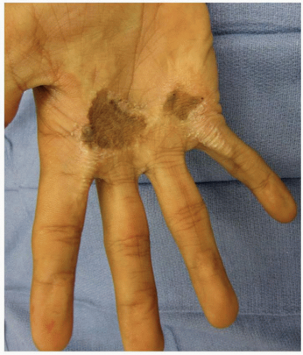
FIGURE 22-2 Hot iron burn. This adolescent male severely burned his hand at 1½ years of age. Split-thickness skin grafts were placed on his palm; predictably, a degree of contraction postreconstruction limited his overall function. The patient’s main stated concern was that he wanted “to be able to open a jar of peanut butter” without his mother knowing. (Courtesy of Andrew C. Krakowski, MD.)


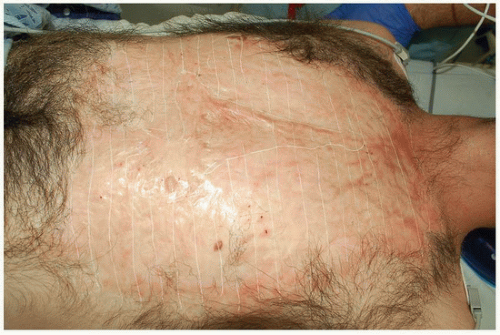
FIGURE 22-3 Large scald burn. This male in his 20s presented with a large burn scar secondary to a scald injury. Note the extensive area of alopecia. Objective evaluation of the scarred areas was performed using high-definition ultrasound, revealing that the majority of the “scar sheet” was approximately 1.5 mm in thickness, with focal areas up to 8 mm. A “grid” pattern was then created over the entire scar sheet using a white surgical marker to help delineate the treatment plan for ablative fractional CO2 laser resurfacing. (Courtesy of Andrew C. Krakowski, MD.)


Medical Conditions

Numerous medical conditions or infections may be associated with scarring (see Chapters 3 and 17).21,22,23,24 Acne, for example, is a frequent source of atrophic facial scars in the adolescent population; however, a larger differential diagnosis for “atrophic scars of cheeks” exists (Table 22-2; Fig. 22-4).21,22 The treatment of childhood cancers may also be associated with scarring alopecia from radiotherapy, for example, or scarring from the placement of chest tubes, catheter lines, and medical ports.25 Iatrogenic surgical scars obtained secondarily to the treatment of underlying medical conditions may also be a frequent source of morbidity for children (Fig. 22-5).


Multimodal, Multidisciplinary Approach

The evaluation and management of scars, like many other areas of medicine, continues to evolve. No longer is a single provider typically able to perform all facets of care required for the successful management of a patient with a complicated medical condition. Within the world of pediatric medicine, the “multidisciplinary model” has shown great promise in treating, for example, cleft lip/cleft palate and vascular malformations. The time has come for us to adopt a similar model for the care of patients with complicated scars—one that allows the complete coordination of services from specialties such as primary care (the “quarterback”), dermatology, plastic/reconstructive surgery, anesthesia, radiology, infectious disease, orthopedics, physical/occupational therapy, psychology, nutrition, and a host of ancillary support services (Figs. 22-6 and 22-7). Crucially, the patients and their caregivers must be
involved in the management plan from the start so that realistic expectations may be established. Payers must also be involved from the beginning, understanding that the right people performing the right treatments will yield the best, most cost-effective results.


Table 22-2 Differential Diagnosis for Atrophic Scarring


Acne vulgaris


Striae distensae


Discoid lupus


Varicella


Molluscum contagiosum


Malignant atrophic papulosis (Degos disease)


Infections (especially Staphylococcus)


Surgery


Trauma


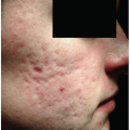
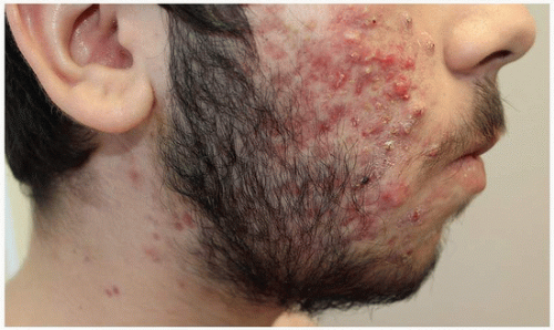
FIGURE 22-4 Severe inflammatory acne frequently leads to atrophic scarring. The notion that an inflammatory skin condition such as acne vulgaris, with the potential to inflict permanent physical disfigurement and psychosocial distress, is “just cosmetic” is antiquated and ignorant. A primary clinical goal of acne vulgaris management should be scar prevention; too many adolescents have been left branded with permanent reminders of missed treatment opportunities. We must evolve our thinking in order to help prevent scars before they form. Ultimately, this is the most efficacious and cost-effective scar intervention we have available today, and insurance reimbursement should reflect this reality. (Courtesy of Andrew C. Krakowski, MD.)


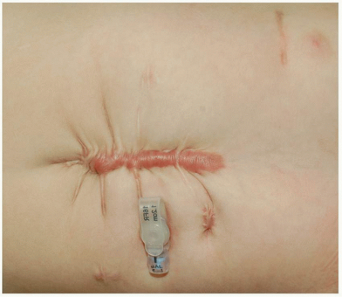
FIGURE 22-5 Iatrogenic hypertrophic scar with scar contractures. This infant with Rubinstein-Taybi syndrome developed a hypertrophic scar after abdominal surgery. The scar itself was pruritic and erythematous. The tension on the wound resulted in multiple “radiating” scar contractures. (Courtesy of Andrew C. Krakowski, MD.)


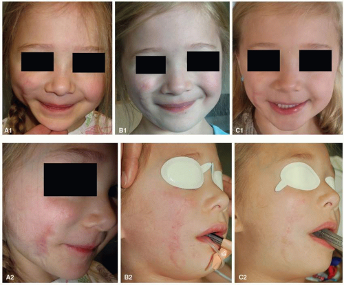
FIGURE 22-6 Multimodal, multidisciplinary treatment of a dog bite scar on the face. A 3-year-old girl presented with a “mixed” (i.e., atrophic and hypertrophic) scar resulting from a dog bite to the right cheek inflicted 21 months earlier. Marked erythema, irregular texture, volume loss, and scar contractures are noted (A1, A2). A total of nine multimodal revision procedures were performed (under general anesthesia because of her age and the extent and location of her injuries). Erythematous portions of the scar were first treated with a 595-nm pulsed dye laser (Vbeam Perfecta, Candela Corporation, Wayland, MA, USA) using a 7-mm spot size, fluence of 8 J per cm2, and 1.5-ms pulse width (clinical endpoint of minimal purpura). The entire scar sheet was then treated using an ablative microfractionated 10,600-nm CO2 laser (UltraPulse, Deep FX; Lumenis, Ltd., Yokneam, Israel) at pulse energy of 15 mJ and density of 15% in a single pass. At the time of her first laser scar revision, the patient’s plastic surgery team performed autologous fat grafting (5 mL) under the depressed scar to help restore volume to her cheek. Serial pulsed dye laser treatments were repeated with fluence settings ranging from 8 to 11 J per cm2 to reach a clinical endpoint of minimal purpura. Additional ablative fractional laser resurfacing treatments were performed with pulse energies ranging from 15 to 25 mJ and corresponding treatment densities of 10% to 5%, respectively. During four early treatment sessions, the patient received triamcinolone acetonide suspension (40 mg per mL) applied topically to the areas of greatest scar hypertrophy immediately after fractional laser treatment. B1 and B2: interim results approximately 10 months after her initial treatment session. C1 and C2: the same patient approximately 19 months after her initial treatment. Marked improvements in scar texture, color, and facial symmetry are noted. Additional enhancements could likely be obtained with repeated interventions such as ablative and nonablative fractional resurfacing and fat grafting. (Admani S, Gertner JW, Gosman A et al. Multidisciplinary, multimodal approach for a child with a traumatic facial scar. Semin Cutan Med Surg. 2015;34:24-27.)

Prevention and mitigation of scar formation are the extremely important “first steps” in the management of scars. Scars located at the shoulder, neck, presternum, and ankle are more prone to hypertrophic scars because of the high tension in these anatomical locations, and certain patients tend to be more prone to pathological scar formation or postinflammatory pigment alteration.26 Therefore, it is imperative for health care providers to identify wounds that may be susceptible to becoming a pathological scar and take action. Selective avoidance of elective surgeries in at-risk individuals, wound closures that minimize tension, the use of proper wound dressings and wound care, optimized nutrition, and the prevention of postoperative infections can help optimize wound healing and reduce scar formation (see Chapters 8 and 9).27


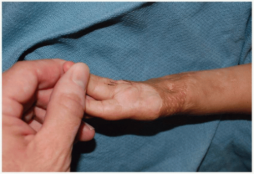
FIGURE 22-7 Contracted split-thickness skin graft and amputation. This 3-year-old boy survived meningococcemia but his body was left ravaged by associated necrosis. He lost several fingers and numerous split-thickness skin grafts were necessary during reconstruction. Unfortunately, many of the skin grafts contracted and caused additional functional deficits. Such a complicated presentation highlights the need for a truly specialized, multidisciplinary scar team that might include the patient’s primary pediatrician (the “quarterback”); infectious disease, orthopedics, plastic surgery, dermatology, nutrition, behavior therapy, psychology specialists, and a patient advocate. (Courtesy of Andrew C. Krakowski, MD.)

A variety of reasonable options exist for the treatment of scars including, but not limited to, intralesional corticosteroids, silicone gel sheets, massage/pressure therapy, cryotherapy, surgical excision, and laser treatment (see Chapters 10 and 13). However, treatment is guided by the characteristics of the specific scar at a particular time point. In complex scars with an array of symptoms (e.g., anxiety, pruritus, pain, dysesthesia, etc.) and signs (e.g., hypopigmentation, erythema, functional deficit, etc.), a multimodal approach is often necessary to achieve optimal results.16


Why Treat?


Associated Comorbidities

The majority of scars are of little consequence for affected patients. However, findings such as erythema, dyspigmentation, pruritus, pain, hyper- or hypohidrosis, hyper- or hypotrichosis, and dysesthesias are not uncommon (Table 22-3).


Color Change

Color changes in scars can be related to pigmentary alteration or underlying vascularity (Fig. 22-8). The initial trauma and the subsequent process of wound healing and scar formation can lead to discrepancies in both the number of melanocytes and melanin density compared to normal skin.28 This may manifest as hypopigmentation, hyperpigmentation, or commonly both. Erythema is also a common finding, especially in young scars. Although it typically fades with time, erythema may sometimes persist for years and can be an indicator of pathological scar formation.28


Table 22-3 Physical Scar Signs and Symptoms to Consider


  • Conspicuous or subtle?


  • Disruption of local cosmetic unit?


  • Erythema/hypervascularity?


  • Skin contracture?


  • Range of motion/functional deficit?


  • Dyspigmentation (hyperpigmentation/hypopigmentation/mottling)?


  • Pruritus?


  • Pain/tenderness?


  • Dysesthesia?


  • Hypertrichosis, hypotrichosis, or alopecia?


  • Hyperhidrosis, hypohidrosis, or anhidrosis?


  • Presence or history of infection (folliculitis, cellulitis, abscess, fasciitis, etc.) within scar area?


  • Presence or history of chronic wound/chronic ulceration within scar area?


  • Presence or history of lymphedema locally or regionally (suggestive of outflow obstruction)?


  • Presence or history of skin cancer (i.e., Marjolin’s ulcer) within scar area?


From Krakowski AC, Totri CR, Donelan MB, et al. State of the art: scar management in the pediatric and adolescent populations. Pediatrics. 2016;137(2):2014-2065, with permission.


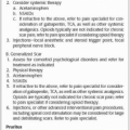
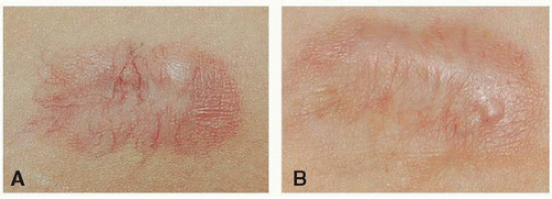
FIGURE 22-8 Tracheostomy scar with prominent telangiectasia. A: This adolescent female’s tracheostomy scar was particularly conspicuous secondary to prominent telangiectases. B: A single treatment with a vascular-specific laser successfully reduced erythema and improved cosmesis, and had the overall effect of reducing the patient’s anxiety levels and enhancing her ability for social interaction. (Courtesy of Andrew C. Krakowski, MD.)


Pruritus

Pruritus is an extremely common symptom (especially in association with hypertrophic and keloid scars), present in up to 87% of those affected.29,30 In one study, more than 86% of patients with pruritus associated with burn scars reported the symptom as unbearable, and up to 100% of patients reported it as bothersome.30 The cause of pruritus in scar tissue is unclear; however, localized inflammation, stimulation of small nerve fibers around the scar, and increased levels of β-endorphin are thought to contribute (see Chapter 11).29,31


Pain

Pain may also be a presenting symptom in existing scars. Several reports of significant and chronic pain, especially after burns, have been reported in the literature.32,33 Even older “mature” scars can cause pain in affected patients. An editor of this textbook (ACK) has himself survived a fall through a plate glass door at the age of 7; some 36 years
later, the sensation of being “stuck with an ice pick” within the deep portions of a resulting hypertrophic scar on his arm wakes him from sleep on a near-monthly basis.


Dysesthesias

Changes in sensation such as anesthesia, paresthesia, perceptions of burning or moisture, and an altered sense of touch are common findings in scar tissue. Tissue damage and the formation of scars in an area can potentially damage peripheral nerve fibers or cause nerve entrapment from aberrant collagen deposition.29


Hyperhidrosis/Hypohidrosis

Hyperhidrosis and hypohidrosis are fairly common findings associated with scars, as the process of wound healing can lead to abnormal tissue composition within a scar. Aberrant formation of the secretory portions of sweat glands and irregular organization of the glands have been found in scar tissue, which can lead to poor temperature regulation and hyperhidrosis or hypohidrosis. This can be particularly problematic in symptomatic patients.34,35,36 Hyperhidrosis may directly interfere with proper fitting of prosthetics, the use of scar camouflage, and the application of silicone gel sheeting.


Hypertrichosis/Hypotrichosis/Alopecia

The anatomical composition of scar tissue differs from that of normal skin (see Chapter 5). The process of scar formation can frequently cause loss or disruption of hair follicles in affected areas, leading to hypotrichosis and alopecia (Fig. 22-3). Interestingly, hypertrichosis has also been reported. It has been hypothesized that this increase in hair growth is a product of increased growth factors and vascularization in healing scar tissue.37


Functional Compromise

Even “normal” scar formation in a particular anatomic location may lead to significant functional impairments for affected patients. Restrictive contractures and adhesions, for example, can be extremely debilitating consequences of scar formation and maturation (Fig. 22-9). These contractures occur through tightening of affected skin overlying a joint or through adhesions with underlying tissues resulting in limited range of motion.38 Decreased ability to perform normal activities of daily living due to scar contractures can lead to significant and long-term negative consequences on a patient’s overall quality of life (see Chapter 19).

Function-limiting scars can be particularly debilitating in pediatric patients where the “use it or lose it” phenomenon is a real concern because of the overlap of the patients’ normal growth and development with the evolution of the scar contracture. Function-compromising scars may directly impact important developmental milestones such as development of right- or left-handedness and the formation of the concepts of self and self-worth. Therefore, it is especially important in the pediatric population to evaluate for any underlying functional compromise associated with scars, as failure to recognize these complications may lead to impairment of normal development.39 Returning affected patients as close to baseline function as possible should be the guiding principle of treatment.


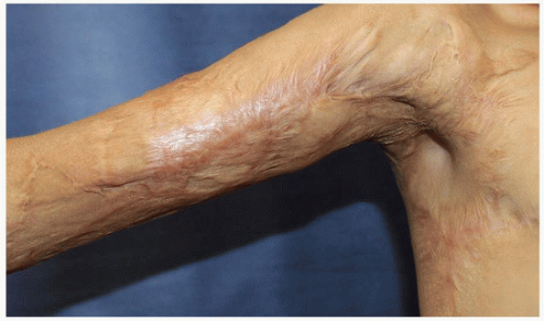
FIGURE 22-9 Scar contracture with functional deficit. This teenage male suffered a gasoline burn injury that resulted in a large hypertrophic scar contracture with the resulting inability to fully abduct his right arm. (Courtesy of Andrew C. Krakowski, MD.)


Psychosocial Aspects

Scars may not only cause functional problems for a patient; they can also have significant deleterious psychosocial consequences, especially for pediatric patients (see Chapter 24). The aesthetic complications of scar formation can lead to depression, posttraumatic stress disorder, anxiety, social withdrawal, and isolation.32,40,41 These psychosocial consequences may even be more incapacitating than the physical sequelae and, in pediatric patients, may negatively influence self-confidence, social interactions, and success in the future.42 Despite these associations, the psychosocial consequences of scars are frequently forgotten, unrecognized, and underreported (Table 22-4).

It may be natural to assume that scar severity would correlate with psychosocial consequences for affected patients. However, the degree of distress a patient experiences does not necessarily correlate with scar origin or size.43 Rather, more consistent predictors of psychosocial distress appear to be scar location and visibility, the patient’s subjective opinion of the scar, the reaction of others to the scar, and the patient’s individual personality traits.42,44,45 Patients with visible scars often also experience significant stigmatization and discrimination from others, leading to low body image, social anxiety, and depression.32 In children and adolescents, this stigmatization can lead to long-term consequences such as difficulty making friends and forming intimate relationships.32 Therefore, it is pertinent for
treating physicians to assess for psychosocial comorbidities when evaluating a patient with scars and, when present, refer for early intervention.


Table 22-4 Psychosocial Scar Comorbidities to Consider


  • Anxiety/stress?


  • Depression?


  • Posttraumatic stress disorder?


  • School, work, or social performance affected?


  • Overall perceived reaction of others to scar?


From Krakowski AC, Totri CR, Donelan MD, et al. State of the art: scar management in the pediatric and adolescent populations. Pediatrics. 2016;137(2):2014-2065, with permission.


Financial Considerations

In addition to the physical and psychosocial sequelae of scars, treatment can lead to a significant financial burden for patients and health care providers. Successful scar management, especially complex scars after extensive injury, begins with professional wound care and often requires multimodal, multisession therapy during rehabilitation. The cumulative costs of initial stabilization and wound care, topical and intralesional agents, pressure garments, conventional surgery, and other adjunctive treatments can be immense.46,47,48 A study investigating the health care cost of treating chronic wounds in the United States estimated that the total annual expenditure for burn scar management is approximately $12 billion.49 These exorbitant costs can cause a significant financial burden for affected patients and their families. Therefore, it is important to consider the financial cost when deciding on treatment plans for patients. It is also imperative that insurance companies recognize the profound importance of scar management, and partner with patients during the treatment course. Objective evidence now exists to support the notion that these treatments can improve and even fully restore functional compromise secondary to scar contractures (Fig. 22-10). As a medical community we should drive these patients to capable experts who can optimize clinical outcomes and minimize unnecessary expense.


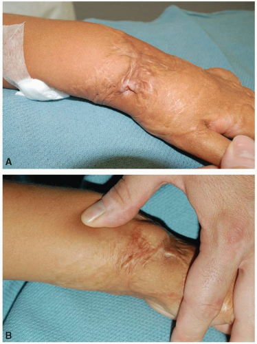
FIGURE 22-10 Chronic wound associated with a burn scar. A: As an infant, this 8-year-old girl had climbed into a bathtub full of hot water and liquid bleach, suffering burns to all four extremities with subsequent associated scar contractures. In one “hot spot” she developed a chronic ulcerated wound that had been present for 8 months despite ongoing traditional wound care. B: One treatment with an ablative, microfractionated 10,600-nm CO2 laser (UltraPulse Deep FX, Lumenis, Ltd., Yokneam, Israel) over the wound and associated contracture bands at pulse energy of 50 mJ and treatment density of 5% helped to stimulate remodeling and relieve the tension on the skin adjacent to the wound, facilitating rapid healing. (Adapted from Krakowski AC, Diaz L, Admani S, et al. Healing of chronic wounds with adjunctive ablative fractional laser resurfacing in two pediatric patients. Lasers Surg Med. 2016;48:166-169.)


Timing of Treatment

For decades (even centuries), the prevailing notion has been that scars should generally not be manipulated procedurally for at least 1 year after creation (i.e., until the scar has had time to “mature”). Because of recent advances in technology and minimally invasive techniques such as fractional laser resurfacing, this notion is no longer universal and requires updating to account for the potential benefits of early intervention to minimize the impact of scars as they form. The use of massage therapy and pressure garments has become fairly routine early in the course after burns to mediate scar tissue formation,50,51,52,53,54,55 and these newer techniques could be considered in a similar (and likely more effective) vein. Additionally, some recent studies suggest early intervention may be more favorable than delayed treatment. For example, in the management of keloids, intraoperative intralesional corticosteroids may be employed to help prevent future recurrence of the lesions.56 Newer therapies such as ablative fractional laser resurfacing (AFR) have also been effective early interventions in mediating scar tissue formation postoperatively.50,57

Only gold members can continue reading. Log In or Register to continue

Stay updated, free articles. Join our Telegram channel

Oct 16, 2018 | Posted by in Dermatology | Comments Off on A Pediatric Perspective

Full access? Get Clinical Tree

Get Clinical Tree app for offline access