Abstract
Vertical scar mastopexy is a reliable short-scar breast lift technique, similar to vertical scar breast reduction. However, instead of resecting the excess parenchyma of the inferior pole, we use this tissue to create a centrally based autoaugmentation flap that can increase superior pole volume and projection without the need for a breast implant. This chapter will review our surgical technique for performing vertical scar mastopexy with autoaugmentation flap in a safe manner with reproducible outcomes.
66 Vertical Scar Mastopexy with Autoaugmentation Flap
Key Points
The technique is similar to vertical scar breast reduction; instead of resecting the tissue in the inferior pole it is used as a centrally based autoaugmentation flap to increase superior pole volume and projection.
It is ideal for patients with grade 2–3 ptosis desiring an improved aesthetic appearance of the breast, but who do not want to increase breast size and/or decline a breast implant.
66.1 Preoperative Steps
Vertical scar mastopexy is a versatile procedure that can be adapted to the majority of patients seeking correction of breast ptosis.
Patients with significant deflation of the breast and insufficient tissue of the inferior pole are not suitable candidates for this technique.
We do not perform this procedure on patients with a body mass index (BMI) ≥35.0 kg/m2 or on active smokers due to a significantly higher risk of complications.
Preoperative mammography is not required prior to vertical scar mastopexy; national or regional screening guidelines for mammography should be followed.
Note: This technique should not be performed in patients with breast implants as the blood supply to the autoaugmentation flap is disrupted during previous placement of the breast implant.
66.1.1 Surgical Markings (Fig. 66.1)
Mark the midline of the chest and the inframammary fold (IMF).
Central axis of the breast is marked by drawing a straight line from the midpoint of the clavicle (7–8 cm lateral to midline) through the nipple-areolar complex (NAC).
Level of the IMF is transposed anteriorly onto the breast and marked; this represents the new location of the superior border of the NAC (point A).
Height of the point A marking is transposed to the contralateral breast to avoid asymmetry of the NAC as a result of IMF asymmetry.
In comparison to breast reduction where there is unweighting of the breast and the NAC is typically located higher than was marked preoperatively, mastopexy does not involve unweighting of the breast so the NAC typically remains where it is marked.
Mark a point 5 to 10 cm superior to point A as a reference for autoaugmentation flap inset.
Inferior extent of the planned skin excision is marked 2 to 4 cm above the IMF (point B).
It prevents migration of the vertical scar onto the chest.
Site for new superior border of the NAC is drawn as a mosque dome starting at point A and extending to points C and D.
It is drawn such that when points C and D are brought together, the mosque dome forms a circle.
Vertical limbs are drawn as curved lines extending from point B to points C and D.
Medial and lateral displacement of the breast can assist in approximating these lines.
Compared to breast reduction where more breast tissue is removed between the pillars necessitating more skin excision, during mastopexy typically 0 to 100 g of breast tissue is removed from the autoaugmentation flap so less skin can be excised between the pillars.
Planned resection can be tested by coning the breast to approximate the planned vertical incisions; it should come together with minimal tension.
Inferior extent of the vertical scar should have a “V” shape to minimize dog-ear formation.
Blocking triangles at points C and D prevent a “teardrop deformity” of the NAC.
Note: The tissue of the inferior pole within the marked vertical limbs will become a centrally based autoaugmentation flap.
Fig. 66.1 Illustration of the skin markings for vertical scar mastopexy with autoaugmentation flap. Point A represents the new location of the superior border of the nipple-areolar complex (NAC). Point B is the inferior extent of the vertical scar, located 2 to 4 cm above the inframammary fold (IMF). Points C and D , the blocking triangles, should create a circle of the mosque-dome pattern when brought together. The tissue of the inferior pole within the marked vertical limbs will become a centrally based autoaugmentation flap.
66.2 Operative Steps
See Video 66.1.
66.2.1 Infiltration
Stab incision made at the inferior extent of the planned vertical scar; tumescent solution (1 L of Ringer’s Lactate + 1 mL of epinephrine 1:1,000) infiltrated under the planned vertical incisions and throughout the superficial breast parenchyma, as well as the lateral chest and axillary roll if liposuction of these areas is indicated.
Avoid infiltration of the inferior pole where the autoaugmentation flap will be created; tumescent fluid distorts the tissues making dissection more difficult and may hydro-dissect the autoaugmentation flap off the pectoralis major muscle (PMM) fascia.
Infiltration performed using the simultaneous separation tumescence (SST) technique with a 4 mm exploded-tip cannula and power-assisted liposuction (PAL).
Avoid use of SST in the central and deep breast parenchyma as this can disrupt the autoaugmentation flap.
Each breast is typically infiltrated with approximately 100 to 200 mL of tumescent solution; more may be infiltrated to the lateral chest and axillary roll if liposuction is required.
66.2.2 Pedicle Selection
Breast tourniquet applied to maintain tension on the skin.
New NAC outlined using a circular metal washer (approximately 44 mm diameter) centered over the nipple.
If any part of the new areola lies superior to an imaginary line drawn between the blocking triangles (points C and D), a superior pedicle is used. If all of the new areola lies inferior to this line, a superomedial pedicle is used (Fig. 66.2a, b).
If the NAC is positioned medially, a superolateral pedicle may be required to allow rotation and insetting.
The selected pedicle is drawn with a 2.5 cm border around the edge of the new areola.
For a superomedial pedicle, a 1:2 width-to-length ratio of the pedicle should be maintained to preserve blood supply to the new NAC.
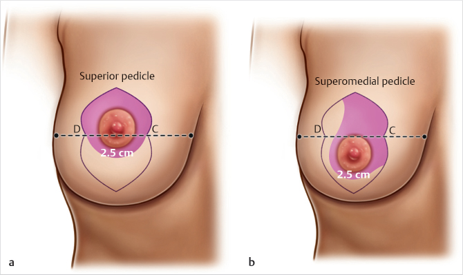
Fig. 66.2 Illustration of dermoglandular pedicle selection. If any part of the new areola lies superior to an imaginary line drawn between the blocking triangles (points C and D), a superior pedicle is used (a). If all of the new areola lies inferior to this line, a superomedial pedicle is used (b). The selected pedicle is drawn with a 2.5 cm border around the edge of the new areola.
Related posts:
Stay updated, free articles. Join our Telegram channel

Full access? Get Clinical Tree


