Abstract
Subfascial breast augmentation is a versatile augmentation mammaplasty technique, wherein a breast implant is placed under the pectoralis major muscle fascia to provide an additional barrier layer between the implant and the breast parenchyma. This technique, similar to subglandular breast augmentation, allows for improvement of breast shape and size in patients with a variety of breast and chest wall morphologies. This chapter will review our surgical technique for performing subfascial breast augmentation in a safe manner with reproducible outcomes.
65 Subfascial Breast Augmentation
Key Points
Subfascial breast augmentation is similar to subglandular techniques; instead of placing the implant behind the breast parenchyma, the implant is placed under the pectoralis major muscle (PMM) fascia.
Theoretically, this provides an additional barrier layer between the implant and the breast gland to reduce biofilm formation.
Smooth surface implants can be safely placed in the subfascial or subglandular plane without a significantly increased risk of complications including capsular contracture.
65.1 Preoperative Steps
65.1.1 Patient Selection
Subfascial breast augmentation is a versatile technique applicable to nearly all patients desiring breast implants.
Subfascial (and subglandular) breast augmentation is particularly beneficial for patients with certain breast and chest morphologies, including:
Patients with a low breast footprint.
PMM is present in the upper one-third of the breast footprint only; this provides minimal additional superior pole coverage but increases the risk of implant malposition and animation.
Patients with poorly defined breast borders.
Subfascial implant placement improves breast border definition without significant implant visibility, palpability, or rippling.
Patients with a wide sternum desiring improved medial cleavage.
Medial pocket dissection is not limited by sternocostal origin of the PMM.
Breast base width is measured and a range of possible implant volumes for the patient to select from is determined by maintaining an implant base width within 5 mm of the breast base width and varying the implant projection.
Smooth, round, optimal shell filled, more cohesive silicone gel implants are routinely used.
With breast implant technology featuring optimal shell fill and more cohesive silicone gel, we have performed subfascial breast augmentation in patients with ≤2 cm superior pole pinch thickness and achieved excellent aesthetic results.
65.1.2 Preoperative Preparation
Twice daily for 3 days prior to surgery, patients cleanse the breasts and chest with chlorhexidine gluconate (4%) and apply mupirocin ointment (2%) to the nose, ears, and umbilicus.
65.1.3 Surgical Markings
The midline of the chest is marked as a vertical line between the sternal notch and the xiphoid process.
Planned borders for implant pocket dissection are drawn based on the footprint of the breast and base width of the breast implant (Fig. 65.1):
Superomedial: The marking is angled to avoid injury to the second and third intercostal perforators during pocket dissection.
Medial: The medial extent of the dissection must not pass beyond the medial breast border to avoid implant edge visibility.
Superolateral: The marking is extended toward the insertion of the PMM; this helps to recruit skin onto the implant and ablate the zone of adherence inferior to the axillary roll.
A 4 to 5 cm incision is planned in the inframammary fold (IMF) for subfascial pocket dissection, starting just lateral to the medial border of the nipple-areolar complex (NAC).

65.2 Operative Steps
Prior to incision, patients receive one dose of intravenous antibiotic:
Cefazolin 1 g if patient weighs <80 kg, 2 g if weight is ≥80 kg.
Clindamycin 600 mg if there is documented penicillin allergy.
Chlorhexidine gluconate solution (4%) is used for surgical field preparation.
A transparent adhesive film dressing (Tegaderm™ transparent film dressing, 3M, St. Paul, MN) is placed over the NAC prior to incision.
Helps to prevent ductal bacterial flora from contaminating the surgical field.
IMF incision and borders of the planned pocket are infiltrated with 20 mL of 1% lidocaine with epinephrine (1:100,000) in the subcutaneous plane to improve hemostasis.
Incision is carried down sharply into the breast gland using a #15 blade scalpel.
Dissection is angled slightly superior toward the inferior origin of the PMM, aided by gentle superior traction with a Miller-Senn retractor.
Two layers of dense superficial fascia can be clearly identified and divided prior to reaching the PMM fascia.
These fascial layers will later provide strength to incision closure.
A hand-switching monopolar electrocautery is used to develop the implant pocket with the aid of a lighted breast retractor; care is taken to ensure prospective hemostasis during dissection.
The implant pocket is developed under the PMM fascia (Fig. 65.2).
Pocket dissection continues up to the preoperative markings of the breast borders.
The superior pocket can be slightly over-dissected to allow for soft tissue redraping over the implant.
Avoid over-dissection medially to prevent visibility of the implant edge.
We prefer to slightly under-dissect the pocket laterally to maximize medial cleavage and reduce the risk of lateral implant malposition.
During pocket dissection, a radial cut of the breast capsule is performed at the medial and lateral ends of the incision.
It allows for expansion of the lower pole of the breast.
Performing this release also aids in retraction and facilitates pocket visualization.
Ensure symmetry between the pockets and confirm hemostasis before proceeding to implant insertion.
Each implant pocket is irrigated with three 60 mL syringes of antibiotic solution; the peri-incisional skin is cleansed with the same solution.
Antibiotic solution (in 500 cc of saline): Cefazolin 1 g, Gentamycin 80 mg, and 50 cc of 10% Povidone-iodine solution.
From this point forward, any instrument inserted into the pocket is cleansed with antibiotic solution prior to insertion.
“Minimal-Touch” technique of implant insertion:
Implant is only handled by the operating surgeon.
Prior to accepting the implant onto the sterile field, the surgeon changes into new sterile gloves.
The implant is kept sealed in its inner sterile package until ready for insertion.
A blunt 18-gauge needle is inserted through the sterile paper packaging and each implant is irrigated with 30 mL of antibiotic solution to reduce electrostatic charge on the implant surface.
Implant is transferred from its package to an implant insertion sleeve (Keller Funnel®2, Allergan Inc., Dublin, Ireland) that has been rinsed with antibiotic solution.
Deaver retractor is inserted into the pocket to assist with implant insertion.
Tip of the implant insertion sleeve is introduced completely into the pocket before expelling the implant to prevent contact with the skin.
Ensure that the implant is inserted with the proper device orientation.
Avoid excessive manipulation of the implant and pocket.
If manual adjustment is required, the surgeon should cleanse their gloves and instruments in antibiotic solution prior to manipulation.
An 18-gauge angiocatheter is inserted into the inferolateral aspect of the pocket under direct visualization.
Incision is closed in multiple layers to ensure a “watertight” closure.
A three-point 2–0 Vicryl Plus (Ethicon, Inc., Somerville, NJ) suture is used to repair the superficial layers of the breast capsule to the deep fascia, thus reconstituting the IMF.
Two or three sutures are typically required.
The superficial breast capsule repair is reinforced using 2–0 Vicryl Plus (Ethicon, Inc., Somerville, NJ) interrupted inverted sutures.
Skin edges are reapproximated using a 3–0 Vicryl Plus (Ethicon, Inc., Somerville, NJ) continuous inverted deep dermal suture.
Skin edges are reopposed using a 4–0 Monocryl Plus (Ethicon, Inc., Somerville, NJ) continuous intradermal suture.
Through the angiocatheter, 10 mL of a solution containing local anesthetic (0.25% bupivacaine with 1:100,000 epinephrine) and anti-inflammatory (ketorolac 15 mg) is infiltrated into each breast pocket.
This helps to minimize postoperative discomfort, limits postoperative opioid requirements, and expedites recovery.
Angiocatheter is withdrawn and nipple shields are removed.
A water-resistant barrier dressing is placed over the incision (Dermabond® Prineo® Skin Closure System, Ethicon Inc, San Lorenzo, PR, USA).

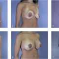
Fig. 65.2 (a–c) A subfascial pocket is created. (Reproduced with permission from Jones GE. Bostwick’s Plastic and Reconstructive Breast Surgery. 4th ed. Thieme; 2020.)
Related posts:
Stay updated, free articles. Join our Telegram channel

Full access? Get Clinical Tree


