2 Facial Fat Compartments
Abstract
Facial fat differs from fat in other regions of the body as it is compartmentalized. Each facial fat compartment exhibits septal boundaries, a regional perforator blood supply, and a specific tendency toward deflation in aging.
Recognition of compartment anatomy is one of the keys to safe subcutaneous dissection of the cheek, as facial nerve branches are often superficially positioned at transition points between compartments. Recognition of compartment-specific deflation provides a guideline for volume restoration in facial rejuvenation.
Key Points
Subcutaneous facial fat is not homogeneous but rather partitioned into a series of compartments separated by specific fibrous septa.
Each facial fat compartment has its own vascular blood supply, thickness and fascial consistency.
Some fat compartments are thin and fibrous, while others typically contain a large volume of easily dissectible fat. The compartmentalization of facial fat explains the regional variation noted in the subcutaneous plane when dissecting from the preauricular area anteriorly.
The facial fat compartments also serve as a model for deflation, confirming the observation that facial deflation in aging is compartment-specific rather than occurring homogeneously throughout the cheek.
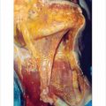
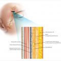
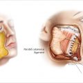
Facial fat compartments exist both superficial and deep to the SMAS (▶ Fig. 2.1a,b and ▶ Fig. 2.2 ).
The superficial facial fat, which lies within the subcutaneous plane, is superficial to the SMAS, and it is this fat which can be manipulated in an SMAS facelift.
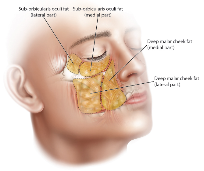
Deep fat compartments, which are situated anteriorly along the orbit, maxilla, zygoma, and pyriform aperture, lie deep to the mimetic muscles and overly the periosteum of the orbit and midface. The deep fat of the cheek is contiguous with that of the lower lid. Deep malar fat along the anterior midface provides anterior cheek volume.
Of note, both the superficial and deep fat compartments deflate over time, and this deflation is responsible for many of the morphologic changes seen in the aging face.
2.1 Compartmentalization of the Superficial Fat Compartments
Superficial facial fat is separated into specific compartments by the terminal extension of the deeper retaining ligaments, which percolate through the cheek from deep to superficial to insert into the skin as retinacular cutis.
Rather than being diffuse in their penetration of the SMAS, the retaining ligaments penetrate the superficial fascia at specific locations and thereby form the fibrous septum which are formed between compartments.
These junctional boundaries also are the location where the vascular perforators to cheek skin penetrate from deep to superficial.
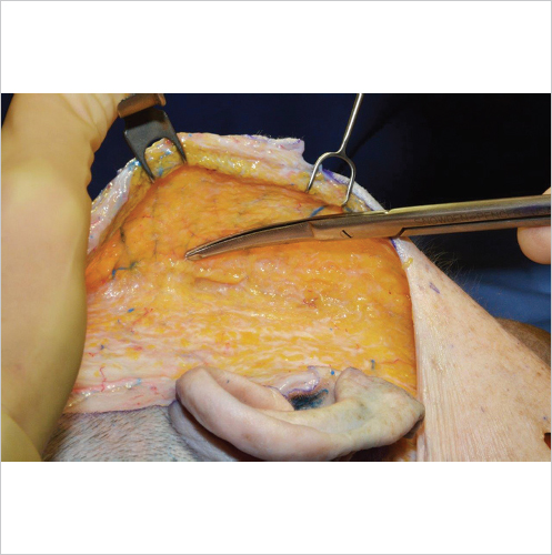
The surgical significance of this is that when encountering numerous perforators while performing subcutaneous dissection, anatomically the dissection is transiting from one superficial fat compartment to another.
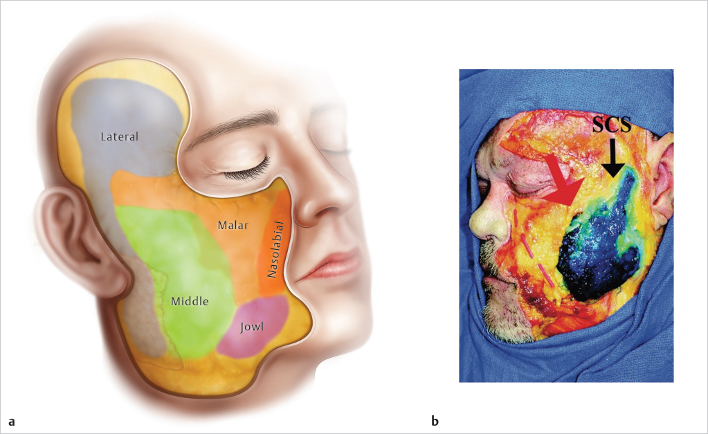
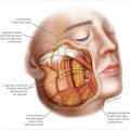
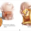
While there are many superficial fat compartments, the five compartments that the surgeon encounters in a facelift include the lateral compartment, middle compartment, superficial malar compartment, nasolabial fold compartment, and jowl compartment.
As subcutaneous dissection proceeds from laterally in the preauricular region medially, if the dissection is performed under direct visualization, it is possible for the physician to recognize both what compartment is being dissected as well as when the transition between compartments occurs (▶ Fig. 2.3 and Video 2.1).
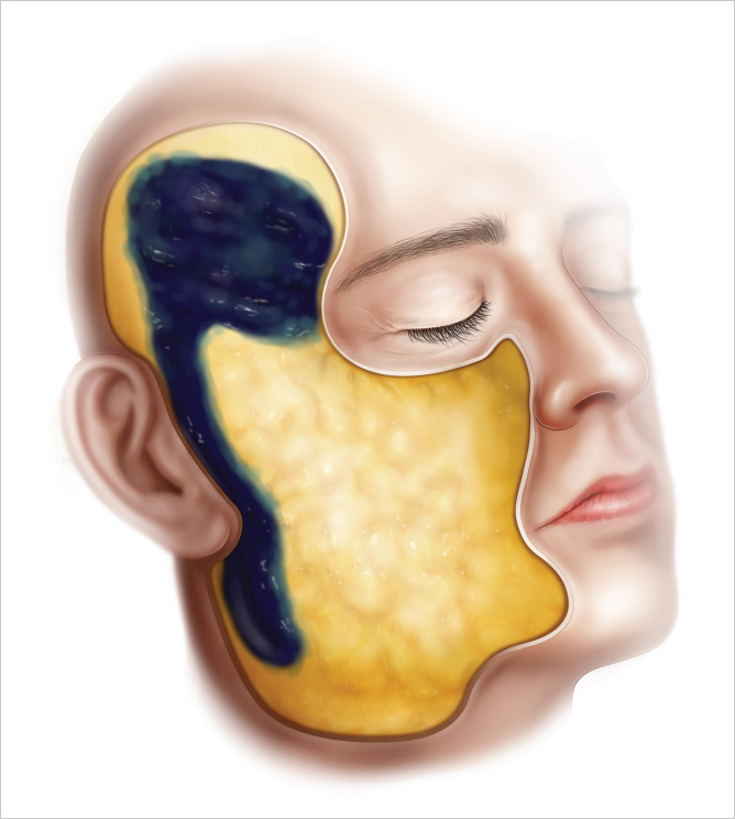
2.1.1 Lateral Compartment
The lateral compartment is located in the preauricular region and tends to be narrow, thin, following the superficial temporal artery cephalically within the temporal region.
Typically, the lateral compartment is only 3 to 5 cm in width and consists of dense, vascular, and fibrous fat.
This compartment is directly situated overlying the parotid gland, and as the dissection proceeds anterior to the parotid, the middle compartment is encountered, and the dissection becomes less fibrous (▶ Fig. 2.4 ).
Related posts:
 1 Overview of Facial Tissue Anatomy
 3 Overview: Facial Nerve Danger Zone
 4 Frontal Branch of the Facial Nerve
 6 Protecting the Marginal and Cervical Branches of the Facial Nerve
 5 Zygomatic and Buccal Branches
 8 Technical Considerations: Extended SMAS Dissection and Lateral SMASectomy/Platysma Window
Stay updated, free articles. Join our Telegram channel

Full access? Get Clinical Tree


