Abstract
White spots in the skin result from decreased melanin pigmentation. This can be caused by a reduction in the number of melanocytes or a decrease in their melanin production. Inflammatory events are frequently responsible for a condition called postinflammatory hypopigmentation, even though the inflammation may not be clinically appreciated. Table 13.1 lists the four most common causes of white spots. Determination of the degree (partial versus complete) of pigment loss and identification of the presence or absence of scale are helpful distinguishing clinical features.
- 1.
Examine for partial versus complete pigment loss and presence or absence of scale
- 2.
A Wood’s light examination accentuates white spots, especially in fair-skinned individuals
- 3.
Vitiligo is a common cause of depigmentation
White spots should be examined for:
- 1.
Partial versus complete pigment loss
- 2.
Presence or absence of scale
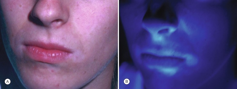
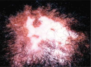
The degree of pigment loss can be assessed roughly with a Wood’s light examination, which helps to accentuate pigment contrast. In a darkened room with a Wood’s light, lesions that are completely depigmented appear almost chalk white. This finding is characteristic in vitiligo. The Wood’s light examination is also helpful in identifying white spots in lightly pigmented individuals; in patients with extremely fair complexions, white spots may not be evident under bright illumination ( Fig. 13.1 ).
White spots are seen more easily by Wood’s light examination.
The admonition, ‘If it scales, scrape it!’ also pertains to white spots. The two common hypopigmentary conditions that scale (i.e., tinea versicolor and pityriasis alba) can be distinguished with a potassium hydroxide (KOH) preparation.
Pityriasis Alba
- 1.
Characterized by hypopigmented, white patches
- 2.
More commonly seen in darkly pigmented children
- 3.
Probably a low-grade “eczematous” reaction
Definition
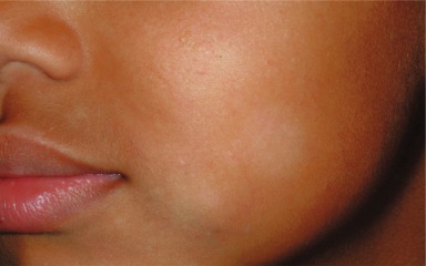
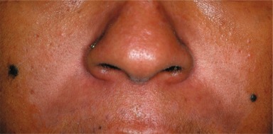
Pityriasis alba is an idiopathic hypopigmentary condition that appears clinically as white (alba) patches surmounted by fine, “bran-like” ( pityron , Greek for bran) scales ( Fig. 13.2 ).
Incidence
The disease is extremely common but usually not sufficiently disturbing for most patients to seek medical attention. It affects mainly children between the ages of 3 and 16 years and is most common (or most noticeable) in dark-skinned individuals. Individuals with atopic dermatitis have a predilection for pityriasis alba.
Pityriasis alba is most commonly recognized and appreciated in darkly pigmented children, but affects children of all skin types.
History
Pityriasis alba is usually asymptomatic, although an occasional patient may complain of mild itching. Patients or parents are most concerned about the appearance of the lesions.
Physical Examination
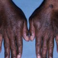
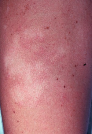
The early lesion is a mildly erythematous, slightly scaling patch with an indistinct margin. Most often, only the subsequent lesion is seen—a 1- to 4-cm white patch with a fine, powdery scale. In children, the face is the most common area of involvement and may have one to several lesions. Pityriasis alba can occur in other locations. Another common area of involvement is the upper arms, especially in children with atopic dermatitis ( Fig. 13.3 ). Rarely, widespread involvement occurs.
Differential Diagnosis
The disease is most often misdiagnosed as tinea versicolor . In temperate climates, adults with tinea versicolor seldom have facial involvement, but in children (in whom the disease is much less common) the face is affected in approximately one-third of cases. Accordingly, a KOH preparation should be performed on all scaling white spots to rule out tinea versicolor. The white spots in vitiligo are distinguished by sharp demarcation, complete depigmentation, and lack of scale. Postinflammatory hypopigmentation from inflammatory dermatoses (e.g., atopic dermatitis or psoriasis) are distinguished by history and extrafacial distribution of skin lesions ( Fig. 13.4 ).
- ●
Tinea versicolor
- ●
Vitiligo
- ●
Postinflammatory hypopigmentation
Laboratory and Biopsy
No specific laboratory test is available to establish the diagnosis. The KOH preparation is negative. The histologic picture is nonspecific, showing slight hyperkeratosis, decreased pigmentation in the basal cell layer, and a mild inflammatory reaction in the upper dermis ( Fig. 13.5 ).
Therapy
Treatment is not often necessary as spontaneous resolution occurs. Emollients can be used for the dry scaling, and 1% hydrocortisone cream is used for the inflammatory reaction. For more severe disease, a trial of triamcinolone 0.1% cream twice daily for several weeks may be beneficial for involvement on the trunk.
Course and Complications
The patient must understand that repigmentation will be slow. In most patients, the disease resolves spontaneously, but this takes months and, sometimes, years. For affected children, the disease rarely persists into adulthood. The disorder has no complications.
Pathogenesis
The origin of this common disorder is unknown. Most investigators believe that the decreased pigment is a postinflammatory phenomenon and that the initial event is a low-grade eczematous reaction. It may be a manifestation of inflammation related to decreased barrier protection from dry skin. The fact that the condition is commonly seen in children with atopic dermatitis lends credence to this dry skin association.
Pityriasis alba may be a low-grade eczema.
Postinflammatory Hypopigmentation
- 1.
Inflammation suppresses or destroys melanocytes
- 2.
Characterized by white macules with or without scale
- 3.
Repigmentation takes months to years
Definition
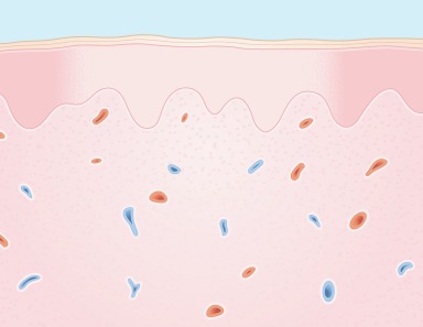
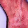
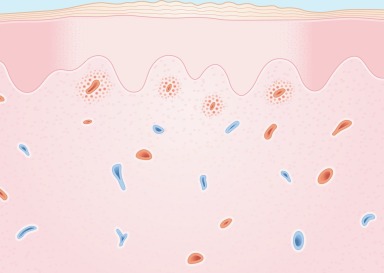
Postinflammatory hypopigmentation is the result of melanocyte destruction or suppressed melanin production secondary to inflammation of the skin ( Fig. 13.6 ). It appears as a hypopigmented or, in severe cases, a depigmented … . macule. The inflammation may be due to physical trauma, a chemical agent, or a primary, inflammatory, skin disease.
Causes of postinflammatory hypopigmentation:
- 1.
Physical trauma
- 2.
Chemicals
- 3.
Inflammatory skin diseases
Incidence
Inflammation-induced white spots are common incidental findings. Occasionally, they are the patienťs primary complaint.
History
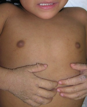
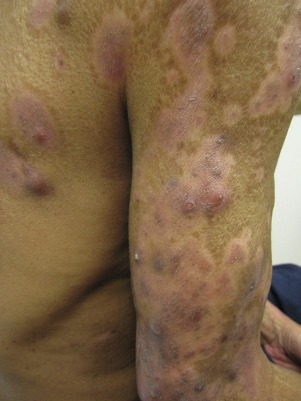
The inflammatory event responsible for the white spots is almost always remembered by the patient. Physical agents that may induce white spots include X-irradiation, frostbite and skin resurfacing lasers. Industrial exposure to chemicals such as phenolic and sulfhydryl compounds can also produce hypopigmentation. Some inflammatory skin diseases can leave residual hypopigmentation. Common examples are discoid lupus erythematosus, eczematous dermatitis (particularly atopic dermatitis and seborrheic dermatitis in a darker-skinned individual), and psoriasis ( Fig. 13.7 ). Uncommon causes are cutaneous sarcoidosis and mycosis fungoides, a form of cutaneous T-cell lymphoma.
Physical Examination
Whitish macules (no scale) or patches (with scale) conform to areas of prior inflammation.
Differential Diagnosis
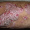
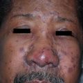
Postinflammatory hypopigmentation may be confused with vitiligo , particularly when hypopigmentation is profound. However, the pigment loss is rarely complete, as it is in vitiligo. Moreover, in vitiligo, the depigmentation is only rarely preceded by recognizable inflammation. It is important to determine the cause of postinflammatory diagnosis, ruling out inflammatory conditions, like atopic dermatitis or psoriasis, and neoplastic conditions, like cutaneous T-cell lymphoma ( Fig. 13.8 ).
- ●
Vitiligo
- ●
Determine primary skin condition
- ●
Inflammatory (e.g., atopic dermatitis)
- ●
Neoplastic (e.g., cutaneous T-cell lymphoma)
- ●
Laboratory and Biopsy
No specific laboratory test is available. The biopsy is not specific, showing decreased pigmentation in the epidermis and occasional mild residual inflammation in the dermis ( Fig. 13.9 ).