Abstract
Induration represents dermal thickening resulting in skin that feels thicker or firmer than normal. Scleroderma is the disease that best exemplifies this process. All of the diseases included in this chapter are uncommon conditions, except for granuloma annulare. Stasis dermatitis, a cause of dermal induration of the lower extremities, has a significant epidermal component and is discussed in Chapter 8 . For all causes of dermal induration, skin biopsy is often necessary to confirm the diagnosis, where the degree of dermal inflammation varies depending on whether the biopsy is performed at an early or late (“burned out”) stage of the disease process. Treatment options are limited and usually have minimal impact on the disease course.
Granuloma Annulare
- 1.
Self-limiting, asymptomatic condition with papules arranged in annular configuration
- 2.
Commonly affects children and young adults
- 3.
Occurs mostly on dorsal hands and feet
Definition
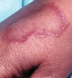
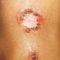
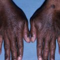
Granuloma annulare is an asymptomatic skin condition characterized by dermal papules (no overlying epidermal change), forming annular plaques and commonly arising on the dorsal hands and feet ( Fig. 18.1 ). Although the center of the plaques becomes depressed, the leading edge of the papules represents dermal induration. Granuloma annulare is most often localized but can be generalized.
Incidence
Granuloma annulare appears most commonly in children and young adults, and affects females more commonly than males.
History
The lesions are usually asymptomatic and come to the attention of the physician because of cosmetic concerns. Some patients mistakenly treat the condition for tinea corporis because of the annular configuration.
Physical Examination
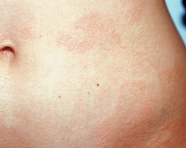
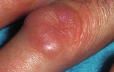
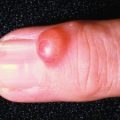
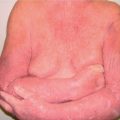
The localized variant appears as shiny dermal papules and annular plaques that are centrally depressed. Granuloma annulare can be skin colored, violaceous, or erythematous. Generalized granuloma annulare can affect the entire body ( Fig. 18.2 ). Other, less common, variants are subcutaneous (deep dermal solitary nodules) ( Fig. 18.3 ) and perforating (papules with central umbilication that is crusted and ulcerated). Both the subcutaneous and perforating variant of granuloma annulare appear on the distal extremities.
Clinical variants of granuloma annulare:
- 1.
Localized
- 2.
Generalized
- 3.
Subcutaneous
- 4.
Perforating
Differential diagnosis
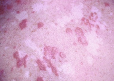
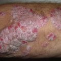
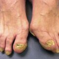
Granuloma annulare can be confused with other, often more serious, conditions. Papular granuloma annulare can appear similar to papular sarcoidosis ( Fig. 18.4 ) and lichen planus . A skin biopsy easily distinguishes between these conditions. Necrobiosis lipoidica diabeticorum (NLD) can appear clinically and histologically similar to granuloma annulare. NLD often has a yellowish hue, and telangiectasias present centrally within the depressed plaques on the lower legs. NLD has a stronger association with diabetes mellitus than does granuloma annulare. Subcutaneous granuloma annulare can appear similar to a rheumatoid nodule, both clinically and histologically. Arthritis is not present with granuloma annulare. Annular granuloma annulare is most often confused with tinea corporis, but lack of scale in granuloma annulare should enable the clinician to distinguish between the two.
- ●
Sarcoidosis
- ●
Lichen planus
- ●
Necrobiosis lipoidica diabeticorum
- ●
Rheumatoid nodule
- ●
Tinea corporis
Annular plaques of granuloma annulare have no scale; tinea corporis has scale.
Laboratory and Biopsy
Laboratory work-up with granuloma annulare is not usually necessary. Diabetes mellitus has been reported to be associated with generalized granuloma annulare. Skin biopsy shows necrobiosis (degenerative collagen) in the dermis with a predominantly histiocytic (i.e., macrophages) and multinucleated giant cell infiltrate on the periphery.
Therapy
Treatment of granuloma annulare is unsatisfactory. For young children, spontaneous resolution occurs. Therefore, no treatment is the best treatment (i.e., benign neglect). For cosmetically disfiguring lesions, treatment with potent topical corticosteroids, such as fluocinonide 0.05% cream, or intralesional triamcinolone 5 to 10 mg/mL may be effective. Skin atrophy is always a concern with prolonged topical or intralesional corticosteroid use. Psoralen plus ultraviolet A radiation (PUVA) and the antimalarial hydroxychloroquine (200 mg b.i.d.) are reserved for patients with generalized granuloma annulare. No well-designed studies favor any systemic therapy.
Localized granuloma annulare often resolves spontaneously.
Course and Complications
After 2 years, approximately 75% of granuloma annulare lesions will disappear. Recurrences are not uncommon. This is a self-limiting disease.
Pathogenesis
The cause remains unknown.
Lichen Sclerosus
- 1.
Sclerotic white plaques
- 2.
Often affects genital skin
- 3.
Pruritic
Definition
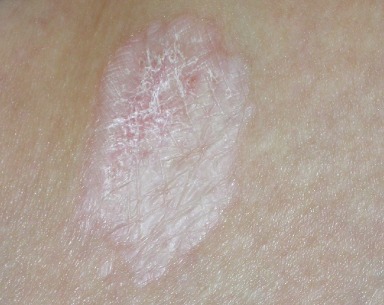
Lichen sclerosus is a chronic inflammatory condition that results in sclerotic white plaques due to thickening of the superficial dermis with overlying, thinned, finely wrinkled epidermis ( Fig. 18.5 ). Genital involvement of lichen sclerosus is more common than nongenital involvement and it can coexist with morphea, suggesting that the two diseases are related. Pruritus is often a major complaint.
Incidence
Lichen sclerosus is an uncommon disease. Females are reportedly more commonly affected, but this is unproven.
History
Lichen sclerosus involving nongenital skin is mostly asymptomatic, but can be dry and pruritic. The lesions can be cosmetically disfiguring. Involvement of the genitalia can result in intractable pruritus, leading to the development of dyspareunia in women and phimosis in men. In young females the initial appearance of lichen sclerosus can have a bruise-like quality (resembling traumatic hemorrhage) and lead to the misdiagnosis of child abuse.
Bruise-like quality of lichen sclerosus in young females can lead to misdiagnosis of child abuse.
Physical Examination
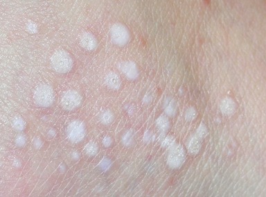
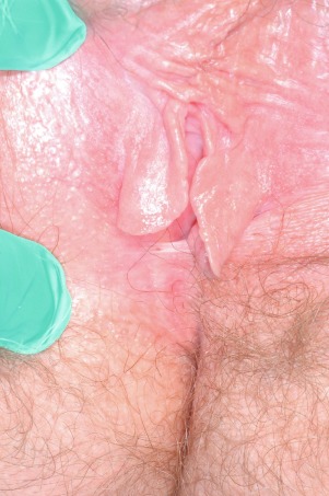
Nongenital disease occurs primarily on the trunk and extremities. The initial lesions appear as guttate (i.e., drop-like), white papules that can coalesce into larger plaques ( Fig. 18.6 ). The sclerotic plaques are covered by a finely wrinkled (often referred to as cigarette paper- or parchment-like) epidermis. In women, the vulva and perianal area are often involved, resulting in a figure-of-eight configuration. The initial erythema evolves into a hypopigmented sclerotic plaque that may erode and scar, making sexual intercourse difficult ( Fig. 18.7 ). The corollary in men is foreskin involvement leading to phimosis.
Guttate white papules are characteristic of early lichen sclerosus.
Differential Diagnosis
The two key differential diagnoses are morphea for nongenital lichen sclerosus and sexual abuse with genital lichen sclerosus in boys and girls. Early squamous cell carcinoma (erythroplasia of Queyrat) can mimic erosive genital lichen sclerosus ( Fig. 18.8 ), and biopsy is mandatory.
Related posts:
Stay updated, free articles. Join our Telegram channel

Full access? Get Clinical Tree


