Abstract
This chapter deals with nodular and cystic “lumps” in the skin ( Table 7.1 ). With the exception of lipomas, the lesions are located in the dermis, often with no alteration in the overlying epidermis. For many patients, common lesions, such as epidermal inclusion cysts, small angiomas, dermatofibromas, and lipomas, are not troubling and are not brought to the attention of a physician. However, these lesions are often found during routine physical examinations, and it is important to be able to distinguish them from malignant dermal growths. The usual question asked by patients who present with a “lump” in the skin is: “Is it cancer?” This concern is appropriate and must always be addressed.
Chapter Contents
Uncommon Dermal and Subcutaneous Growths
- 1.
Biopsy nodules of uncertain origin
- 2.
Suspect cancer for hard dermal nodules
Color and consistency are helpful distinguishing features.
Color and consistency are helpful distinguishing clinical features. The color of the lesion sometimes reflects the nature of the proliferating elements. Vascular lesions, for example, have hues ranging from red to purple. Consistency often distinguishes a nodule from a cyst. A cyst is usually fluctuant or malleable. For nodules, a soft consistency lends reassurance that the lesion is benign, a firm consistency is of intermediate concern, and a hard consistency should lead to suspicion of a possible malignant process. Sometimes, the diagnosis can be made only with biopsy. This is particularly important for firm to hard nodules in which the clinical diagnosis is uncertain and malignancy needs to be ruled-out.
For any skin nodule of uncertain origin, a biopsy is indicated to rule out malignancy.
| Laboratory Test (Biopsy) | |||||
|---|---|---|---|---|---|
| Frequency a | Physical Examination | Differential Diagnosis b | Diagnostic But Usually Not Necessary | Diagnostic and Necessary | |
| Angioma | 100% in elderly | Bright red macule or papule | Petechiae Blue nevus Melanoma | ✘ | |
| Dermatofibroma | 0.2 | Tan to brown, firm, flat to slightly elevated papule; “dimples” with lateral pressure | Nevus Melanoma Dermatofibrosarcoma, protuberans (rare) | ✓ | |
| Epidermal inclusion (follicular) cyst | 0.5 | Flesh-colored, firm, but malleable nodule | Pilar cyst Lipoma Malignant dermal tumor | ✓ c | |
| Hemangioma | 0.8 | Red or purple (often blanchable ) soft-to-firm macule, papule, or nodule | Vascular malformation Rapidly involuting congenital hemangioma Noninvoluting congenital hemangioma | ✓ | |
| Kaposi’s sarcoma | < 0.1 | Purple macules, plaques, or nodules | Bruise Angioma Bacillary angiomatosis | ✓ | |
| Keloid | 0.2 | Pink, firm, elevated scar | Hypertrophic scar Dermatofibrosarcoma, protuberans (rare) | ✓ | |
| Lipoma | 0.2 | Flesh-colored, rubbery, subcutaneous nodule | Epidermal inclusion cyst Angiolipoma Malignant tumor | ✓ | |
| Neurofibroma | 0.1 | Flesh to brown, soft, and often compressible (“buttonhole” sign) papule or nodule | Skin tag Dermal nevus | ✓ | |
| Xanthoma | < 0.1 | Yellow papules and nodules Hard subcutaneous tendon nodules | Sebaceous gland hyperplasia Juvenile xanthogranuloma Rheumatoid nodules | ✓ | |
| Malignant dermal tumors | < 0.1 | Flesh, red, or purple, hard nodules | Any of the above | ✓ | |
a Percentage of new dermatology patients with this diagnosis seen in the Hershey Medical Center Dermatology Clinic, Hershey, PA.
b A malignant tumor should be in the differential diagnosis for all dermal growths.
c Incision and drainage reveals cheesy, foul-smelling material.
Angioma
- 1.
Very common in older adults
- 2.
Typically cherry red color, but can be dark red or purple
Definition
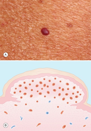
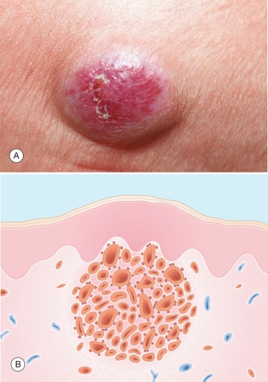
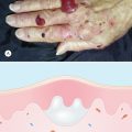
An angioma is a common benign vascular growth in older adults. It is also referred to as cherry angioma because of its bright red color ( Fig 7.1A ).
Incidence
Angioma are very common and increase in number as one ages. Virtually all elderly individuals have angioma.
History
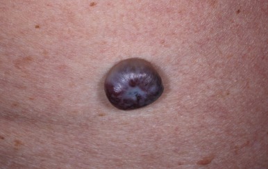
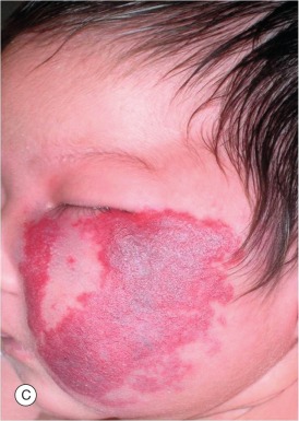
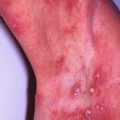
Angioma are frequently an incidental finding on the skin examination. They are asymptomatic unless traumatized. Occasionally, the patient or physician is concerned that an angioma may be a melanoma if deep purple color ( Fig 7.2 ).
Physical Examination
Angioma are easily recognized by their smooth surface and red color. There may be few to numerous angiomas occurring most commonly on the trunk but can be on the head and extremities. They vary from being pinpoint macules resembling petechiae to papules or small plaques. Angiomas usually are bright red but may be shades of deep red to purple.
Differential Diagnosis
Angioma is easily recognized most of the time. Petechiae are sometimes confused with numerous small flat angiomas. The occurrence on the trunk and presence for years in a healthy individual makes angioma the diagnosis. When deep purple, a blue nevus or melanoma should be considered. Examination with the dermatoscope is very helpful in visualizing the vascular globules seen in an angioma. If uncertain, a biopsy should be performed.
- ●
Petechiae
- ●
Blue nevus
- ●
Melanoma
Laboratory and Biopsy
The diagnosis of angioma is almost always made clinically. If in doubt, a biopsy reveals a well-demarcated collection of small blood vessels in the dermis ( Fig 7.1B ).
Therapy
In most cases, no therapy is needed. If irritated or cosmetically unattractive, they can be shaved off, electrodesiccated, or lasered.
- ●
None
- ●
Shave biopsy
- ●
Electrodesiccate
- ●
Laser
Course and Complications
The number of angiomas increase with age. They are usually of no concern other than for their cosmetic appearance. They are chronic and not associated with complications other than bleeding if traumatized.
Dermatofibroma
- 1.
Dermal fibrotic papule or small nodule
- 2.
Chronic, asymptomatic, and stable
Definition
A dermatofibroma is an area of focal dermal fibrosis, often accompanied by overlying epidermal thickening and hyperpigmentation. It appears clinically as a brown papule or small nodule, often more indurated than elevated. The origin is unknown. However, it is probably a response to trauma.
Incidence
Dermatofibromas are common and are often found incidentally during cutaneous examinations. Occasionally, they cause a patient to seek medical advice. They are seen most often in young adults.
History
Dermatofibromas usually are asymptomatic. The patienťs concern, if any, is over the possibility of malignancy.
Physical Examination
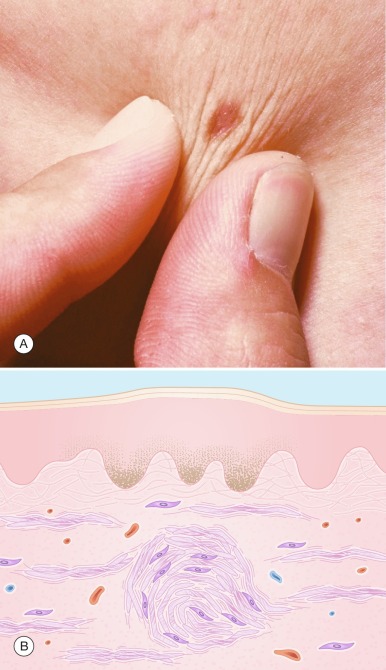
Typical dermatofibromas are approximately 5 mm in size and are slightly elevated. They vary in color from light tan to dark brown. The fibrotic nature of the lesion is best appreciated by palpation, which reveals a firm consistency. A helpful diagnostic test is the “dimple sign,” in which pinching results in central dimpling ( Fig. 7.3A ). Most dermatofibromas exhibit this sign; it is rarely seen with any other skin lesion. Dermatofibromas may occur anywhere, but the thighs and legs are the most common locations. One or several lesions may be present.
The “dimple sign” is characteristic of dermatofibroma.
Differential Diagnosis
With its brown color, a dermatofibroma may be confused with a nevus . Nevi, however, are usually softer and do not exhibit the dimple sign. Darker dermatofibromas may raise a clinical suspicion of melanoma . Dermatofibromas are usually purely brown (a benign color), whereas a nodular melanoma usually has shades of dark gray or blue. If any doubt exists, however, a biopsy should be performed.
Enlarging or atypically colored lesions should undergo biopsy to rule out malignancy.
Dermatofibrosarcoma protuberans is a low-grade malignant fibrous tumor that grows slowly but persistently, and rarely metastasizes. It is a rare tumor and is distinguished clinically from a dermatofibroma by its larger size, irregular shape, and continued growth.
- ●
Nevus
- ●
Melanoma
- ●
Dermatofibrosarcoma protuberans
Laboratory and Biopsy
The diagnosis is usually made clinically. If any doubt exists, a biopsy should be performed to rule out malignancy. The histologic picture is diagnostic and shows a focal proliferation of densely packed collagen bands that are twisting and intertwining ( Fig. 7.3B ). Fibroblasts are interspersed and increased in number. Increased pigmentation of the slightly thickened overlying epidermis accounts for the frequently brown color of these lesions.
Therapy
Therapy is usually not indicated. If desired, a simple excision is sufficient for removal and histologic examination.
- ●
None
- ●
Excision if desired
Course and Complications
Dermatofibromas are chronic and usually stable in size. They are not associated with any complications.
Pathogenesis
Although the origin is unknown, trauma (e.g., an insect bite) may be an initiating factor for some of these lesions. The proliferation of fibroblasts and subsequent fibrosis may represent an exuberant healing response to injury. However, most patients do not recollect a history of trauma in the area.
Two other lesions are considered within the spectrum of a dermatofibroma. A histiocytoma (an aggregate of histiocytes in a focal area within the dermis) probably represents an early phase in the formation of a dermatofibroma. A sclerosing hemangioma , as the name suggests, shows more of a vascular component, but the end result is that of dermal fibrosis as well.
Epidermal Inclusion Cyst
- 1.
Central pore and cheesy, foul-smelling discharge is diagnostic
- 2.
Origin is from the hair follicle
Definition
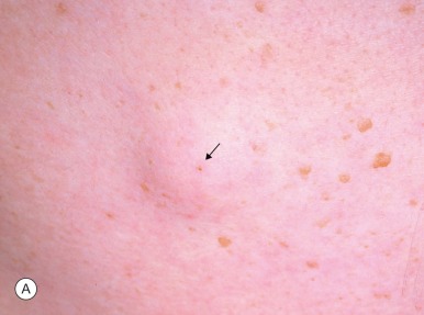
An epidermal inclusion cyst ( Fig. 7.4A ) is derived from the upper portion (infundibulum) of the epithelial lining of a hair follicle and is located in the mid and lower dermis. It is also called an epidermoid cyst. Clinically, it appears as a flesh-colored, firm, but often malleable, solitary nodule in the skin.

Incidence
These lesions are common, but usually are not brought to the attention of a physician, so the exact incidence is not known. They may occur at any age.
History
Epidermal inclusion cysts are usually asymptomatic, slow growing, and most frequently are found incidentally by either the patient or the examining physician. Occasionally, they are the primary complaint in a patient concerned about the possibility of malignancy. Another reason for medical attention is rupture of the cyst or secondary infection, either of which produces inflammation, pain, and drainage of foul-smelling material.
Physical Examination
Characteristically, the lesion is a flesh-colored, dome-shaped nodule that feels firm (but not hard). On palpation, it often feels slightly malleable, a finding that suggests the contents are semi-solid. This is a helpful diagnostic aid, as is the finding of a central pore , which represents the opening of the follicle from which the cyst originated. Lesions range in size from 0.5 to 5 cm. They may be located anywhere, but occur most frequently on the head and trunk. If the central pore is patent, the diagnosis is sometimes confirmed by squeezing the lesion and expressing some of the whitish, cheesy, foul-smelling material that is trapped within. This material represents macerated keratin.
A central pore is characteristic of an epidermal inclusion cyst.
Differential Diagnosis
Pilar (trichilemmal) cysts arise from the middle third (isthmus) of the follicular canal. They occur most frequently on the scalp, where they are the most common type of cyst. In other locations, they are less common than epidermal inclusion cysts, but the two may be indistinguishable clinically, and histologically, some cysts may have elements of both. The difference is not critical: both are benign. A lipoma is usually deeper than an epidermal inclusion cyst, and although a lipoma may feel rubbery, it is usually not malleable. When the diagnosis is uncertain, particularly if the lesion feels firm, a malignant tumor must be considered.
- ●
Pilar cyst
- ●
Lipoma
- ●
Malignant tumor
Laboratory and Biopsy
Usually, the diagnosis can be made clinically. If desired, confirmation can be obtained by incising and draining the lesion, which reveals the cheesy, foul-smelling, keratinous contents. A biopsy is equally confirmatory, but usually not necessary ( Fig. 7.4B ).
Therapy
Frequently, no therapy is requested or needed. If removal is desired, the entire cyst should be excised with its lining, to prevent recurrence. This is accomplished by incising the skin overlying the cyst without disrupting the cyst wall and then bluntly dissecting the entire cyst, along with its wall. If the cyst breaks, a curette can be used to remove the remaining contents and cyst wall. Elliptical excision is usually required for removal of cysts that have previously ruptured and scarred.
To prevent recurrence, the entire cyst, with its lining, should be removed.
- ●
None
- ●
Incision and drainage
- ●
Excision
Course and Complications
Untreated, most epidermal inclusion cysts reach a stable size, often in the range of 1 to 3 cm, rarely larger.
Complications are rare and usually limited to occasional rupture or infection. Rupture or infection results in redness and tenderness of the cyst and, on examination, increased fluctuance. If this occurs, the lesion should be treated as an abscess with incision and drainage, and occasionally oral antibiotics.
Multiple epidermal inclusion cysts are a feature of Gardner syndrome , an uncommon, autosomal-dominant, heritable disorder manifested by multiple epidermal cysts, fibromas, osteomas, and intestinal polyps. The intestinal polyps often undergo malignant degeneration.
Pathogenesis
Epidermal inclusion cysts arise from the upper portion (infundibulum) of a hair follicle. The epidermal lining of the cyst is identical to that of the surface epidermis and produces keratin, which, having no place to shed, accumulates and forms the cystic mass.
Infantile Hemangioma
- 1.
Benign vascular tumor in infancy
- 2.
Superficial and subcutaneous involvement
- 3.
Although most regress spontaneously, treat early if aesthetic or functional impairment anticipated
Definition
An infantile hemangioma ( Fig. 7.5A ) is a benign proliferation of blood vessels in the dermis and subcutis. The vascularity imparts a red, blue, or purple color to these lesions, depending on the size and depth of the proliferative vessels.
Incidence
Infantile hemangiomas are the most common soft tissue tumor of infancy, occurring more frequently in females, premature, and Caucasian infants. They are likely to be brought to the attention of a physician because of their rapid growth or cosmetic concerns.
History
Most arise in the first few weeks of infancy. Infantile hemangiomas are usually asymptomatic, except when they ulcerate, or cause local obstruction – fortunately an uncommon occurrence.
Physical Examination
Superficial infantile hemangiomas have a bright red color, whereas the deeper subcutaneous forms have a bluish hue. Mixed infantile hemangiomas are bright red, dome-shaped nodules.
Types of infantile hemangioma:
- 1.
Superficial (strawberry)
- 2.
Subcutaneous (cavernous)
- 3.
Mixed
Related posts:
Stay updated, free articles. Join our Telegram channel

Full access? Get Clinical Tree


