Abstract
The approach to a patient with skin disease does not differ markedly from the approach to any other patient. Data are collected from a history and physical examination (and sometimes from the laboratory), a differential diagnosis is generated, and the best diagnosis is selected.
- 1.
Morphologic appearance is critical in making the diagnosis
- 2.
Skin diseases can be divided into growths and rashes
Steps in dermatologic diagnosis:
- 1.
History
- 2.
Physical: identify the morphology of basic lesion
- 3.
Consider clinicopathologic correlations
- 4.
Configuration or distribution of lesions (when applicable)
- 5.
Laboratory tests
In history taking, a modified format is suggested. Instead of beginning with an exhaustive interrogation, it is more efficient to divide the history into a preliminary and a follow-up format. You should sit, face the patient, let the patient talk, listen, show empathy, and then clarify with questions (location, duration, symptoms, and prior treatment)
The most important part of the physical examination is inspection. Dermatology is a visual specialty, and diagnosis rests heavily on skin inspection. Unfortunately, although the skin is the most visible organ of the body, in a routine physical examination it often is the one most overlooked. Skin lesions need to be looked for , not at . Just as the examiner hears only the subtle heart sounds for which he or she listens, so will a clinician see on the skin only the lesions for which he or she searches. We need to train our eyes to see the skin lesions before us and ultimately be able to recognize them.
Dermatologic diagnosis depends on the examiner’s skill in skin inspection.
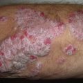
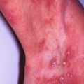
We have divided skin disorders into two broad categories: growths and rashes. A growth is a discrete lesion resulting from proliferation of one or more of the skin’s components. A rash is an inflammatory process that usually is more widespread than a growth. For both skin growths and rashes, the most important task is to characterize the clinical appearance of the basic lesion, that is, to identify its morphology. The pathophysiologic processes responsible for the clinical lesion must then be considered. These clinicopathologic correlations are emphasized in the diagnostic approach presented in this book. For skin rashes, important diagnostic information can sometimes also be obtained by noting the manner in which the lesions are arranged or distributed.
After the history and physical examination have been completed, laboratory tests may be indicated. In dermatology, these are usually simple office procedures that can provide valuable information needed either to confirm or to establish a diagnosis in selected disorders.
History
- 1.
Establish rapport.
- 2.
Let the patient talk uninterruptedly in the beginning
- 3.
Clarify location, duration, symptoms, and prior treatment
- 4.
Expand the history based on the differential diagnosis
In medicine, the traditional approach is to take the history before performing the physical examination. Some dermatologists prefer to reverse this order. We find it most useful to ask questions both before and after the examination. With this approach, a preliminary history is taken, in which several general questions are asked of all patients. Depending on the physical findings, more selective questions may be asked subsequently. For example, a history of sexual contacts would be inappropriate for an 82-year-old invalid complaining of an itching scalp, but would be indicated for a patient with an indurated ulcer on the penis.
Preliminary History
In addition to its diagnostic value, a preliminary history also helps to establish rapport with the patient. The short-cut of examining the skin without expressing an interest in the person will often be found wanting, especially by the patient. This initial history is composed of two parts that correlate with the chief complaint and the history of the present illness in the standard history format.
The initial history can be abbreviated by asking four general questions:
- 1.
How long?
- 2.
Where affected?
- 3.
Does it itch or other symptoms?
- 4.
How have you treated it?
Chief Complaint
In eliciting the chief complaint, one can often learn much by asking an open-ended question, such as, “What is your skin problem?” This is followed by four general questions regarding the history of the present illness.
History of the Present Illness
The general questions concern onset and evolution of the condition, distribution, symptoms, and treatment to date.
Onset and Evolution
“When did it start? Has it gotten better or worse?” Answers to these questions determine the duration of the disorder and how the condition has evolved over time. For most skin conditions, this is important information.
Symptoms
“Does it bother you?” is an open-ended way of asking about symptoms. For rashes, the most common symptom is itching. If the patient does not respond to the general symptom question, you may want to ask specifically, “ Does it itch?” Questions concerning systemic symptoms (e.g., “How do you feel otherwise?”) are not applicable for most skin diseases and are more appropriately reserved until after the physical examination.
Treatment to Date
The question, “How have you treated it?” results in an incomplete response from almost all patients. For skin disease, one is particularly interested in learning what topical medications have been applied. Many patients do not consider over-the-counter preparations important enough to mention. The same applies for some systemic medications. Providing the patient with specific examples of commonly used topical and systemic medications, such as calamine lotion and aspirin, may jog a patienťs memory enough to recall similar products that he or she may have used. It is important to inquire about medications, not only because they cause some conditions, but also because they may aggravate many others. For example, contact dermatitis initially induced by poison ivy may be perpetuated by contact allergy to an ingredient in one of the preparations used in treatment.
After the skin examination, one may need to return to the treatment question if any suspicion exists that a medication is causing or contributing to the disorder. Interestingly, a patient often recalls using pertinent medication only when he or she is asked the question again.
Persistence is often required in eliciting a complete medication history.
Finally, at the end of the visit, when one is ready to prescribe medications for the patient, it is helpful to know what medications have already been used. This approach avoids the potentially awkward situation in which a patient replies to your enthusiastic recommendation of your favorite therapy with, “I’ve already tried that and it didn’t work!”
Follow-Up History
After the initial history and physical examination, it is hoped that a diagnosis, or at least a differential diagnosis, has been formulated. With a diagnosis in mind, more focused questions may be necessary. This questioning may include obtaining more details about the history of the present illness or may be directed toward eliciting specific information from other categories of the traditional medical history, including past medical history, review of systems, family history, and social history. The following serve only as examples for the use of focused questions.
Past Medical History
After the physical examination, one may want to learn more about the patienťs general health. For example, in a patient with suspected herpes zoster, a past history of chickenpox would be of interest. We have discussed how topically applied and systemically administered medications often contribute to skin conditions. Skin findings may encourage further pursuit of these possibilities. For example, in a patient with a generalized erythematous rash or hives, systemic drugs should be high on the list of possible causes. Because drugs can cause virtually any type of skin lesion, it is useful to consider drug eruptions in the differential diagnosis of almost any skin disease. It may also be helpful to ascertain whether the patient has any known allergies, in order to determine whether any medications are currently being used that could produce a cross-reaction.
Drugs can cause all types of skin rash.
Review of Systems
In a patient with a malar rash, a diagnosis of systemic lupus erythematosus should be considered, and the examiner will want to question the patient further for symptoms of additional skin or other organ involvement, including Raynaud’s phenomenon, photosensitivity, hair loss, mouth ulcers, and arthritis. In a patient with a generalized maculopapular eruption, the two most common causes are drugs and viruses, so the physician will want to inquire about both medication use and viral symptoms, such as fever, malaise, and upper respiratory or gastrointestinal symptoms.
Family History
In certain cutaneous conditions, some knowledge of the family history may help in diagnosis. Innumerable inherited disorders have dermatologic expression. The following serve only as examples:
- ●
In a child with a chronic itching eruption in the antecubital and popliteal fossae, atopic dermatitis is suspected. A positive family history for atopic diseases (atopic dermatitis, asthma, hay fever) supports the diagnosis.
- ●
In a youngster with multiple café-au-lait spots, a diagnosis of neurofibromatosis is considered. A positive family history for this disorder, substantiated by examination of family members, helps to support the diagnosis of this dominantly inherited disease.
- ●
Inherited disorders have numerous skin findings.
Knowledge of the family’s present health is also important when considering infectious diseases. For example, impetigo can occur in several family members, and this knowledge may help in considering the diagnosis; it would certainly be important for treatment. Likewise, in a patient with suspected scabies, it is important to know, for both diagnostic and therapeutic purposes, whether other family members are itching.
Social History
In some disorders, knowledge of the patienťs social history may be important. For example, a chronic skin ulcer from persistent herpes simplex infection is a sign of immunosuppression, particularly acquired immune deficiency syndrome (AIDS). Therefore, a patient with such an ulceration should be asked about high-risk factors for acquiring AIDS, including sexual behavior, intravenous drug abuse, and exposure to blood products.
For persistent skin infections, consider the possibility of AIDS.
Another common occasion for probing into a patienťs social history is when the patient is suspected of having contact dermatitis; this aspect of the social history could be subtitled the skin exposure history . Patients encounter potentially sensitizing materials both at work and at play. Industrial dermatitis is a leading cause of workers’ disability. For chronic hand dermatitis, questions about occupational exposure are important and should be directed particularly to materials and substances the patient contacts either by handling or by immersion. Similarly, a patient presenting with an acute eruption characterized by streaks of vesicles should be queried regarding recent outdoor activities resulting in exposure to poison ivy or poison oak. Contact dermatitis is a common and challenging problem. On the part of the physician, it often requires painstaking efforts in a detective-type search to elicit from the patient an exposure history that fits the dermatitis.
A complete “skin exposure history” is required whenever contact dermatitis is suspected.
Some harbor the misconception that in dermatology, one needs only to glance at the skin to arrive at a diagnosis and that talking with the patient is superfluous. Although this is occasionally true, we hope that the previous examples serve to illustrate that this frequently is not the case. In fact, in some instances (and contact dermatitis is a good example), detailed historical information is essential to establish a diagnosis.
Physical Examination
- 1.
Complete skin examination is recommended at the first visit
- 2.
Good lighting is critical
- 3.
Describe the morphology of the eruption
The physical examination follows the preliminary history. For the skin to be inspected adequately, three essential requirements must be met: (1) an undressed patient, clothed in an examining gown; (2) adequate illumination, preferably bright overhead fluorescent lighting; and (3) an examining physician prepared to see what is there.
One should do hand hygiene prior to and after touching the patient.
Examine the entire mucocutaneous surface, but patients will be more firmly convinced of your sincere interest in their particular problems if you start by examining the affected areas before proceeding with the more complete examination.
At least for the initial examination, the patient needs to be disrobed so that the entire skin surface can be examined. Busy physicians who tend to overlook this rule will miss much. An occasional patient may be reluctant to comply, saying, “My skin problem is only on my hands; why do you need to look at the rest of my skin?” We tell such patients that we have at least two reasons:
- 1.
Other lesions may be found that “go along with” the lesions on the hands, and help to confirm the diagnosis. For example, in a patient with sharply demarcated plaques on the palms, the finding of a few scaling plaques on the knees or a sharply marginated intergluteal plaque will help to substantiate a suspicion of psoriasis.
- 2.
An important incidental skin lesion may be found. The finding of a previously undetected malignant melanoma on a patienťs back is an example. We studied the yield from a complete skin examination in 1157 consecutive new dermatology patients and found an incidental skin malignancy in 22. Some 20 of these patients had basal cell carcinoma, one had melanoma, and one had Kaposi’s sarcoma that served as the presenting manifestation of AIDS. A subsequent study of 874 patients reported an incidental skin cancer detection rate of 3.4%.
- 1.
The entire skin surface is examined for:
- 1.
Lesions that may accompany the presenting complaint
- 2.
Unrelated, but important incidental findings
For the skin to be examined adequately, it must be properly illuminated. Natural lighting is excellent for this purpose, but is difficult to achieve in most offices and hospital rooms. The alternative is bright overhead fluorescent lighting, supplemented with a movable lamp that is usually wall mounted. One additional illuminator that is often useful is a simple penlight. Either this or the movable lamp can be used as side lighting to detect whether a lesion is subtly elevated. For this technique, the light is directed onto the lesion from an angle that is roughly parallel to the skin. If the lesion is elevated, a small shadow will be thrown, and the relief of the skin will be appreciated. The penlight also is useful for examining the mouth, an area that is sometimes overlooked but in which one may detect lesions that are helpful in diagnosing a cutaneous disorder.
Another piece of examination equipment that is occasionally useful is the Wood’s light, a long-wavelength ultraviolet “black” light. Contrary to some popular misconceptions, this light does not enable one to diagnose most skin fungal infections; it detects fluorescence of affected hairs only in some, now uncommon, types of tinea capitis. The Wood’s light is, however, still used to accentuate pigmentary alterations in the skin, such as vitiligo.
Except for provision of adequate illumination, minimal equipment is needed for examining the skin. A simple hand-held lens can be helpful. Enlarging the image may improve diagnostic accuracy. However, on some occasions, such as clarifying a burrow in scabies or detecting Wickham’s striae in a lesion of lichen planus, a hand-held lens can be useful. For diagnosing pigmented growths, dermatologists often employ a dermatoscope. This is an illuminated hand-held magnifying device intended to help the clinician to diagnose melanoma and other growths clinically.
A dermatoscope is useful in diagnosing growths, especially melanoma.
An adequate examination of the skin should actually be called a mucocutaneous examination so that one is reminded to include an examination of the mouth. Similarly, the scalp and nails should not be overlooked. Because both cutaneous and systemic diseases may be expressed in the nails and nail beds as well as in the mouth, these areas should be inspected in every cutaneous examination.
The scalp, mouth, and nails should not be overlooked.
Physical examination depends largely on inspection, but one should not neglect the opportunity to palpate the skin as well. The two major purposes for this are (1) to assess the texture, consistency, and tenderness of the skin lesions; and (2) to reassure patients that we are not afraid to touch their skin lesions–that they do not have some dreadful contagious disease. Nothing is more disquieting to a patient than to be cautiously approached with a gloved hand. For anogenital, mucosal, and all weeping lesions, gloving is necessary and expected, but for most other lesions, the physician learns more and the patient is less frightened if the touching is done without gloves. Palpation is the major method by which we evaluate not only the consistency (e.g., softness, firmness, fluctuance) but also the depth of a lesion.
Palpation helps to:
- 1.
Assess texture and consistency
- 2.
Evaluate tenderness
- 3.
Reassure patients that they are not contagious
After the patient is properly gowned and perfectly illuminated, for what do we inspect and palpate? The first and most important step is to characterize the appearance (i.e., identify the morphology) of each skin lesion. After the morphology of a lesion is identified, its clinicopathologic correlation can be considered.
The most important task in the physical examination is to characterize the morphology of the basic lesion.
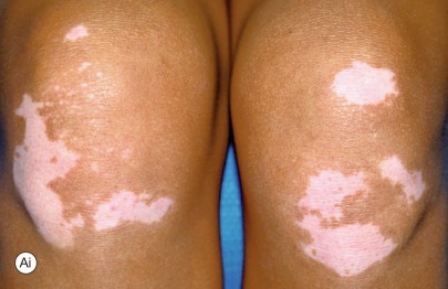
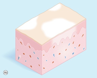
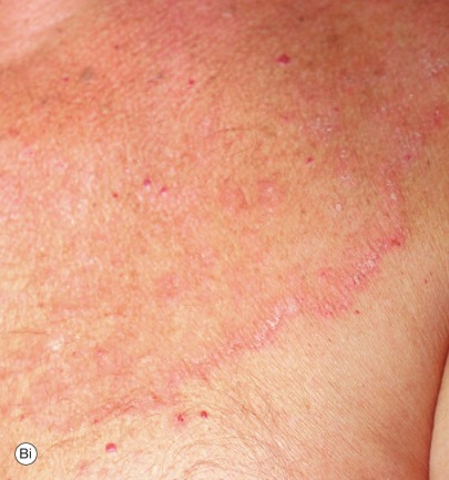
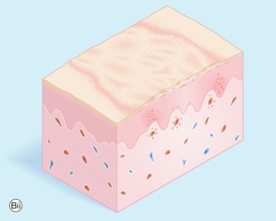
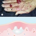
Terminology of Skin Lesions
- 1.
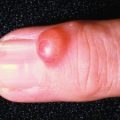
Primary lesions include macule, patch, papule, plaque, nodule, cyst, vesicle, pustule, ulcer, wheal, telangiectasia, burrow, and comedo
- 2.
Secondary lesions include scale, crust, oozing, lichenification, induration, fissure, and atrophy
A special vocabulary is used in describing the morphologic appearances of skin lesions. These terms are illustrated and defined in Fig. 3.1 .