Urticaria
Susan J. Huang
Arturo Saavedra-Lauzon
Acute and Chronic Urticaria
 |
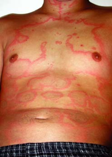
Mary is a 21-year-old student who presents to the university health services. She complains that she developed numerous itchy hives all over her body a half hour ago after a clambake. She reports a history of hives after eating shellfish. On examination, there are multiple well-circumscribed pink plaques over the arms, trunk, and legs (Fig. 21-1). She has no involvement of the lips or around the eyes. Her vital signs are stable and she has no other findings. You recommend avoidance of shellfish to prevent future episodes and prescribe fexofenadine for daytime use and, as necessary, diphenhydramine at bedtime. You instruct her to return to the clinic if her symptoms progress or last for more than 24 hours.
Background/Epidemiology
Urticaria is more commonly referred to as hives or wheals. It is a fairly common condition, affecting up to 20% of the general population at some time in their lifetime. There is no predominant gender for acute urticaria, but more females have the chronic form of urticaria. Its distribution is worldwide.
Pathogenesis
Urticaria is frequently caused by allergic triggers, which lead to mast cell degranulation of histamines, cytokines, and other vasoactive substances. While angioedema and anaphylaxis are separate entities, all three conditions reflect edema caused from leaky endothelium of differing depth and with differing distribution of involvement. Urticaria involves edema of the epidermis and dermis and affects the skin only. Individual lesions are usually pruritic, marked by rapid onset and resolve in less than 24 hours. Systemic symptoms including fatigue, sweats, chills, and joint pain may accompany severe attacks of urticaria.
Angioedema involves edema of the dermis, subcutaneous tissues, and/or submucosal tissues. Unlike urticaria, lesions usually are not pruritic, but rather, are painful. Lesions favor the eyelids, lips, genitalia, palms, and soles; commonly last between 24 and 48 hours.
Causes of angioedema include food exposures, or medications including nonsteroidal anti-inflammatory drugs (NSAIDs), sulfa medications, penicillins, or anticholinesterase (ACE) inhibitors. Angioedema is frequently accompanied by urticaria. Angioedema without urticaria should increase suspicion of C1 esterase inhibitor deficiency. Anaphylaxis involves edema of the skin and mucosa and involves multiple organ systems. It may lead to cardiovascular and respiratory decompensation. It is caused by a systemic allergic reaction, most commonly to a food, medication, insect bite, or other exposure. Primary evaluation of the patient’s vital signs is essential.
Key Features
“Hives” are common, affecting up to 20% of the population during a lifetime.
Most episodes resolve within 6 weeks (acute urticaria). Continued episodes beyond 6 weeks are defined as chronic urticaria.
Cause of urticaria is identified in acute urticaria (40%–60%) more commonly than in chronic urticaria (10%–20%).
Therapy is with oral antihistamines, topical antipruritics, and oral immunosuppressive medications. Ideally, the cause is identified and the etiology appropriately treated or exposure eliminated.
For patients with anaphylaxis, consider prescribing an EpiPen (epinephrine autoinjector).
Clinical Presentation
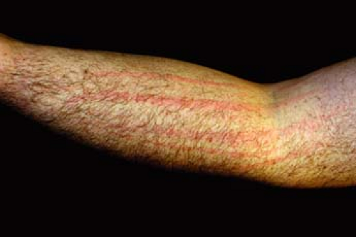
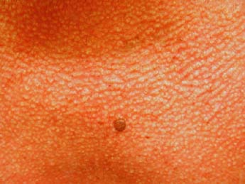
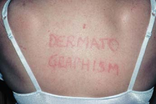
In this section, we will discuss two patterns of urticaria: acute and chronic (Table 21-1) (Figs. 21-2 to 21-6). We will discuss physical urticaria in the next section. Acute urticaria lasts for under 6 weeks, whereas chronic urticaria is marked by onset of lesions for more than two times per week for 6 or more weeks without treatment. The most common known causes of acute urticaria and chronic urticaria are presented in Table 21-2 (Figs. 21-7 to 21-9). Some important causes are upper respiratory tract infections medications (Table 21-3), and foods. However, many cases are idiopathic or autoimmune. Chronic urticaria can have a profound negative affect on a patient’s quality of life. The cause of urticaria is identified in acute urticaria (40%–60%) more commonly than in chronic urticaria (10%–20%).
Given the prevalence of idiopathic cases, this condition is often difficult to treat. Conditions associated with chronic urticaria include autoimmune
hypothyroidism, parasitic and Helicobacter pylori infections, systemic lupus erythematosus, and hematologic malignancies. It is estimated that approximately 50% of chronic urticaria is autoimmune related.
hypothyroidism, parasitic and Helicobacter pylori infections, systemic lupus erythematosus, and hematologic malignancies. It is estimated that approximately 50% of chronic urticaria is autoimmune related.
Table 21-1 Classification of Urticaria* | ||||||||||||||||||||||||||||||||
|---|---|---|---|---|---|---|---|---|---|---|---|---|---|---|---|---|---|---|---|---|---|---|---|---|---|---|---|---|---|---|---|---|
| ||||||||||||||||||||||||||||||||
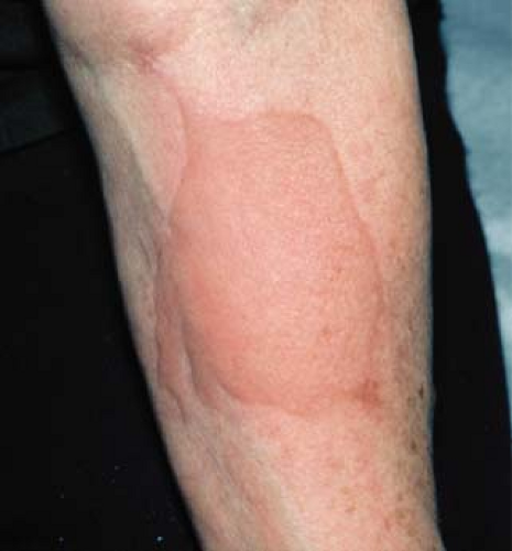 Figure 21-6 Cold urticaria–wheal forming after exposure to an ice cube. From Goodheart HP. Goodheart’s Photoguide to Common Skin Disorders.3rd ed. Philadelphia: Lippincott Williams & Wilkins, 2009. |
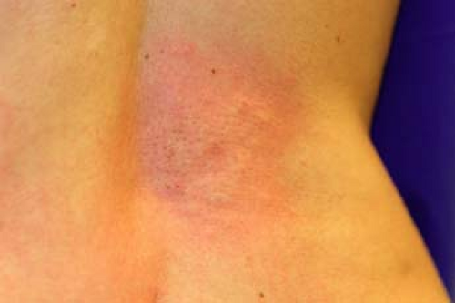
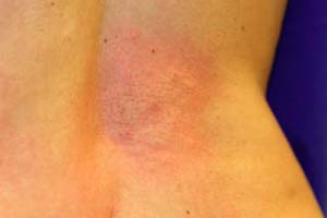
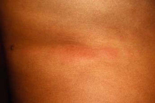 Figure 21-8 Wheal on the back of a woman with active systemic lupus erythematosus.
Stay updated, free articles. Join our Telegram channel
Full access? Get Clinical Tree
 Get Clinical Tree app for offline access

|
