10 The Punch Biopsy
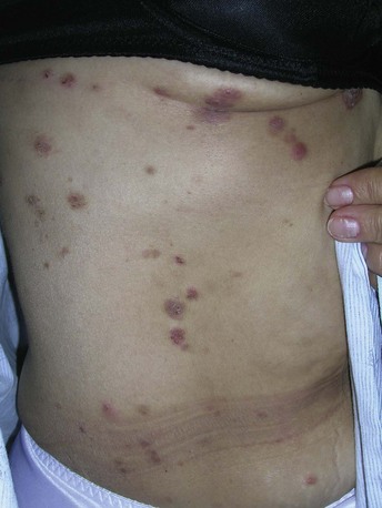
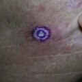
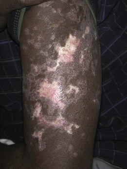
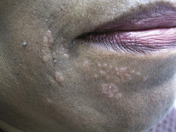
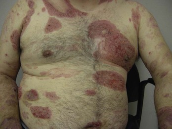
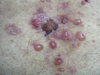
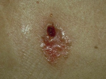
Flat lesions that are amenable to punch biopsy include inflammatory skin conditions such as drug eruptions, dermatoses, psoriasis, and cutaneous lupus (Figure 10-1). Infiltrative skin conditions such as sarcoidosis and granuloma annulare also can be diagnosed with a punch biopsy (Figure 10-2). In addition, a punch biopsy may be used to diagnose all types of skin cancers including melanoma and cutaneous lymphomas (Figure 10-3).
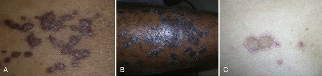
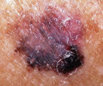
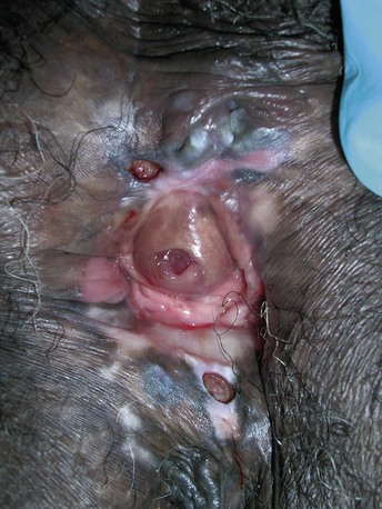
A punch biopsy is one option in the diagnosis of melanoma if the entire lesion is large, making it too difficult to remove the whole lesion at the time of biopsy. In this case, the diagnostic yield will generally be best if a biopsy is performed on the darkest, most elevated, and/or most suspicious areas (Figure 10-4). Using a dermatoscope may help identify a suspicious area for the punch biopsy (see Chapter 32, Dermoscopy). If the suspicion for melanoma is high, excising the entire lesion is preferred, when possible, to improve the diagnostic yield. There are also times when a broad scoop shave may provide better tissue for the pathologist. The highest risk of using a punch biopsy to diagnose a melanoma is the risk of a false-negative result. If the lesion remains suspicious for melanoma and a punch biopsy was performed with a negative result, the remainder of the lesion should be excised for histology (see Chapter 8, Choosing the Biopsy Type).
Indications
A punch instrument can be used to create an opening in an epidermal inclusion cyst for a minimally invasive cyst removal. Some clinicians use a punch incision to remove small lipomas (see Chapter 12, Cysts and Lipomas).
Relative Contraindications and Cautions
Punch biopsy is a more invasive biopsy technique than needed for most BCCs or SCCs, which can be diagnosed by shave biopsy. In one study there was no significant difference in the accuracy rate for histologic classification of BCCs with both the shave and the punch biopsy.1 Punch biopsies generally bleed more than shave biopsies and the risk of infection is somewhat higher than for a shave biopsy.
A punch biopsy does have certain risks that are greater than those of a shave biopsy, including the possibility of cutting larger blood vessels and nerves. Therefore, clinicians must be familiar with the underlying anatomy. Fortunately, most major nerves and blood vessels are deeper than a punch instrument, but digital nerves and the temporal branch of the facial nerve are more superficial and care needs to be taken in these areas (Figure 11-2 of Chapter 11). Punch biopsies over the digits or the eyelid margins are generally to be avoided. When possible, it is also prudent to avoid doing a punch biopsy over superficial arteries such as digital or temporal arteries. Caution should also be exercised over areas where there is little soft tissue between the skin and the bone (over the tibia, digits, and ulna) because the punch can cut through the underlying bone.
Advantages of A Punch Biopsy
The advantages of a punch biopsy for the clinician include the following:
The following advantages of a punch biopsy benefit the patient:
Disadvantages of A Punch Biopsy
The disadvantages of a punch biopsy for the clinician include the following:
For the patient, the disadvantages of a punch biopsy include the following:
Making A Diagnosis
It helps to have a good idea of the differential diagnosis before choosing the biopsy type and location. For most punch biopsies, a clinician should choose a punch size that will result in excision of the whole lesion or will provide a sample of the portion of the lesion that appears to have the worst pathology. However, for bullous disorders such as pemphigus or pemphigoid, it is best to punch the edge of the bulla to include the perilesional skin (Figure 10-10). A scoop shave under an intact bulla or at the border of a bulla may yield an equally good specimen. The goal is to keep the epidermis attached to the dermis at the edge of the bulla.
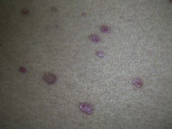
FIGURE 10-10 Bullous lichen planus on the back. A punch biopsy should include a whole intact bulla or the edge of a bulla.
(Copyright Richard P. Usatine, MD.)
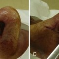
When performing a biopsy on an ulcerative lesion of unknown origin, it is helpful to remove tissue from the edge of the ulcer rather than the center portion. For example, if pyoderma gangrenosum is suspected, the biopsy should include the edge of the lesion, with some perilesional skin (Figure 10-11).
FIGURE 10-11 Pyoderma gangrenosum on the leg, with diagnosis confirmed by punch biopsy of an active edge.
(Copyright Richard P. Usatine, MD.)