Flaps

13 Flaps


The proper development and implementation of the skin flap is a key skill in reconstruction of skin defects, especially facial defects, following trauma or the removal of benign skin lesions or skin cancers. Although the surgeon should always consider primary, linear closure of a skin defect, there are many circumstances in which a skin flap may be the ideal choice for reconstruction.1,2


Skin flaps may be chosen instead of a primary linear closure to:


In addition, flaps can also perform very well in situations that would not be ideal for skin grafts; flaps can be employed to fill deeper defects that would lead to poor contours with skin grafting or to cover anatomic structures that may prevent the use of grafts (cartilage or tendon).


It is critical to understand the advantages and disadvantages of the different types of flaps rather than apply a “cookbook” approach to flap planning. When selecting the best choice of flap for a particular defect, the relative benefits and risks of each flap should be considered beforehand. A deep understanding of both flap theory and anatomy can lead to the best results.


Terminology


Flaps can be classified by their blood supply into axial pattern flaps, which have a larger, typically named artery supplying their vascular needs (such as the paramedian forehead flap, which depends on the supratrochlear arteries of the medial lower forehead), and random pattern flaps, which rely on the unnamed vasculature of the dermis, subcutaneous fat, and in some cases, the superficial musculature of facial structures. Because the blood supply of the skin and soft tissues of the scalp, face, and neck is so rich, the random pattern flaps enjoy very high rates of success. These local, random pattern flaps are by far the more commonly employed flaps and will be the focus of this chapter.


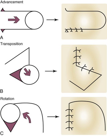
The random pattern flap is further classified by its movement. The three basic types of movement are (Figure 13-1):


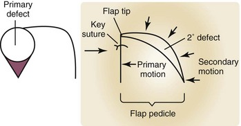
The initial wound that is to be reconstructed is the primary defect, and here it refers to the result of the removal of a skin tumor (Figure 13-2). Once a flap has been advanced, transposed, or rotated into position to close the primary defect, the wound that remains behind at the donor site of the flap is the secondary defect. The primary motion of a flap is the movement that the flap makes when it advances, transposes, or rotates into and closes the primary defect (Figure 13-2). The secondary motion is the movement that the tissue adjacent to and surrounding the flap makes when the final defect is closed. Both the primary and secondary motion of a flap are important in proper flap design. The very end of the flap that fills the primary defect, furthest from the donor site is the flap tip. The flap pedicle is that portion of the flap that connects it to the surrounding skin, and is the conduit for the vascular supply of the flap. The key suture of a local flap is the location in which the first suture is typically placed to provide the initial correct alignment of a flap to fill the primary defect and to direct additional placement of sutures to close the secondary defect. Standing cutaneous cones (dog ears) are areas of local tissue redundancy that occur in wound closure, but that can be minimized, eliminated, or moved to more cosmetically advantageous locations by the correct choice of and meticulous planning for a flap.


Planning the Flap


Careful planning is essential for successful flap reconstruction. Following confirmed tumor clearance of the surgical margins, the primary defect is lightly infiltrated with local anesthetic so that it may be manipulated without patient discomfort. It is important for the surgeon not to proceed immediately with an anticipated flap procedure before carefully considering the ramifications of tissue movement for surrounding structures. At this point the defect should be evaluated for surrounding tissue laxity and any potential reservoirs of loose skin that may be recruited for repair. This may be done by pinching the skin with gloved fingers. If the fingers are inadequate in more inflexible areas, the skin hook is a very useful surgical instrument for gauging tissue movement without tissue damage even in large defects of inflexible skin. Careful flap planning will then include not only the evaluation of possible sources of skin for flap creation, but also the consideration of nearby anatomic structures that could be distorted by either the primary or secondary movement of the flap. Especially vulnerable areas are the free margins of the eyelids, lips, and the nasal tip. The vectors of flap movement should be placed perpendicular to such structures, lest tissue movement distort their anatomy.


Tissue texture, and color should be matched whenever possible; a flap from similar adjacent skin will lead to the best results. A defect on the nose should be filled with similar-appearing skin from the nose instead of skin from a flap on the cheek. Finally, anticipated tissue redundancies from wound closure should be considered, with thought to how these redundancies or dog ears may be best camouflaged along anatomic borders. The proposed flap should always be drawn out prior to incision, and this may be done with a surgical marker.


Advancement Flap


Conceptual


The primary motion of the advancement flap is the sliding of the flap, usually in a single linear vector as it advances into the primary defect. This sliding motion may be unidirectional (single advancement flap) or bidirectional (double advancement flap).3 Although there are several variations on this type of flap, the two main benefits of the advancement flap are as follows:
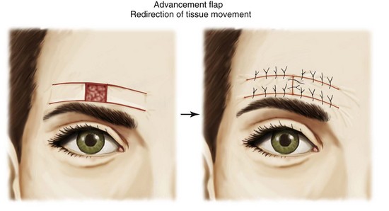
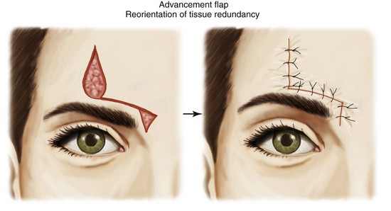


Note that the purpose of the advancement flap is not to increase laxity to close a wound under high tension. Although an advancement flap may appear to provide superior tissue movement over a primary linear closure, this is rarely the case. In preoperative planning, if a defect cannot be approximated primarily by pinching the skin or mobilizing the skin edges with hooks, an advancement flap will not be the best option because the amount of additional tissue movement that it provides over primary closure is often minimal.


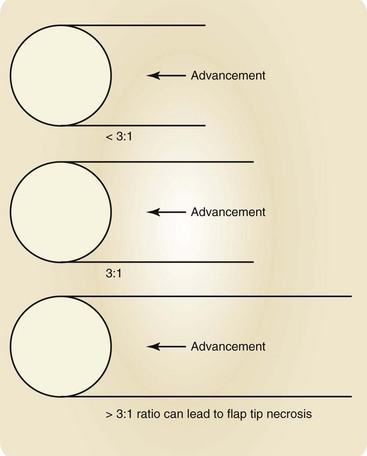
Advancement flaps are also limited by their blood supply. The pedicle of skin and soft tissue must not be compromised. A length:width ratio should ideally not exceed 3 : 1. A long, thin advancement flap in violation of this ratio runs a high risk of distal flap tip necrosis (Figure 13-5).


Mar 12, 2016 | Posted by in General Surgery | Comments Off on Flaps

Full access? Get Clinical Tree

Get Clinical Tree app for offline access