Abstract
Within the pediatric age group, orbital fractures are among the most common facial fractures. Although pediatric craniofacial trauma remains relatively uncommon when compared to the adult population, it continues to cause significant morbidity and mortality. Orbital trauma can be caused by a range of mechanisms from low-energy falls to high-energy trauma caused by motor vehicles or sporting injuries. While orbital floor fractures are a common occurrence in both children and adults, orbital roof fractures are found disproportionately more often in children.
Keywords
pediatric upper third, frontal bone fractures, orbital roof fractures
Background and Incidence
Within the pediatric age group, orbital fractures are among the most common facial fractures. Although pediatric craniofacial trauma remains relatively uncommon when compared to the adult population, it continues to cause significant morbidity and mortality. Orbital trauma can be caused by a range of mechanisms from low energy falls to high energy trauma caused by motor vehicles or sporting injuries. While orbital floor fractures are a common occurrence in both children and adults, orbital roof fractures are found disproportionately more often in children.
It is well described in the literature that the relative frequency of orbital floor to roof fractures is dependent on age. The fracture patterns of the orbit are affected by the differing craniofacial shape among children by age. After birth the cranium grows faster than the face, reaching 80% of its final size by the age of 2 years. Brain and ocular growth are nearly complete by age 7 years; however, facial growth then continues well into the teenage years, eventually resulting in a final cranium:facial ratio of 2 : 1. Orbital roof fractures occur primarily in younger children, and are associated with high-energy mechanisms and less commonly need surgical intervention. On the other hand, orbital floor fractures occur more commonly after the age of 7 years as a consequence of the proportionally larger cranium and the lack of maxillary sinus pneumatization in younger children. In contrast, due to the lack of frontal sinus pneumatization in these young children, impact to the superior aspect of the orbital rim cannot be dissipated by the frontal sinus, and is thus transmitted through to the orbital roof. However, as the frontal sinus pneumatizes into the superior orbital rim as the child becomes older, it serves as a buffer to shield the direct transmission of energy from the superior orbital rim into the orbital roof, and the incidence of orbital roof fractures decreases.
Surgical Anatomy
The orbital roof is formed almost completely by the frontal bone, though it extends laterally to the greater wing of the sphenoid and zygoma. The lesser wing of the sphenoid forms the most posteromedial portion of the orbital roof as it extends upwards to the superior orbital fissure and optic canal. Medially, the orbital roof extends to the orbital plate of the ethmoid, posterior lacrimal crest, and the frontal process of the maxilla. The orbital roof is thin with comparable structure to the orbital floor and serves to separate the brain and anterior cranial fossa from the orbit and ocular structures. The anteromedial portion of the orbital roof borders the frontal sinus in children in whom it is developed, separating the orbit from the frontal sinus and communication with the nasal cavity.
Clinical Presentation
Examination of patients with upper third facial fractures begins with a complete craniomaxillofacial trauma history and physical. Younger children may be more difficult to examine and indirect measures must sometimes be resorted to, particularly for extraocular movement evaluation and visual acuity examination.
For frontal bone fractures, examination of the skull contour for irregularity should be performed. Any evidence of CSF leak should be noted, through the nose, ear or from lacerations. Overlying lacerations are relatively common and can sometimes be used as access approaches for limited surgical repair. For orbital roof fractures, evidence of vertical dystopia or proptosis should be looked for, as well as visual acuity and full range or limitation extraocular movements. Evidence of an afferent pupillary defect (Marcus Gunn pupil) is also a highly concerning sign. At our institution, in addition to the surgical team survey most patients with orbital fractures receive comprehensive ocular examination by the ophthalmology service.
Radiological Evaluation
In most centers, plain skull films have largely been supplanted by computed tomography (CT) with multiplanar reconstruction (MPR). Evaluations of axial, sagittal, and coronal views are all valuable given the complex frontal/orbital anatomy and significant symptomatology that can be caused by relatively small bone fragments. Three-dimensional reconstructions can sometimes offer added value, particularly to contextualize the significance of skull fractures to overall cranial contour.
Radiation dose from imaging is an important consideration, especially in younger children. Our radiology department, as a tertiary care center performing a significant number of pediatric head/face CTs, has developed special scanning protocols to offer lower dose scans of the head and face in children (e.g., angling cuts away from the brain, taking slightly thicker cuts in less essential areas, etc.) while still maintaining quality. Unnecessary scans should be avoided and, when scans are indicated, dose should be minimized to the extent possible.
In patients with developed frontal sinuses, evaluation of the integrity of the anterior and posterior tables should be performed, as well as the status of the nasofrontal duct. Orbital fractures should be carefully examined to identify both the integrity of the orbital contents and the position of any dislodged bony fragments that may lead to ophthalmic symptoms.
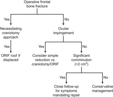
Surgical Indications
Indications for surgery in these fractures are undoubtedly the most controversial aspect of their management. Consideration must be given to the overall status of the patient, possibility of future growth, alterations, and risk of long-term complications. Concomitant neurosurgical pathology and need for neurosurgical intervention are often an important adjunctive consideration.
Orbital Roof Fractures
In our review of 159 pediatric patients with orbital roof fractures, we found that most can be managed nonoperatively. Only 14 (9%) of the patients who sustained orbital roof fractures required surgical management. In four of these patients only the orbital floor was repaired. Three patients were treated for isolated orbital roof fractures (with a mean fracture size in these cases of 3.9 cm 2 ). One of these patients received an orbitotomy with fragment reduction only while the other two patients underwent craniotomies with open reduction and internal fixation (ORIF) and pericranial flaps.
Seven patients presented with both frontal bone and orbital roof fractures. Three of these patients had the orbital roof directly addressed via craniotomy and wiring or plating and/or onlay alloplastic implant (SynPor). Three patients underwent frontal bone ORIF. One patient underwent frontal sinus obliteration.
In our cohort, most orbital roof fractures were noncomminuted and were treated nonoperatively. Management of operative orbital roof fractures will be dictated by the concomitant frontal bone or neurosurgical injuries. If a craniotomy is already necessary, it is a relatively straightforward decision to reduce and repair the roof or place an implant or split cranial bone graft, external to the roof defect. Similarly, in older children who have involvement of the frontal sinus that necessitates intervention, treatment should first address reconstruction of the orbital rim and the frontal sinus, including the possible need for obliteration or cranialization (see below). In our experience isolated roof fractures <2 cm 2 are unlikely to develop orbital encephalocele or leptomeningeal cysts.
Our analysis strongly supports the management concept that displacement direction in orbital roof fractures is significant, with inferiorly displaced “blow-in” fractures representing a substantially worse prognostic sign than superior displacement. Despite representing nearly half of fractures, none of the 10 patients who had operative roof repair had superior displacement. The magnitude of displacement was also predictive of need for surgery but only for fractures displaced inferiorly, lending further support to this concept. Two patients who had superiorly displaced roof fractures did have ORIF of the orbital floor performed but nothing done to their orbital roof fractures, suggesting that the “blow-out” mechanism can lead to simultaneous roof-floor fracture but rarely causes roof fractures that need repair. All other patients requiring surgery had significant inferior displacement of the roof bone fragments.
Direct comparison of “defect size” with roof fractures can be misleading as a concept because a 2 cm 2 defect in an infant is far different proportionally than the same area in a teenager. However, it does seem likely that there is a size below which herniation of the leptomeninges will not occur and thus fracture size should be a consideration in repair. We followed long-term outcomes on 9 patients with sub-2 cm 2 defects and 14 patients with sub-4 cm 2 defects, and none of these patients developed late complications. We therefore believe that roof fractures that measure less than 2 cm 2 in size and do not have a significant frontal bone component are unlikely to develop encephalocele and can be regarded with less concern. Fig. 2.2.1 represents our overall algorithm for management of pediatric orbital roof fractures. In the absence of any symptoms or concomitant injuries, we generally recommend close follow-up rather than prophylactic intervention even in larger defects.