Abstract
The management of fractures of the midface has undergone significant evolution as practitioners have gained a more in-depth understanding of the interplay with the remainder of the craniofacial skeleton. This chapter addresses the history, classification, diagnosis, work-up, techniques for management, and complications associated with the treatment of complex Le Fort level fractures.
Keywords
midface fractures, Le Fort I, Le Fort II, Le Fort III
Background
Injury to the midface can have significant aesthetic and functional sequelae. The bony skeleton serves as a framework that aids in respiratory, ocular, vocal, olfactory, and digestive functions. Normal anatomy and symmetry of the midface is integral to social recognition and perception.
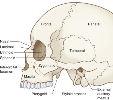
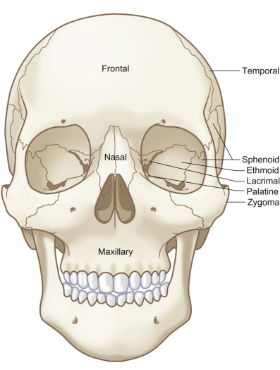
Injury to the midface can involve a complex constellation of the skeletal anatomy: the maxillary and zygomatic processes of the frontal bone, nasal bones, bones of the orbit, zygomas, ethmoids, vomer, pterygoid plates, maxilla, and palate. Both conceptually and practically, a fundamental understanding of the horizontal and vertical structural pillars of the midface skeleton is critical to understanding the diagnosis and management of these injuries. Failure to restore the structural pillars after injury can lead to inadequate projection, height, and width, resulting in a short, retruded, and widened face. The anatomical patterns if these structural pillars are compromised following the application of frontal or lateral injury forces at varied levels within the midface have been shown to be predictable, and are termed Le Fort fractures.
Etiology
Maxillary fractures are most commonly associated with motor vehicle and motorcycle collisions, followed by assault. They often occur in conjunction with facial lacerations, other facial fractures, spinal and neurological injuries, and polysystem trauma.
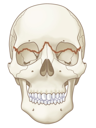
Specifically, Le Fort I fracture type patterns occur secondary to a force vector directed at or below the infraorbital foramen and above include the maxillary arch, resulting in a floating palatal and maxillary segment containing the alveolus and teeth. Le Fort II type fractures occur in the setting of a force directed at the level of the nasal bones. The fracture leads to mobility of the central midface through the orbits in a pyramidal pattern. Concomitant brain injuries are more commonly observed than in laterally directed forces. Finally, the Le Fort III type fractures result from a force vector delivered at the orbital level frequently laterally, resulting in craniofacial dysjunction from the skull base. Le Fort III type injuries are frequently more extensive and complete on one side.
Who Was René Le Fort?
In 1901, René Le Fort published his landmark work in a three-part experiment using 32 cadavers. The cadaver heads were subjected to various traumatic force vectors, the soft tissue envelope removed and fracture patterns observed within the craniofacial skeleton. First, Le Fort observed that the skull was rarely fractured if the face was fractured. Secondly, he observed that facial fracture patterns were reproducible, and occurred across three weak levels, or “linea minoris resistentiae” of the midface skeleton when the injury force was directed in an anterior to posterior vector. The most common patterns were termed Le Fort I, II, and III fractures, occurring in “weak areas” of the midface, defining the original Le Fort classification system. The classification scheme established a simple and efficient vocabulary promoting improved dialogue amongst practitioners. Though the frequency of multivector high-energy injuries has increased, resulting in midface fractures of less predictability which are more comminuted and frequently vary from side to side, these classically described major fracture patterns of injury are still applicable today.
Difference Between Surgical Le Fort Osteotomies and “Impure” Le Fort Type Injuries
Pure Le Fort fractures (single fragment and bilaterally symmetrical) are rare in the present day due to the increase in frequency of multivector high-energy mechanisms of injury. Presently, these classically described bilateral symmetric patterns form a loose classification system, and were not even purely observed in Le Fort’s experiments, and are almost relegated to surgically planned exacting osteotomies performed for correction of midface craniofacial dyscrasias. In contrast, in the traumatic setting, Le Fort fractures are often asymmetric and associated with other midface fractures. They may even occur unilaterally. Single fragment (greensticked and incomplete) Le Fort III fractures are occasionally observed, and display minimal displacement and malocclusion. Though René Le Fort did not describe a “hemi-Le Fort” injury pattern in his original classification scheme, the nomenclature serves to provide expedient and comprehensible discourse between radiologists and surgeons alike.
Midfacial Buttresses and Their Role in Craniofacial Reconstruction
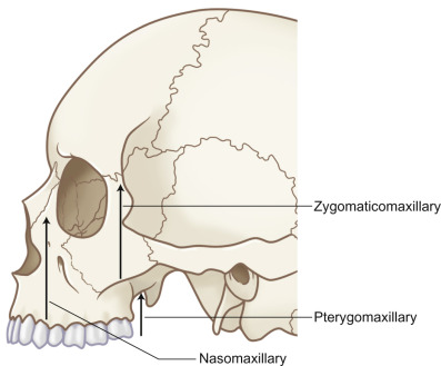
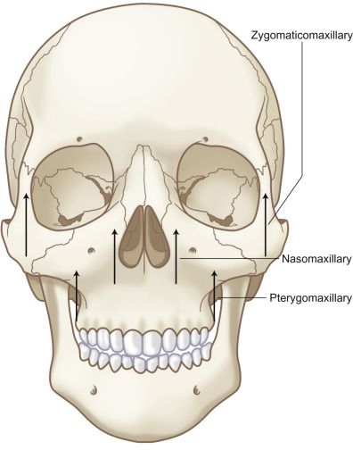
The midface “buttress” system transmits and absorbs the kinetic energy of injury forces when applied to the facial skeleton. The midface serves an important role functionally and cosmetically. The vertical pillars, which are comprised of the pterygomaxillary, nasofrontal, and zygomaticomaxillary buttresses, primarily provide protection from vertically directed force or stress vectors ( Figs. 1.13.1 and 1.13.2 ). Masticatory forces are transmitted through the skull base, mainly through the vertical buttresses. The horizontal pillars, which are comprised of the supraorbital bar, infraorbital rims, and zygomatic arches, support the transverse facial dimension and contribute supplemental orthogonal support to the buttress vertical pillars ( Fig. 1.13.3 ). These major buttresses are further supported by the thin lateral nasal walls, nasal septum, and the maxillary walls, and although weak, they serve to resist frontal and laterally directed forces. The buttress system resists external forces and prevents disruption of the facial skeleton until a critical force level is reached, resulting in fracture. Posterior to the buttress system are the skull base superiorly and the medial and lateral pterygoids inferiorly, completing a craniofacial framework with minimal sites of anatomic weakness and therefore predictable resistance to fracture patterns.
An in-depth understanding of the concepts of the buttress support system will aid in the: (1) diagnosis and treatment planning of complex facial fractures; (2) simplification of the complex interrelationship of the facial bones; and (3) placement of screws and plates in regions of adequately dense bone, allowing for the restoration of facial dimensions and profile, width and height and projection, establishing support for the nose, teeth, and globes.
Surgical Anatomy
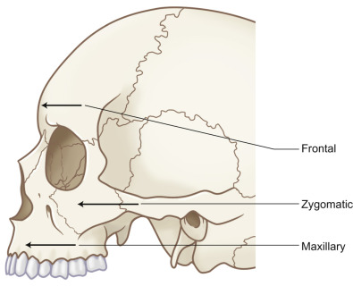
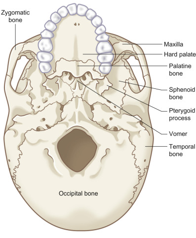
The skeletal anatomy of the cranial and midface skeleton is depicted in Figs. 1.13.4–1.13.6 .
Clinical Presentation
History
The diagnostic evaluation and work-up for the midface starts with a thorough history. Special attention to past medical history may reveal previous trauma, bone disease, osteoporosis, neoplasms both primary and metastatic, preexisting dental pathologies and interventions, nutritional and metabolic disorders, endocrinopathies, and psychiatric conditions which might influence both the etiology, timing of surgery, and choice of treatment stratagem for the relevant fracture types.
Ascertaining the etiology of the injury, specifically its force and velocity, can help the practitioner confirm the ultimate midface fracture pattern. Similarly, the vector of injury forces often serves as a predictive indicator of the ultimate fracture profile. For example, traumatic forces directed anteriorly may result in the classically described bilateral, symmetric midface Le Fort II fractures, whereas multidirectional or laterally oriented injury forces may lead to a nonclassic, asymmetric fracture pattern.
Often overlooked and frequently underappreciated is the impact of the patient’s premorbid dental history and occlusion on surgical decision-making and outcomes. Old photographs of the patient’s facial profiles, contours, lip–tooth relations and prominence of the eyes are essential. The surgeon should be aware of any history of dental interventions, extractions, cosmetic and reconstructive dental rehabilitation, use of orthodontic appliances, significant carious and/or periodontal disease. Perturbations of the premorbid occlusion should be initially screened by asking if the patient feels that their bite is abnormal. If available, premorbid photographs can be reviewed to better elucidate the patient’s baseline bite. In the patient who is completely or partially edentulous, the status of their dentures should be ascertained. For the patient engaged in phased orthodontic care, a preoperative discussion with the orthodontist may help guide treatment modalities.
Physical
Given the force that is often required to create significant midface fracture patterns and the potential for airway compromise and associated polysystem trauma, the assessment begins always with the ABCs of trauma care and Advanced Trauma Life Support protocols. Once the patient is stabilized, facial symmetry should be evaluated to distinguish the obvious deformities of the skull, soft tissue injuries, and lacerations. Presence of rhinorrhea or otorrhea should be assumed to be cerebrospinal fluid (CSF) until proven otherwise. If a patient is able to answer questions, any metallic or salty-tasting discharge can be indicative of CSF drainage.
Bimanual palpation of the entire craniofacial skeleton should be performed to identify any step-offs or bony irregularities. Signs of orbital trauma such as periorbital ecchymosis subconjunctival hematoma and edema are suggestive of orbital fracture and should be appreciated. “Raccoon eyes” or a “Battle” sign may be present and are indicative of anterior, middle or posterior cranial base fractures. Visual acuity, extraocular muscle function, the presence and extent of subconjunctival hemorrhage, diplopia, strabismus, pupil size, shape, and reaction to light should all be evaluated. In a study by Al-Qurainy et al. it was shown that 90% of patients with midface fractures presented with some degree of ocular injury, 15% had decrease in vision, 12% had severe injuries, and 16% had moderate injuries. Thus, facial fractures involving the bony orbit should have an ophthalmological evaluation.
The integrity and stability of medial canthal ligament should be evaluated by palpation. Physical findings of medial canthal disruption include epiphora, increased intercanthal distance, and rounding of the eyelid commissure and lacrimal lake. If detachment is suspected, a bow-string test can initially be performed by placing one finger on the medial canthal tendon followed by pulling the eyelid laterally with the clinician’s other hand. Lack of resistance or movement of the underlying bony platform upon lateral stress is indicative of a fracture of the area and loss of medial canthal integrity. Paskert and Manson advocated an alternate version of the bimanual exam characterized by placement of a Kelly clamp intranasally while assessing the stability of the central fragment by a finger placed externally in the region of the medial canthal attachment. The provider should also consider that soft tissue avulsion/laceration in region of the medial lid and canthus may portend an underlying injury to the lacrimal apparatus that will need to be formally evaluated.
Maximum incisal opening should be assessed to determine the extent of zygoma involvement. Decreased incisal opening can be a result of trismus, however it can also be caused by impingement on the coronoid process by a zygoma fracture. Intraoral examination should include assessment of soft tissue integrity, status of dentition and periodontium with evaluation of the teeth for subluxation, malposition dental fracture, and most importantly, occlusion. Gingival lacerations predict tooth, alveolar, and palate fractures, and contribute to malocclusion. A number of malocclusive bite patterns can be seen, the nature of which is largely dependent on the pattern of and laterality of presenting midface fracture.
Le Fort I
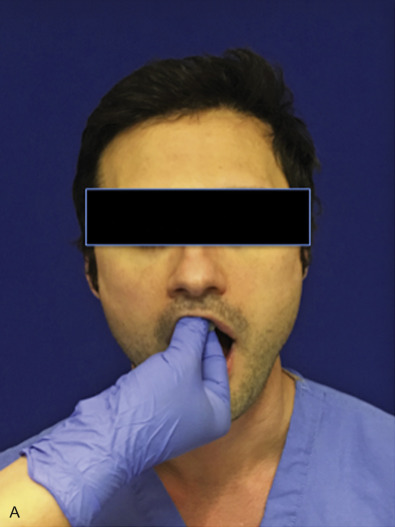
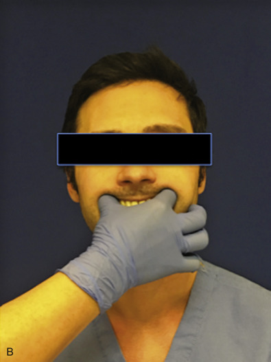
Malocclusion and segmental mobility are the most obvious indicators of an isolated Le Fort I level or palatal or alveolar fracture. Fractures of the maxilla are typically displaced posteriorly, resulting in premature contact of posterior teeth and an anterior open-bite ( Fig. 1.13.7 ). The maxilla should be assessed by placing an index finger and thumb as posteriorly as possible on the maxilla, ideally posterior to the maxillary tuberosities, followed by attempting to displace the maxilla in all three dimensions ( Fig. 1.13.8B ). Mobility of the maxilla in the superior and inferior direction can also be assessed by firmly grasping the premaxilla, holding and supporting the skull to prevent excessive movement ( Fig. 1.13.8A ). Transverse discrepancies in the arch should also be evaluated by attempting to expand and compress the maxillary arch and comparing these dimensions to the mandibular arch width. Palatal ecchymosis is a common finding in maxillary fractures. Pharyngeal lacerations should be assessed for retropharyngeal bleeding and are common in palatal fractures.
Le Fort II
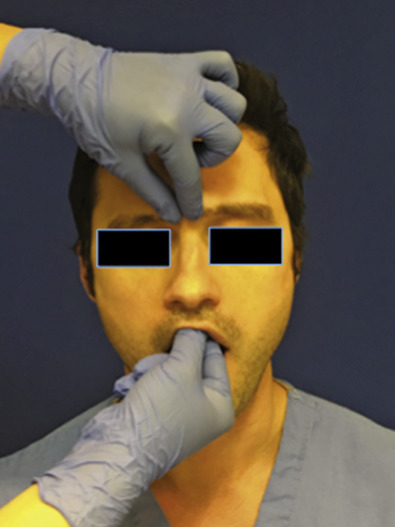
The Le Fort II fracture pattern is typically more grossly clinically apparent. Significant periorbital, perinasal, malar, maxillary, and upper lip swelling may be present. Fractures at the nasofrontal region may present with CSF rhinorrhea or epistaxis. Involvement of the orbital floor and rim may result in significant soft tissue ecchymosis, dystopia, entrapment, and conjunctival or globe injury. Extension through the maxillary wall may result in infraorbital nerve paresthesia. Gingivobuccal vestibular soft tissue swelling and ecchymosis is common. The injury complex can be evaluated by mobilizing the maxilla with thumb and forefinger as previously discussed for Le Fort I level injury, while concomitantly palpating the nasofrontal junction and orbital rims ( Fig. 1.13.9 ). Le Fort II fracture patterns are either high through the nasofrontal junction centrally, or low through the mid/lower nose.
Le Fort III
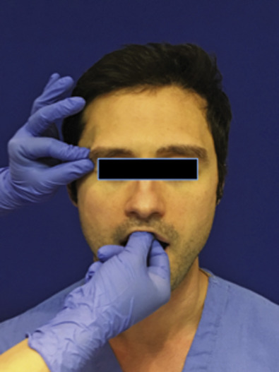
Le Fort III level injuries can result in complete craniofacial dysjunction and be associated with raccoon eyes on exam. A strong clinical suspicion for concomitant skull base injury should be maintained. CSF rhinorrhea or otorrhea may be present and should be evaluated with beta-2 transferrin as the most specific diagnostic modality. A Le Fort III fracture can be assessed with mobilization of the maxilla as previously described while simultaneously palpating the nasofrontal and zygomaticofrontal sutures for instability ( Fig. 1.13.10 ).
Radiological Evaluation
There is limited applicability for plain film radiography in the evaluation of midface Le Fort level fractures. If utilized for craniofacial skeletal evaluation outside of the midface, nonspecific radiographic opacifications of the maxillary sinuses on Waters and Caldwell views may be seen.
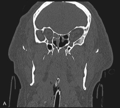
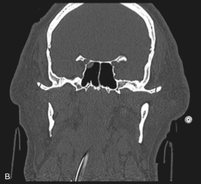
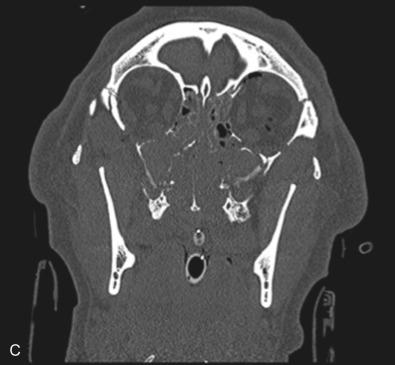
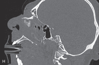
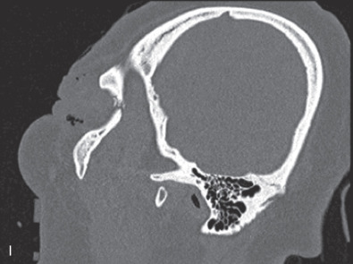
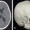
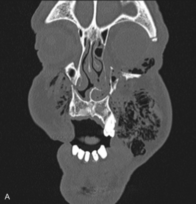
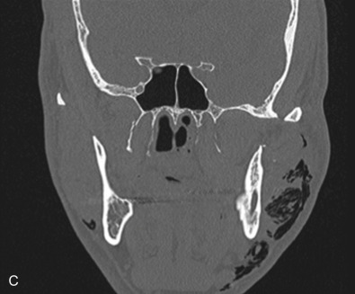
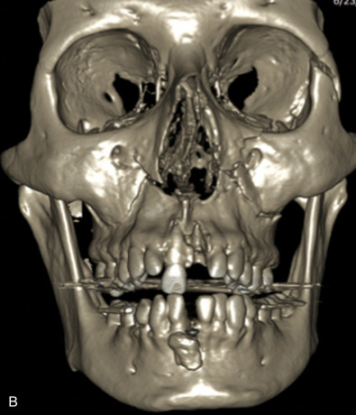
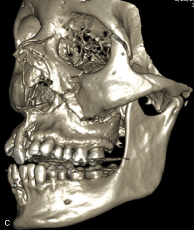
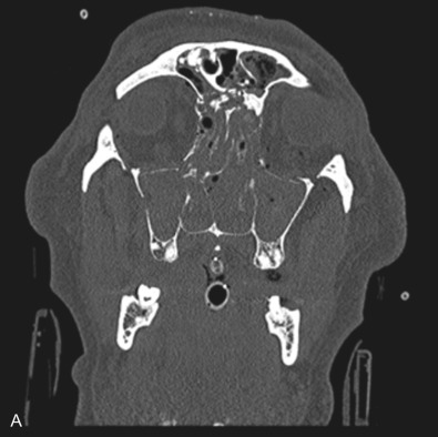
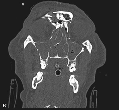
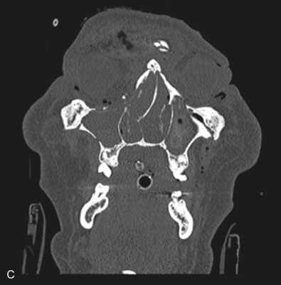
The gold standard for radiographic evaluation is thin-slice helical computed tomography (CT). Midface fractures are confirmed by axial, coronal, and sagittal views. The degree of comminution, bone loss, and detailed images of the fracture patterns can be assessed and juxtaposed to surrounding soft tissue structures. 3D reconstruction, when utilized, can aid in visualizing the complex 3D anatomical orientation of fracture fragments that occur in Le Fort injuries and facilitate reconstructive planning.
Classification
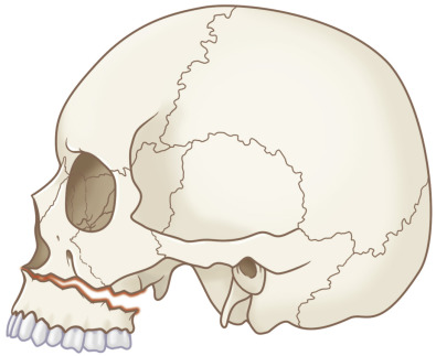
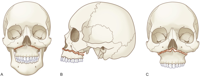
Le Fort I pattern fractures are characterized by a transverse fracture extending from the pyriform aperture, propagating laterally across the maxillary wall involving medial, anterior, and lateral components, and ending posteriorly at, or through, the level of the pterygoid plates ( Figs. 1.13.11–1.13.13 ). This results in mobilization of the lower third of the midface, whilst the upper two-thirds remains intact. The force is typically delivered above the maxillary teeth, causing a palatal/alveolar separation from the upper maxilla.
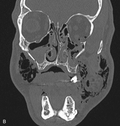

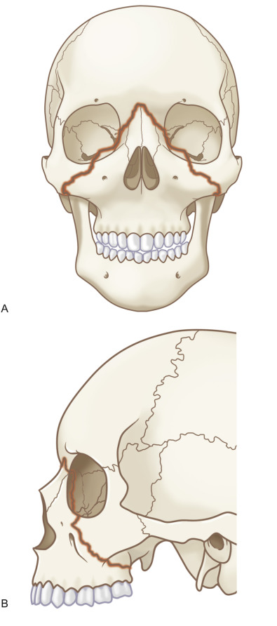
Le Fort II fractures are pyramidal in shape, involving the central portion of the midface while the lateral orbits and zygoma remain intact. The line of fracture extends bilaterally through the nasofrontal junction, medial orbital wall, inferior orbital rim, along the maxilla, through the dental alveolus anteriorly and posteriorly at the level of the maxillary tuberosity into the pterygoid plates ( Figs. 1.13.14 and 1.13.15 ). Only Le Fort II fractures violate the inferior orbital rim, causing the highest incidence of infraorbital nerve hypesthesia due to the proximity to the infraorbital foramen. Bones of the maxilla below the Le Fort II line of fracture can be intact, however they are often comminuted with other fracture patterns occurring in the Le Fort II segment. The force is typically delivered centrally at the level of the nasal bones, resulting in the separation of the central maxilla from the surrounding facial skeleton. Brain injuries are more frequent in central Le Fort II injury patterns.
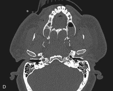
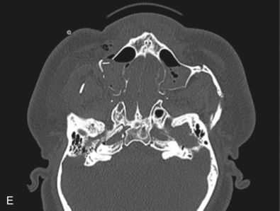
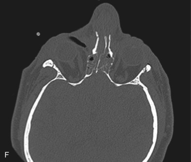
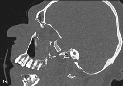
Le Fort III fractures generally consist of a combination of fractures that involve the palatine bones, the maxilla, the pterygoid plates, the nasal bones, lacrimal bone, and zygomas; they essentially separate the face along the base of the skull. The fracture pattern extends through the nasofrontal suture along the medial wall of the orbit, through the inferior orbital fissure and the lateral orbital wall to the zygomaticofrontal suture. In addition, the zygomaticotemporal suture is separated. The fracture extends across the sphenoid bone resulting in dysjunction at the pterygoid plates ( Figs. 1.13.16 and 1.13.17 ). The septum is separated from the cribriform plate of the ethmoid. Pure Le Fort III fractures are rare, and in actuality, most are ZMC fractures in conjunction with Le Fort I and II fractures lending the appearance of a comminuted “Le Fort III.” The force is delivered from the orbital level, resulting in craniofacial dysjunction. The fracture is generally more comminuted and more extensive on the side of force application.