Superficial Bacterial Infections, Folliculitis, and Hidradenitis Suppurativa
Overview
Bacteria such as Corynebacterium, Brevibacterium, Acinetobacter, and some Staphylococcus species, are commonly found on normal skin and are not pathogenic. Propionibacterium bacteria reside in hair follicles and contribute to acne.
The two gram-positive cocci, Staphylococcus aureus and the group A β-hemolytic streptococci, account for the vast majority of cutaneous infections. They may colonize normal skin or be secondary invaders of cutaneous ulcers, surgical or traumatic wounds, as well as in eczematous dermatitis, a skin disorder in which the barrier function of the skin is defective.
Methicillin resistant Staphylococcus aureus (MRSA) is the term used for bacteria of the S. aureus group that are resistant to the traditional antibiotics used against them. Problems arise because antibiotic choice becomes very limited.
 Impetigo
 Folliculitis
Staphylococcal folliculitis
Pseudomonas (hot tub) folliculitis
 Folliculitis: other types
Irritant, frictional, or chemical folliculitis
Steroid-induced acne and rosacea (see also Chapter 1, “Acne and Related Disorders”)
Eosinophilic pustular folliculitis (see also Chapter 24, “Cutaneous Manifestations of HIV Infection”).
Pityrosporum folliculitis
Majocchi’s granuloma
Viral folliculitis: herpes virus folliculitis (see Chapter 6, “Superficial Viral Infections”)
 Furunculosis (“BOILS”)
 Hidradenitis suppurativa (ACNE INVERSA)
Impetigo
Basics
Impetigo is a primary superficial bacterial infection of the superficial layers of the epidermis. Traditionally, impetigo has been divided into two forms—bullous and nonbullous (crusted). These conditions are clinically more or less indistinguishable; therefore, it is probably less confusing to use the term impetigo to describe both of them.
Impetigo is a common, highly contagious finding in preschoolers. The incidence of impetigo in children younger than 6 years of age is higher than it is in adults; however, the condition may occur in persons of all ages. Impetigo rarely progresses to systemic infection, although poststreptococcal glomerulonephritis is a rare complication.
Impetigo is caused most often by Staphylococcus aureus; less often, group A beta-hemolytic streptococci (GABHS) may be the primary pathogen. In fact, both organisms can be present at the same time in the affected sites. In recent years, methicillin-resistant S. aureus (MRSA) has been noted as a cause of impetigo; this infection is observed more commonly with the nonbullous form of impetigo than the bullous form.
 5.1 Impetigo. This child has a mixture of intact bullae and drying crusts. |
Secondary Impetigo (Impetiginization)
Impetigo can, and often does, emerge as a secondary infection of preexisting skin disease or traumatized skin; it is then referred to as secondary impetiginization. Examples include impetiginized eczema. Patients who have atopic dermatitis or other inflammatory skin conditions often have skin colonized by S. aureus.
Other conditions that may lead to impetiginization include the following:
Stasis dermatitis
Herpes simplex and varicella infections
Scabies and insect bites
Lacerations and burns
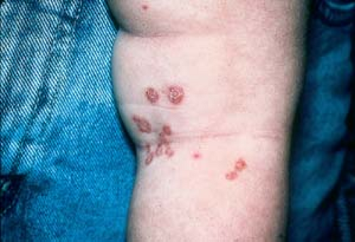 5.2 Impetigo. In this 2-year-old child, intact blisters are not present; only the flaccid remains (scaly collarettes) of bullae are seen. |
Description of Lesions
Impetigo begins as a crust or thin-roofed, fragile vesicle or bulla that ruptures and often leaves a peripheral collarette of scale or a darker, hemorrhagic, crusted border. Intact bullae are not usually present because they are very fragile; rather, they often demonstrate a collarette of scale or the flaccid remains of bullae (Figs. 5.1 and 5.2).
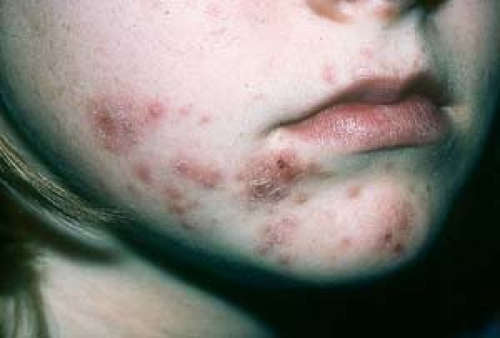
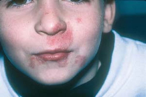
Oozing serum dries and gives rise to the classic golden-yellow, “honey-crusted” lesion. Lesions appear to be stuck on (Fig. 5.3).
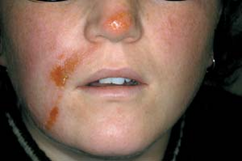
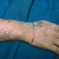
In time, a varnishlike crust (Fig. 5.4) develops centrally that, if removed, reveals a moist red base.
 5.3 Impetigo. Oozing “honey-crusted” lesions in a typical location. |
Distribution of Lesions
In children, the face is commonly involved, particularly in and around the nose and mouth, along with other exposed parts of the body (e.g., arms, legs), sparing the palms and soles.
In adults, lesions may occur anywhere on the body.
Clinical Manifestations
Spread of lesions is by autoinoculation.
Lesions are usually asymptomatic; occasionally they may itch.
The infection is self-limiting—even without treatment—and generally spontaneously resolves after a few weeks. It also typically clears with topical or oral antibiotics; only rarely do serious complications occur (see below).
Healing takes place without scarring, but it may cause temporary postinflammatory hyperpigmentation in dark-skinned persons.
Recurrent or persistent impetigo may indicate a carrier state in the patient or the patient’s family.
 5.4 Impetigo (gladiatorum). This is a college wrestler (see also discussion of herpes gladiatorum in Chapter 6). |
Diagnosis
Diagnosis is usually made on clinical grounds.
Bacterial culture and sensitivity testing are recommended if standard topical or oral treatment does not result in improvement.
A bacterial culture of the nares may be obtained to determine whether a patient is a carrier of S. aureus.
Urinalysis is necessary to evaluate for acute poststreptococcal glomerulonephritis if the patient develops edema or hypertension. Hematuria, proteinuria, and cylindruria are indicators of renal involvement.
Tinea Corporis
The potassium hydroxide examination or fungal culture is positive.
Central clearing of lesions is noted.
Honey-colored crusts are absent.
Other Diagnoses
Rare considerations include primary bullous diseases such as bullous dermatosis of childhood and bullous pemphigoid.
Rare Complications
Cellulitis, lymphangitis, scarlet fever, erysipelas, bacteremia with subsequent pneumonitis, septic arthritis, and septicemia may develop; rarely, bacterial endocarditis may also occur.
Staphylococcal scalded skin syndrome occurs more commonly in younger children. (See Chapter 9, “Bacterial Exanthems”)
Acute glomerulonephritis develops in 2% to 5% of individuals with impetigo caused by GABHS, most often in children aged 2 to 4 years. The onset is usually 10 days after the lesions of impetigo first appear, but it can occur from 1 to 5 weeks later. Transient proteinuria and hematuria may occur during impetigo and resolve before renal involvement develops.
Ecthyma, a deep dermal infection, can result, after which subsequent scarring can occur.
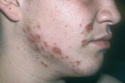
 5.5 Tinea corporis (faciale). This young boy was initially treated with topical antibiotics for presumptive impetigo. |
Antibacterial soaps such as povidone-iodine (Betadine) or chlorhexidine (Hibiclens) are used twice daily.
Gentle débridement of lesional crusts is done with a washcloth and the antibacterial soap.
Mupirocin 2% (Bactroban, Centany) ointment or cream applied three times daily may be used alone to treat very limited cases of impetigo. It is used until all lesions are cleared. Topical application of these preparations has been shown to be as effective as oral antibiotics.
For widespread involvement, an oral staphylocidal penicillinase-resistant antibiotic, such as cephalosporin, dicloxacillin, or erythromycin, may be used alone or in conjunction with topical antibiotics.
If bacterial cultures reveal MRSA, tetracyclines, trimethoprim/sulfamethoxazole (Bactrim), clindamycin, or linezolid are effective oral antibiotics.
In patients who have been determined to be chronic nasal carriers and have recurrent impetigo, and for those with MRSA, mupirocin 2% cream or ointment can be applied inside the nostrils three times daily for 5 days each month to reduce bacterial colonization in the nose.
Patients who are chronic nasal carriers also can be treated with rifampin (see later in this Chapter).
Rarely, poststreptococcal glomerulonephritis (but not rheumatic fever) has been reported to follow impetigo caused by certain strains of streptococci.
Family members should be evaluated as potential nasal carriers of S. aureus and treated, if necessary.
Chronic or recurrent impetigo should alert the clinician to the possibility of an impaired immune status.
Strains of S. aureus are usually resistant to penicillin and may be resistant to erythromycin.
An emerging problem is MRSA, which may appear in both immunocompromised and immunocompetent patients. If infectious skin lesions do not improve during treatment intended for methicillin-sensitive S. aureus, the diagnosis of MRSA should be considered, and definitive antibacterial therapy should be determined on the basis of in vitro antibiotic sensitivity.
Folliculitis
Basics
Folliculitis, in its broadest sense, may be defined as a superficial or deep infection or inflammation of the hair follicles. It has multiple causes: various infections, physical or chemical irritation, occlusive dressings or clothing, and the use of topical or systemic steroids. Hereditary forms of folliculitis such as follicular eczema are generally classified as atopic dermatitis (see Chapter 2, “Eczema”). The deeper forms of inflammatory folliculitis that involve the entire follicular structure, such as folliculitis decalvans, occur most commonly in black men and women (see Chapter 10, “Hair and Scalp Disorders Resulting in Hair Loss”).
Folliculitis may also be seen as a secondary infection in conditions such as eczema, scabies, and excoriated insect bites. It is more commonly found in patients who are diabetic, obese, or immunocompromised. Viral folliculitis may be seen in patients with herpes simplex infections, particularly in patients with human immunodeficiency virus (HIV) infection.
Related posts:
Stay updated, free articles. Join our Telegram channel

Full access? Get Clinical Tree


