Benign Skin Neoplasms
Overview
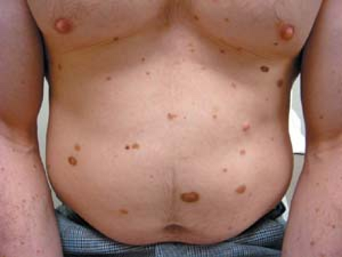
Benign lesions such as melanocytic nevi, skin tags, seborrheic keratoses, cherry angiomas, and epidermoid cysts are expected consequences of the skin’s normal, age-appropriate, often hereditary, maturation process.
As with most skin lesions, familiarity breeds recognition. This chapter, along with Chapter 22, presents the various common benign and malignant neoplasms in their diverse clinical guises.
Skin lesions—particularly pigmented skin lesions—often present a difficult and puzzling conundrum for the nondermatologist health care provider. Questions such as “Am I missing a melanoma?” “Is this mole suspicious?” and “Is this a skin cancer?” may arise. In reality, the answer is not always apparent. In fact, the decision of whether a lesion is benign or malignant is often a challenge for many dermatologists as well. Distinguishing between a benign pigmented lesion such as melanocytic nevus or a seborrheic keratosis and a potentially fatal skin cancer such as melanoma creates the most concern among health care providers.
 Melanocytic nevi
Junctional melanocytic nevus
Compound melanocytic nevus
Dermal melanocytic nevus
Blue nevus
Halo nevus
Spitz nevus
Congenital melanocytic nevus
Atypical nevus (dysplastic nevus, Clark’s nevus)
 Seborrheic keratosis
Stucco keratosis
Dermatosis papulosa nigra
Sign of Leser-Trèlat
 Skin tags
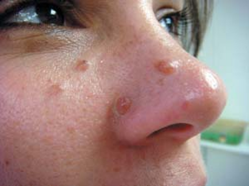
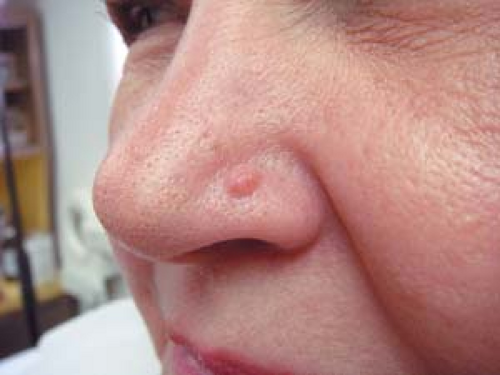
 Sebaceous hyperplasia
 Cysts
Cysts
Epidermoid
Pilar
Milium
 Lipoma
 Chondrodermatitis nodularis chronica helicis
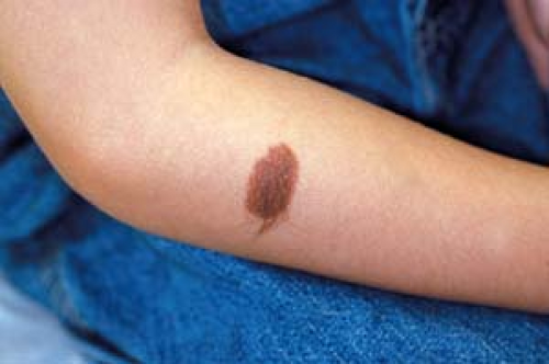
 Dermatofibroma
 Fibrous papule
 Hypertrophic scar
 Keloid
 Common angiomas
Cherry angioma
Venous lake
Angiokeratomas
Spider angioma
Pyogenic granuloma
Melanocytic Nevi
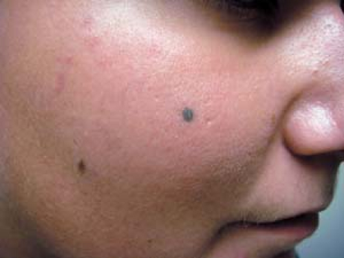
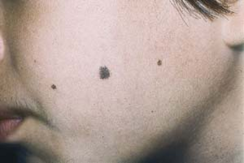 21.1 Junctional melanocytic nevi. These small, flat, lesions are uniform in color. |
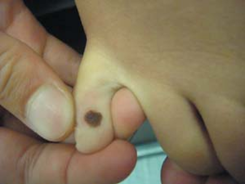 21.2 Junctional melanocytic nevus (congenital). This small lesion is extremely unlikely to become malignant. |
Basics
Melanocytic nevi (MN), commonly called moles or “beauty marks,” are, most often, benign proliferations of normal skin components. They are composed of nevus cells that are derived from melanocytes, the pigment-producing cells that colonize the epidermis.
The acquisition of MN is greatest in childhood and adolescence. In addition to an hereditary predisposition, it has been suggested that their onset is a response to sun exposure.
During adulthood, the development of new lesions tapers off, and many existing lesions gradually lose their capacity to form melanin and become skin-colored or disappear completely.
MN may be congenital or acquired, and they are much more often seen in patients with light or fair skin than in blacks or Asians.
Acquired MN are sometimes associated with melanoma; however, the frequency of transformation into a melanoma is not known. Congenital nevi, on the other hand, especially when very large, hold the greater risk of malignant transformation (see later discussion).
Junctional Melanocytic Nevi
These small, macular (flat), frecklelike lesions are uniform in color. Individual lesions may be brown to dark brown to black (Fig. 21.1).
Histologic examination reveals melanocytic nevus cells located at the dermoepidermal junction.
Whether acquired or congenital, junctional MN are most prevalent on the face, arms, legs, trunk, genitalia, palms, and soles (Fig. 21.2).
The most important lesions in the differential diagnosis of junctional MN include:
Dysplastic nevus (atypical mole; see later discussion)
Lentigo maligna (see Fig. 22.47)
Melanoma (see Chapter 22)
Freckles (Ephelides)
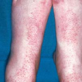
These small, tan macules appear on the sun-exposed skin of fair-skinned people (Fig. 21.3).
They darken after sun exposure and lighten when they are no longer exposed to the sun.
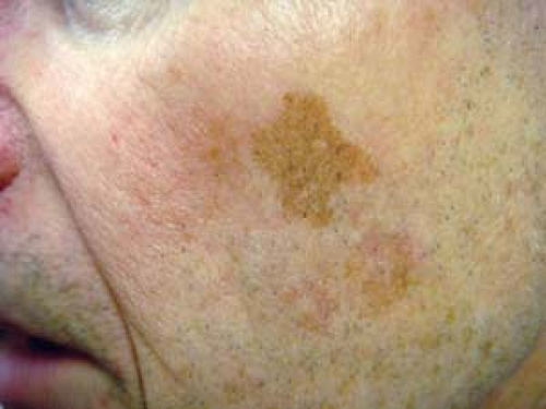
Lentigo (Plural: Lentigines) or “Liver Spot”
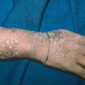
These small, acquired tan macules occur on sun-exposed areas during middle and elderly age. They are uniform in color (Fig. 21.4). Most often, they appear on the face, dorsal hands (Fig. 21.5), extensor forearms, and anterior legs.
 |
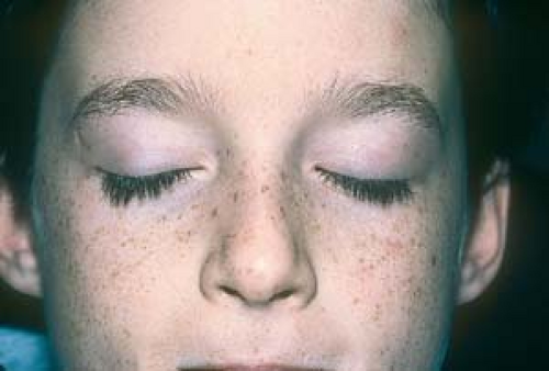 21.3 Freckles (ephelides). Freckles come and go with sun exposure. They are most often seen in people who have fair skin. |
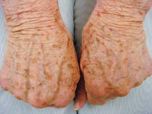 21.5 Solar lentigines. These extremely common tan macules arise on sun-exposed areas during middle age. |
 21.6 Compound melanocytic nevi. Elevated, dome-shaped papules that are most often seen on the face, arms, legs, and trunk. These lesions tend to lose pigmentation as the patient ages. |
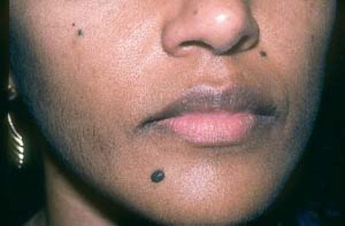 21.7 Compound melanocytic nevi. In individuals with dark skin, such lesions tend to be more intensely pigmented. |
Dermal nevi and compound nevi often resemble one another as well as the following:
Atypical nevus
Neurofibromas
Skin tags (acrochordons)
Basal cell carcinomas
Seborrheic keratoses
Angiofibroma (fibrous papules)
Nodular melanoma
Compound Melanocytic Nevi
Compound MN are elevated, dome-shaped papules or papillomatous nodules.
Uniformly brown to dark brown, they may contain hairs and are seen most often on the face, arms, legs, and trunk (Figs. 21.6 and 21.7).
Their histologic structure combines features of junctional and dermal nevi (see next section).
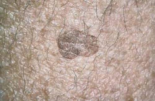
Dermal Melanocytic Nevi
Dermal MN may be elevated and dome-shaped, wartlike, or pedunculated.
Most often skin-colored, they may be tan or brown, or they may be dappled with pigmentation. Lesions tend to lose pigmentation with age and become skin-colored (Fig. 21.8).
They are most often seen on the face and neck.
Microscopy reveals that dermal MN cells are located in the dermis.
Clinical Manifestations of Dermal Nevi and Compound Nevi
Dermal nevi and compound nevi are asymptomatic unless they are irritated or inflamed.
Very rarely do they transform to malignant melanoma.
Diagnosis
The diagnosis is based on clinical appearance or, if necessary, a histopathologic evaluation after removal.
 21.8 Dermal melanocytic nevus. This lesion was pigmented when the patient was in her teens and twenties. |
Clinical Variants
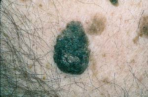
Blue Nevi
These lesions are a benign variant of dermal MN that are heavily pigmented. They occur as blue-gray or blue-black macules, papules, or nodules (Fig. 21.9) and are rarely malignant. The dark brown pigment that creates a bluish color to these lesions is caused by the Tyndall phenomenon.
Like the more common dermal and compound MN, blue nevi usually begin to appear in adolescence or early adulthood.
Halo Nevi
Halo nevi are MN that are encircled by a white halo of depigmentation. The halo represents a regression of a preexisting nevus caused by a lymphocytic infiltrate. Frequently, the entire nevus disappears, and the area regains normal pigmentation.
Most often, halo nevi are initially seen on preadolescents; they usually appear on the trunk (Fig. 21.10).
If a halo nevus is seen on an adult, two very rare possibilities should be considered: the lesion may be malignant, or a melanoma may be present elsewhere on the body. Biopsy and removal are indicated in this situation. Also bear in mind that in a patient of any age, a biopsy should be performed if the nevus has an atypical clinical appearance.
Spitz Nevi
These nevi are a distinctive variant of MN. In the past, they were referred to as “juvenile melanomas,” but now they are recognized as benign. In children, the lesions may appear as pink papules; sometimes they are heavily pigmented and are jet black in color.
A heavily pigmented, small, spindle-cell variant of Spitz nevus may be seen on the legs of women.
It is prudent to completely excise these lesions to minimize the risk of recurrence and possible confusion with a malignant lesion.
 21.9 Blue nevus. This is a variant of melanocytic nevus. Note the blue-gray color caused by the Tyndall effect. Compare to the tan-colored nevus on this boy’s face. |
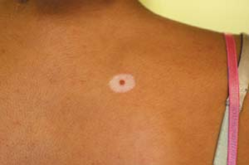 21.10 Halo nevus. An inflamed compound nevus has lost some of its original tan pigmentation. It is encircled by a white halo of depigmentation. Ultimately, the nevus may disappear, and the area will regain normal pigmentation. |
 21.11 Small to medium-sized congenital hairy nevus. This lesion is evenly pigmented and symmetric and probably has little, if any, malignant potential. |
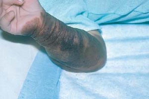 21.12 Giant pigmented hairy nevus. A patient with this lesion may have a greater than 6% lifetime risk of melanoma. |
Congenital Melanocytic Nevi
Often generating great concern in both patents and pediatricians, congenital melanocytic nevi (CMN) are MN that are present at birth or arise during the first year of life.
By definition, CMN are present at birth or soon thereafter, although some small CMN are clearly so-called “tardive” and may appear as late as up to 2 years of age.
CMN occur in about 1% of children.
On histopathology, in contrast to acquired MN described above, the nevus cells of CMN tend to be deeper in the dermis and subcutaneous fat and are often present in the skin appendages such as nerves and hair follicles.
CMN vary considerably in size and are commonly classified as small (under 1.5 cm), intermediate (1.5 to 19.9 cm) (Fig. 21.11), or large/giant (20 cm). (These figures are based on the predicted final adult size of lesions.)
Physical findings include the following:
CMN are generally relatively evenly pigmented and tan or brown, especially those that are thin.
Some lesions can have an array of colors.
The malignant potential of small or medium-sized CMN is controversial. However, many experts believe that a small nevus does not significantly increase the lifetime risk of developing melanoma.
CMN are of the greatest concern when they are 20 cm or larger in diameter. These nevi, which are often called giant pigmented hairy nevi, “garment nevi,” or “bathing trunk nevi,” may develop into melanoma—the lifetime risk is estimated to be between 3.3 and 6.0% (Fig. 21.12).
All MN should be carefully examined and biopsy should be considered, particularly if there is any suggestion of clinical atypia.
However, for most MN, biopsy is not indicated. Persons with numerous MN, particularly atypical nevi (see later discussion), are at greater risk for developing malignant melanoma.
Indications for Removal
Atypical appearance
Cosmetic reasons
Repeated irritation by clothing, such as a bra strap
Persistent discomfort (a lesion that itches, hurts, or bleeds)
Methods of Removal
Lesions can be removed by shave excision (which is often followed by electrodesiccation) or by elliptic excision (see Chapter 26, “Diagnostic and Therapeutic Procedures”).
Shave (tangential) excision: This method is fast and economical, and it generally provides satisfactory cosmetic results. Its disadvantage is that it often results in only partial removal of the lesions, which infrequently necessitates a second excisional procedure.
Elliptic excision: This technique is performed with the intent of removing lesions completely. Surgical margins can be identified. However, elliptic excision is slower than shave excision. It also requires suturing and suture removal, and it results in linear scars that may not be as cosmetically pleasing as scars that result from shave excisions.
Any pigmented lesion that changes rapidly in size or color or that has an atypical appearance should be removed for biopsy.
A primary care physician should have a low threshold for referral to a dermatologist if there is any concern regarding the diagnosis and management of a pigmented lesion.
All MN that are removed should be submitted for microscopic evaluation.
Large CMN have a low but real risk of malignant transformation and the development of melanoma.
Atypical Nevus (Dysplastic Nevus, Clark’s Nevus)
 21.13 Atypical nevus (dysplastic nevus). Note the raised center and indistinct border; such a nevus is sometimes called a “sunny-side-up egg lesion.” It is generally larger than a common mole. |
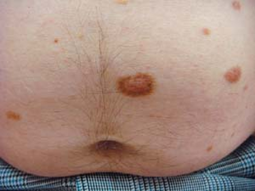
 21.14 Multiple dysplastic nevi. Note the characteristic distribution on the trunk. |
Basics
The atypical nevus, which is also called dysplastic nevus, an atypical mole, or Clark’s nevus, is a controversial and confusing lesion. Even among dermatopathologists, there is no consensus regarding the histopathologic criteria for its diagnosis.
Some individuals have only a few atypical nevi, and their risk of melanoma may not be much higher than those individuals without such nevi.
This much is agreed: When a patient has numerous atypical nevi and there is a positive family history of melanoma, the potential for melanoma in that patient and in his or her family is extremely high. Such dysplastic nevi may be inherited as an autosomal dominant trait (see discussion of familial atypical mole syndrome).
Atypical nevi are rarely seen in black, Asian, or Middle Eastern populations.
Description of Lesions
Atypical nevi have some or all of the following features:
They are usually larger than common moles and frequently measure 5 to 15 mm in diameter.
Their borders are usually irregular, notched, and ill-defined.
They have a macular appearance, but the centers may be raised (for this reason, they are sometimes called “sunny-side-up egg lesions”) (Fig. 21.13).
Their coloration (tan, brown, black, pink, or red) is irregular.
Distribution of Lesions
Atypical nevi are most often found on the trunk, legs, and arms; generally, the face is spared.
Clinical Manifestations
The exact risk of an individual atypical nevus developing into a melanoma is uncertain.
Unlike dermal and compound nevi, these lesions often continue to appear into adulthood.
Differentiating them clinically from melanoma is often difficult.
Clinical Variants
Sporadic Atypical Nevi
A patient with an isolated atypical nevus and no family history of multiple atypical nevi or melanoma probably carries little risk of developing melanoma and should not necessarily be identified as prone to melanoma.
Multiple Atypical Nevi
The exact risk of an individual nevus developing into a melanoma is uncertain (Fig. 21.14).
In certain situations, atypical nevi are considered possible precursors to, as well as potential markers for, the development of melanoma that may occur de novo without evolving from a precursor dysplastic nevus.
Familial Atypical Mole Syndrome
Those persons who meet the following criteria are considered to have an extremely high potential for developing malignant melanoma:
Patients with a first-degree (e.g., parent, sibling, or child) or second-degree (e.g., grandparent, grandchild, aunt, uncle) relative who has a history of malignant melanoma have heightened risk.
Many nevi—often more than 50—are present, and some of them are atypical moles.
Moles show certain dysplastic features when examined under the microscope.
Other MN (see earlier discussion)
Malignant melanoma
Pigmented basal cell carcinoma
The method chosen for removing suspected atypical nevi depends on the purpose of treatment.
If melanoma is suspected, complete excision should be performed.
If melanoma is not suspected, the lesion can be removed and prepared for biopsy with a shave or punch biopsy technique.
Prevention
Patients with many atypical nevi should avoid excessive sun exposure and should routinely use a sunscreen with a sun protective factor of 15 or greater.
Patients who meet the criteria for familial atypical mole syndrome should examine their own skin every 2 to 3 months, in addition to having a full body examination and regular screening visits performed by a dermatologist.
High-risk patients and their families should be taught self-examination to detect changes in existing moles and should be given printed material with photographs to help them recognize the features of malignant melanoma.
The risk of melanoma is greatly increased in patients with multiple atypical nevi and a personal or family history of melanoma.
Once a diagnosis of multiple atypical nevi is established, other family members should be examined.
A person with an isolated atypical nevus probably carries little risk of developing melanoma and should not be identified as melanoma prone.
Melanoma risk is greater in those persons who have one relative with melanoma than in those with no affected relative. The lifetime risk of melanoma may approach 100% in persons with atypical nevi who are from melanoma-prone families (i.e., individuals having two or more first-degree relatives with melanoma).
Patients with the familial atypical multiple mole and melanoma syndrome (also known as the dysplastic nevus syndrome) should be monitored vigilantly.
Despite the fact that patients with many dysplastic nevi are at a very high risk of developing a melanoma, the notion of removing all of their dysplastic nevi to reduce their risk of melanoma is generally believed to be ill advised. To consider these nevi to be precursors of melanomas creates undo anxiety for patients but does not appear to decrease their potential for developing melanomas.
Seborrheic Keratosis
 21.15 Seborrheic keratoses. The largest, darkest, lesion has a warty, rough-surfaced, tortoise shell-like, “stuck-on” appearance. The lesions in the background are also SKs; these lesions are unusual before 30 years of age. (Compare with images of melanoma in Chapter 22: Figs. 22.35, 22.36, 22.37, 22.38, 22.39, 22.40, 22.41, 22.42 and 22.43.) |
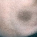
 21.16 Seborrheic keratoses. This lesion is almost flat. Lesions are found on the arms and legs. |
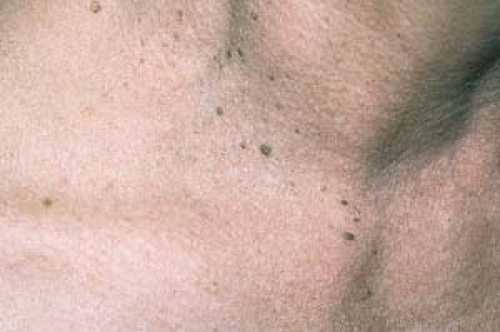 21.17 Seborrheic keratoses. Multiple pigmented papules, some of which are clinically indistinguishable from skin tags, are evident. (See Fig. 21.22.)
Related posts:Stay updated, free articles. Join our Telegram channel
Full access? Get Clinical Tree
 Get Clinical Tree app for offline access

|