Illustrated Glossary of Basic Skin Lesions
Lesions
Primary Lesions
 |
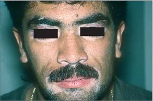
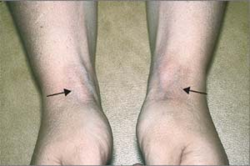
Macules are small, flat, nonpalpable changes in skin color (you cannot feel macules and, if you close your eyes, they “disappear”). They occur in various shapes and sizes.
Examples include tattoos, flat nevi, postinflammatory hyperpigmentation, postinflammatory hypopigmentation, erythema, purpura, and freckles.
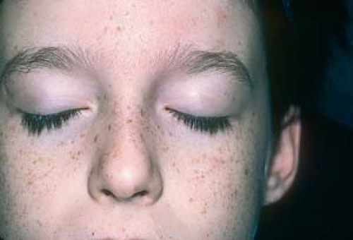 Macule. Freckles (ephelides). |
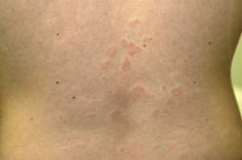
Patches are large macules.
Examples include melasma and vitiligo. There is some confusion regarding patches; some dermatologists refer to a patch as a large macule, whereas others refer to patches as macules with overlying fine scale (e.g., the scaly patches seen in pityriasis rosea and tinea versicolor).
 Patch. Vitiligo. |
 |
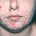
Papules are small, solid lesions that are generally 1 cm or less in diameter.
Examples include, molluscum contagiosum, warts, and palpable nevi (moles).
Note: “Maculopapule” is a contradiction in terms, and the use of this term should be abandoned (an eruption may be described as being macular and papular, rather than “maculopapular”).
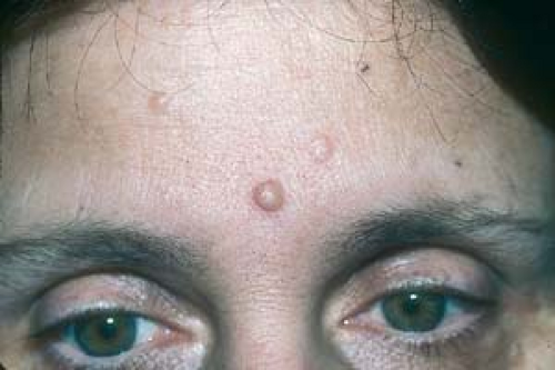 Papule. Multiple nevi |
 |
Nodules are firm, solid palpable lesions that are generally 1 cm or more in diameter. They may be seen as elevated lesions or can be palpated without any elevation of the skin.
Examples include erythema nodosum, lipoma, rheumatoid nodules, basal cell carcinoma, and keloid.
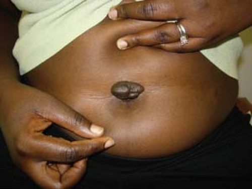 Nodule. Keloid |
 |
Vesicles (small blisters) are clear, fluid-filled lesions generally 1 cm or less in diameter.
Examples include herpes simplex, acute vesicular tinea pedis, and early chickenpox.
 Vesicle. Chickenpox. |
 |
Bullae (large blisters) are clear, fluid-filled lesions generally 1 cm or more in diameter.
Examples include second-degree burns, herpes zoster, and insect bite reactions.
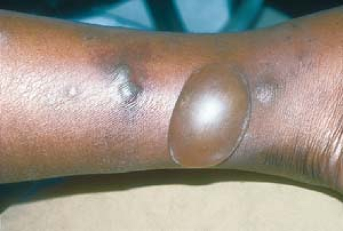 Bulla. Bullous insect bite reaction. |
 |
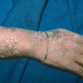
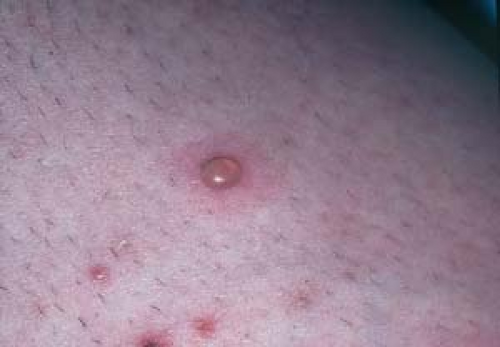
Pustules are superficial lesions that contain purulent, cloudy material.
Examples include evolving chickenpox, folliculitis, and pustular acne.
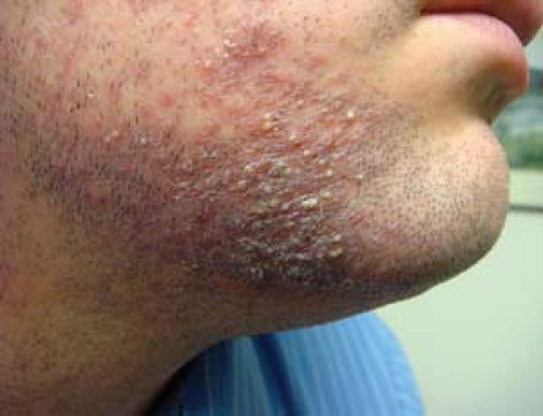 Pustule. Folliculitis barbae. |
 |
Plaques are solid, elevated, or depressed (atrophic) flat-topped, plateaulike lesions that cover a fairly large area; they may arise from papules that coalesce or arise de novo.
Examples include chronic eczematous dermatitis and psoriasis.
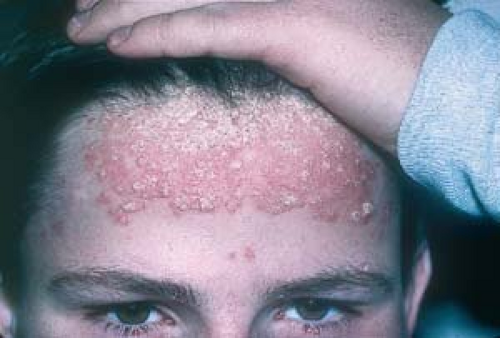 Plaque. Psoriasis vulgaris. |
 |
Atrophic plaques include discoid lupus erythematosus, morphea (localized scleroderma), and steroid atrophy.
 Atrophic plaque. Atrophy caused by intralesional cortisone injections. |
 |
Wheals are raised flesh-colored or erythematous papules or plaques that are transient lesions. They generally last less than 24 hours, during which time they may change shape and size.
Examples include urticaria (hives) and angioedema.
 Wheal. Urticaria.
Related posts:Stay updated, free articles. Join our Telegram channel
Full access? Get Clinical Tree
 Get Clinical Tree app for offline access

|