Superficial Fungal Infections
Overview
Superficial fungi are capable of germinating on the dead outer horny layer of skin. They produce enzymes (keratinases) that allow them to digest keratin, with resulting epidermal scale (e.g., tinea pedis, tinea versicolor); thickened, crumbly nails (onychomycosis); and hair loss (tinea capitis). In the dermis, an inflammatory reaction may result in erythema, vesicles, and, infrequently, a more widespread autoeczematous eruption known as an “id” reaction.
Infection may be acquired by the following means:
Person-to-person contact
Animal contact, especially with kittens and puppies
Contact with inanimate objects (fomites)
Environmental and hereditary factors leading to fungal infections are as follows:
Warm, moist, occluded environments such as the groin, axillae, and feet
Family history of tinea infections
Lowered immune status of the host, such as seen in patients with acquired immunodeficiency syndrome (AIDS), diabetes, collagen vascular diseases, or long-term systemic steroid therapy
Diagnosis can often, but not always, be made on clinical grounds.
A direct potassium hydroxide (KOH) examination or a fungal culture is necessary to make a definitive diagnosis.
Periodic acid–Schiff stain on biopsy specimens can be helpful.
Wood’s lamp examination may be useful in some cases of tinea capitis and tinea versicolor.
 Dermatophyte Infections
The term tinea refers to an infection by dermatophytes. Tinea is named according to the location on the body:
Tinea pedis and tinea manuum (feet and hands)
Tinea cruris (inguinal folds)
Tinea capitis (scalp)
Tinea corporis and tinea faciale (body and face)
Tinea unguium (onychomycosis) (nails)
 Yeast Infections
Tinea versicolor
Tinea versicolor is an exception; in fact, it is not caused by a dermatophyte but rather by a yeastlike organism. Tinea versicolor is referred to as pityriasis versicolor by many authors.
Candidiasis
Cutaneous candidiasis is also caused by yeast.
Tinea Pedis (“Athlete’s Foot”)
Basics
Tinea pedis is an extremely common problem seen mainly in young men. Ubiquitous media advertisements for athlete’s foot sprays and creams are testimony to the commonplace occurrence of this annoying dermatosis.
Most cases are caused by Trichophyton rubrum, which evokes a minimal inflammatory response, and less often by T. mentagrophytes, which may produce vesicles and bullae; less frequently, Epidermophyton floccosum may be responsible. There are three clinical types of tinea pedis: type 1: interdigital; type 2: chronic plantar; and type 3: acute vesicular.
Type 1: Interdigital Tinea Pedis
Basics
This is the most common type of tinea pedis. It is seen predominantly in men between the ages of 18 and 40 years.
Description of Lesions
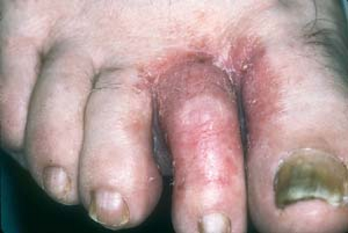
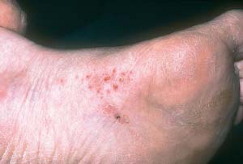
Scale, maceration, and fissures are characteristic (Fig. 7.1).
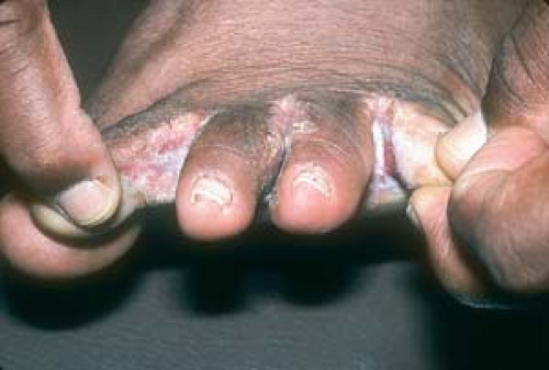 7.1 Interdigital tinea pedis (toe web infection). Note fissuring and maceration. |
Distribution of Lesions
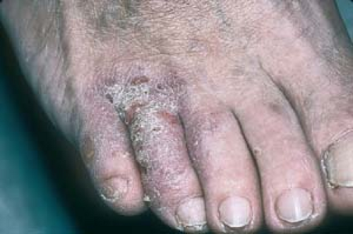
Toe web involvement is seen, especially between the third and fourth and the fourth and fifth toes; however, any web space may be involved (Fig. 7.2).
Clinical Manifestations
It is often asymptomatic; however, it may itch intensely.
Marked inflammation and fissures suggest secondary bacterial superinfection.
There may be coexistent yeast or saprophytic fungi present.
Diagnosis
A positive KOH examination or fungal culture is diagnostic.
 7.2 Interdigital tinea pedis. Here the lesions are more inflammatory. |
Atopic Dermatitis
Atopic dermatitis and dyshidrotic eczema may be clinically indistinguishable from tinea pedis (see Chapter 2, “Eczema”).
There is a positive atopic history.
It is seen especially in children on the dorsal or plantar surface of the feet (tinea pedis is unusual in preteens).
Contact Dermatitis (Fig. 7.4)
This occurs most often on the dorsum of the feet.
 7.3 Atopic dermatitis. Note the lichenification on the dorsal toes. The distinction from tinea pedis was based on a negative KOH and a positive atopic history. |
 7.4 Contact dermatitis. In this case it was determined that the patient was allergic to a component of his shoe leather. |
For acute oozing and maceration, Burow’s solution compresses are used two to three times daily. (See handout Burow’s Solution)
Broad-spectrum topical antifungal agents such as ketoconazole (Nizoral), ciclopirox (Loprox), or clotrimazole (Lotrimin) are applied once or twice daily. (See Table 7.1.)
Prevention consists of maintaining dryness in the area by:
Using a hairdryer after bathing.
Applying powders, such as Zeasorb-AF, that contain miconazole as an active ingredient.
Table 7.1 Antifungal Drug Formulary | ||||||||||||||||||||||||||||||||||||||||||||||||||||||||||||||||||||||||||||||||||||||||
|---|---|---|---|---|---|---|---|---|---|---|---|---|---|---|---|---|---|---|---|---|---|---|---|---|---|---|---|---|---|---|---|---|---|---|---|---|---|---|---|---|---|---|---|---|---|---|---|---|---|---|---|---|---|---|---|---|---|---|---|---|---|---|---|---|---|---|---|---|---|---|---|---|---|---|---|---|---|---|---|---|---|---|---|---|---|---|---|---|
| ||||||||||||||||||||||||||||||||||||||||||||||||||||||||||||||||||||||||||||||||||||||||
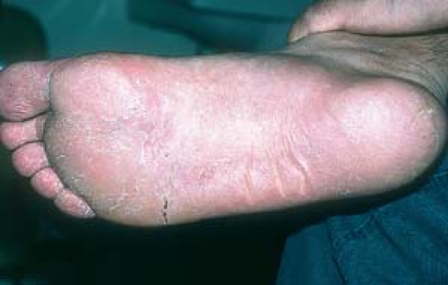
Type 2: Chronic Plantar Tinea Pedis
Basics
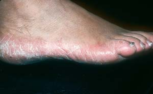
Tinea pedis (“moccasin” type) is relatively common.
Distribution of Lesions
The entire plantar surface of the foot is usually involved.
Borders are distinct along the sides of the feet.
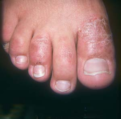
There is often nail involvement.
 7.5 Tinea pedis. Chronic scaly infection of the plantar surface of the foot in a “moccasin” distribution. Note involvement of the nails. |
Clinical Manifestations
Symptoms are minimal, unless painful fissures occur.
Diagnosis
The KOH examination or fungal culture is positive.
“Two Feet, One Hand” (Palmar/Plantar) Tinea
Tinea can present on one or both palms (tinea manuum). Not infrequently, it appears in a “two feet, one hand” distribution. This is pathognomic for tinea (Fig. 7.6).
Management is similar to that for chronic tinea pedis.
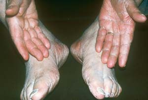 7.6 “Two feet, one hand” variant of tinea pedis. The scale is present on one hand only. Note the nail involvement. These findings are pathognomonic. |
Atopic Dermatitis
Atopic dermatitis usually is pruritic, whereas chronic plantar tinea pedis is generally asymptomatic.
There is a positive atopic history.
 7.7 Psoriasis. A well-demarcated plaque that spares the instep is seen here. |
This is the most difficult type of tinea pedis to cure, because topical agents do not effectively penetrate the thickened epidermis. Treatment generally requires oral antifungal agents such as the following (Table 7.1):
Terbinafine (Lamisil) 250 mg once daily for 30 days or longer, if necessary
Itraconazole (Sporanox) 200 mg once daily for 30 days or longer, if necessary
Fluconazole (Diflucan) 150 to 200 mg once daily for 4 to 6 weeks, if necessary
Nail involvement requires longer treatment because nails may serve as a reservoir for reinfection and take much longer to grow out normally (see later in this chapter).
Type 3: Acute Vesicular Tinea Pedis
Basics
This is the least common clinical variant.
Description of Lesions
Vesicles and bullae generally occur on the sole, great toe, and instep of the foot.
Clinical Manifestations
Acute vesicular tinea pedis is pruritic (Fig. 7.8).
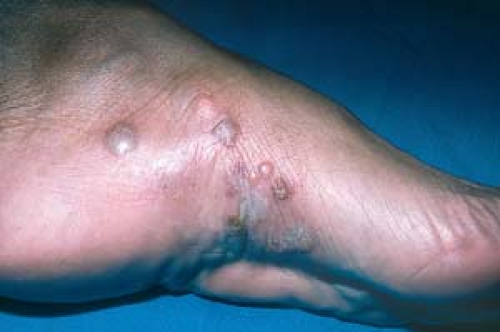 7.8 Acute vesicular tinea pedis. A KOH culture specimen is obtained from under the roof of a vesicle. |
Diagnosis
For diagnosis, the specimen should be obtained from the inner part of the roof of the blister for KOH examination or culture.
Dyshidrotic eczema is easily confused with acute tinea pedis (Fig. 7.9).
Patients often have a positive atopic history.
It is KOH negative.
 7.9 Dyshidrotic eczema. Note the small vesicles and the similarity to acute tinea pedis. |
See Table 7.1.
For symptomatic relief of severe inflammation and itching, a potent topical steroid, such as triamcinolone acetonide cream, may be used for 4 to 5 days. The resultant anti-inflammatory effect also helps to increase the yield of obtaining organisms on KOH examination or culture.
Treatment is similar to that of type 1, although systemic as well as topical antifungals are often necessary.
Prevention involves decreasing wetness, friction, and maceration. Absorbent powders, such as miconazole (Zeasorb-AF) powder, should be applied after the eruption clears to prevent recurrence.
Point to Remember
Not all rashes of the feet are fungal. In fact, if a child younger than 12 years has what appears to be tinea pedis, it is probably another skin condition, such as eczema.
When the diagnosis is in doubt, a potent topical steroid may be applied—for a week or so only—to relieve the acute itch and burning.
To increase positive yields, KOH examination or fungal cultures should be obtained only after the patient has not used topical therapy for at least 24 to 48 hours.
Positive results of KOH examination and fungal cultures are not necessarily proof of pathogenesis, because some organisms, especially yeasts and molds, may be saprophytes, or “contaminants.”
When there is a rash on the palms, the feet should always be examined.
A common error is automatically to assume that a rash of the feet, or “athlete’s foot,” is fungal in origin. Often, these conditions are mistakenly treated with topical antifungal preparations alone or in combination with topical steroids with a “shotgun” approach. Careful observation and a positive KOH examination reveal the true nature of the problem. This caveat also applies to tinea cruris (see following section).
Type 2 (Chronic Plantar and/or Palmar) Only:
In patients in whom oral antifungal agents are contraindicated, a keratolytic agent such as Salex cream or lotion can be used to remove scale, followed by a topical antifungal agent such as ketoconazole cream.
Clinical Variant
Uncommonly, an id reaction (dermatophytid) may occur. This is considered to be a hypersensitivity to fungal elements. Clinically, lesions consist of itchy, sterile (KOH and culture negative) vesicles on the hands similar to dyshidrotic eczema; this resolves when the primary acute process on the feet resolves.
Tinea Cruris
Basics
Tinea cruris (“jock itch”) is a common infection of the upper inner thighs that most often occurs in postpubertal male patients. It is generally caused by the dermatophytes T. rubrum and E. floccosum. In contrast to candidiasis and lichen simplex chronicus, it generally spares the scrotum.
Description of Lesions
Lesions are bilateral, fan-shaped, or annular plaques (plaques with central clearing), with a slightly elevated scaly “active border” (Fig. 7.10).
Distribution of Lesions
Lesions may involve the upper thighs, the crural folds, and possibly the pubic area and buttocks (see Fig. 3.12).
It generally spares the scrotum and penis.
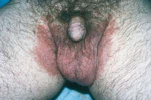
 7.10 Tinea cruris. Note the scalloped shape with an “active border.” |
Clinical Manifestations
Generally, the lesions are pruritic, “burning,” or irritating.
Frequently, the patient also has tinea pedis.
The condition may be chronic or recurrent, depending on environmental factors and exercise.
The likelihood of the spread of tinea cruris between sexual partners appears to be very small.
Diagnosis
A positive KOH examination or fungal culture is found most easily by sampling from the borders of the lesions.
Lichen Simplex Chronicus (Eczematous Dermatitis)
Figure 7.11 (see also Fig. 2.45).
It often involves the scrotum.
KOH examination and fungal cultures are negative for fungus.
Often, there is an atopic history.
Lesions are confluent (no central clearing).
Lichenification occurs as the condition becomes chronic.
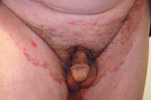
 7.11 Lichen simplex chronicus (chronic eczematous dermatitis). Lichenification of scrotal and inguinal skin results from relentless scratching and rubbing. |
Inverse Psoriasis
Figure 7.12 (see also Fig. 3.24.)
It often involves the scrotum.
Evidence of psoriasis may be noted elsewhere.
KOH examination and fungal cultures are negative for fungus.
Lesions are confluent (no central clearing).
Other Diagnoses
Seborrheic Dermatitis, Candidiasis, Intertrigo, and Irritant Dermatitis
All of these conditions may be clinically indistinguishable from one another.
 7.12 Inverse psoriasis. Note the well-demarcated plaques with no “active border.
Related posts:Stay updated, free articles. Join our Telegram channel
Full access? Get Clinical Tree
 Get Clinical Tree app for offline access

|