Abstract
Urticaria and erythema multiforme are characterized by lesions that are so distinctive that they are assigned special names. In urticaria, the lesion is a hive (or wheal), which is defined, in Chapter 3 , as a papule or plaque of dermal edema, often with central pallor and an irregular, erythematous border. The target lesion , when present, is diagnostic of erythema multiforme and is characterized by three concentric zones of color. The third disease described in this chapter is erythema migrans, an expanding annular erythematous skin lesion found in Lyme disease. Additional features of these disorders are outlined in Table 16.1 and in the discussion that follows.
- 1.
The distinctive morphology of the specialized erythemas is key to the diagnosis (e.g., erythema multiforme)
- 2.
Specialized erythemas represent a reactive pattern to an underlying cause (e.g., erythema multiforme caused by herpes simplex infection)
| Frequency (%) a | Etiology | History | Physical Examination | Differential Diagnosis | Laboratory Test | |
|---|---|---|---|---|---|---|
| Erythema migrans | < 0.1 | Tick-borne spirochete ( Borrelia burgdorferi ) | Constitutional symptoms accompany rash Prior tick bite | One or more expanding red, annular macules at least 5 cm in diameter | Cellulitis Tinea corporis Granuloma annulare Fixed drug reaction Other insect bite reaction | Serology Skin biopsy |
| Erythema multiforme | 0.3 | Drugs Infection Idiopathic | Constitutional prodrome Prior herpes simplex infection | Erythematous plaques Bullae Target lesions Mucous membrane involvement | Urticaria Viral exanthem Drug reaction Blistering disease | May be indicated: Chest radiography Skin biopsy |
| Urticaria | 2 | Ingestants Drugs Foods Infection Physical agents ?Emotions Idiopathic | Lesions last < 24 h | Wheals Generalized distribution | Erythema multiforme Drug reaction Urticarial vasculitis Erythema marginatum | – |
a Percentage of new dermatology patients with this diagnosis seen in the Hershey Medical Center Dermatology Clinic, Hershey, PA.
Erythema Migrans
- 1.
Hallmark sign of Lyme disease, the most common tick-borne disease in the United States
- 2.
Caused by Borrelia burgdorferi , which is transmitted by a tick, Ixodes spp.
- 3.
Treatment with antibiotics avoids late complications, notably arthritis
Definition
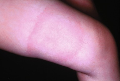
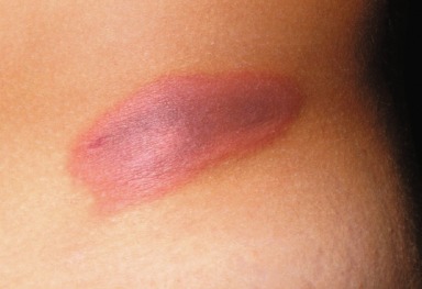
Erythema migrans represents the skin lesion associated with Lyme disease, a tick-borne illness caused by the spirochete Borrelia burgdorferi . Erythema migrans begins as a small, erythematous macule or papule that expands slowly over days to weeks. It must achieve a diameter of at least 5 cm to qualify as erythema migrans ( Fig. 16.1 ). Erythema migrans occurs in 60% to 80% of patients with Lyme disease. The late manifestations include involvement of the musculoskeletal, nervous, or cardiovascular system.
The size criterion for erythema migrans is a diameter of 5 cm.
Erythema migrans occurs in 60% to 80% of patients with Lyme disease.
Incidence
Lyme disease was first described in 1977, when it was diagnosed in a cluster of children living near Lyme, Connecticut, who were initially thought to have juvenile rheumatoid arthritis. Since then, the numbers of reported cases and their geographic distribution have increased steadily. Although cases have been reported from nearly every state in the United States, most cases occur in the northeast, midwest, and west coast. Cases are also seen in Central Europe and Scandinavia. Lyme disease is the most frequently reported arthropod-borne disease in the United States. Most cases occur between May and September.
Lyme disease is the most frequent arthropod-borne disease in the United States.
History
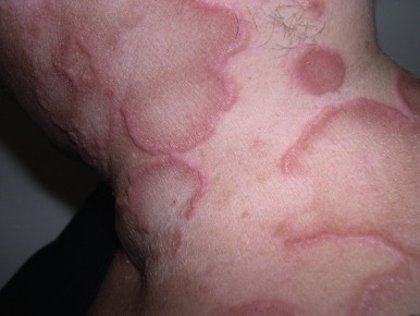
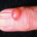
Erythema migrans begins 3 to 30 days after a tick bite ( Fig. 16.2 ). Because the tick is so small, many patients do not recall having received a bite. Most patients, however, do have a history of recent exposure to potential tick habitats such as woodlands or grassy areas. Many patients with erythema migrans have accompanying systemic symptoms such as fever, myalgia, arthralgia, headache, malaise, or fatigue. The skin lesion itself is usually asymptomatic but is noted by the patient to expand slowly over time.
A history of tick bite is often lacking.

Physical Findings
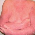
The erythema migrans lesion is located at a body site favored by a feeding tick, such as the waistband and intertriginous areas, as well as the extremities ( Fig. 16.3 ). The diameter of the lesion must be at least 5 cm to qualify as erythema migrans. From reported cases, the average diameter has been found to be 15 cm, but a diameter of 68 cm has been reported.
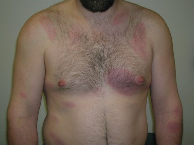
A central punctum from the tick bite may be evident but often is not. Typical erythema migrans has a macular border and a clearing center, but less classic features are common and include a papular border, alternating rings of erythema and clearing, and a center that is intensely erythematous, vesicular, purpuric, necrotic, or even ulcerated. However, all erythema migrans lesions have in common an expanding border. Multiple skin lesions occur in 15% of patients ( Fig. 16.4 ).
Erythema migrans lesions expand slowly over days to weeks.
Differential Diagnosis
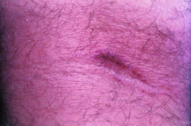
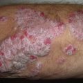
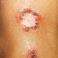
The differential diagnosis includes cellulitis, tinea corporis, granuloma annulare, fixed drug reaction, and other insect bite reactions. Except for cellulitis, these conditions are not accompanied by systemic symptoms. In addition, compared with erythema migrans, the cellulitis is more tender and usually more acute, warmer, and redder; tinea corporis has a scaling border that is potassium hydroxide-positive for fungal elements and is more chronic; granuloma annulare, an idiopathic dermal granulomatous process, has a firm, elevated border and persists for months to years ( Fig. 16.5 ); fixed drug eruption has no central clearing, is violaceous, and characteristically recurs in the same spot within hours of ingestion of the offending agent ( Fig. 16.6 ); other insect bite reactions often have more prominent central puncta, are smaller, and usually more transient than erythema migrans.
- ●
Cellulitis
- ●
Tinea corporis
- ●
Granuloma annulare
- ●
Fixed drug reaction
- ●
Other insect bite reactions
Laboratory and Biopsy
Lyme disease is diagnosed clinically, especially in endemic areas. Serologic tests (e.g., enzyme-linked immunosorbent assay; ELISA) for IgM and IgG anti- Borrelia burgdorferi antibodies are commonly used, but this test is neither sensitive (antibodies do not appear until after the first 2–4 weeks of illness) nor specific (false-negative results in infected individuals and false-positive results in persons with other diseases including systemic lupus erythematosus and rheumatoid arthritis). Positive or equivocal tests should be followed up with a standardized Western blot. An unequivocal diagnosis is established by culturing Borrelia . This is a low-yield procedure. Polymerase chain reaction (PCR) testing for Borrelia DNA is best done on cerebrospinal or synovial fluid, but is expensive and not widely available.
Serologic tests have limited usefulness.
Therapy
The ideal treatment is prevention, including tick avoidance, protective clothing, DEET ( N,N -diethyl-m-toluamide) repellent, and prompt removal of ticks within 24 hours. All patients with erythema migrans are defined as having Lyme disease and thus require antibiotic therapy. The preferred agent is doxycycline, 100 mg twice daily for 10 to 21 days. Alternatively, amoxicillin 500 mg three times daily for 10 to 21 days can be used. Erythromycin 250 mg four times daily for 10 to 21 days is a third but less preferable alternative. Manifestations and therapy of early disseminated infection and late persistent infection are discussed below.
Course and Complications
The course and manifestations of Lyme disease share many similarities with syphilis. In Lyme disease, even without therapy, the erythema migrans lesion usually resolves spontaneously within a month. However, without treatment of early localized infection (stage 1), the disease may progress to stage 2 or 3.
Stages:
- 1.
Localized
- 2.
Early disseminated
- 3.
Late persistent
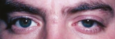
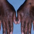
In stage 2 (early disseminated infection), the B. burgdorferi spirochete is spread hematogenously to distant sites. The skin is affected in approximately 50% of patients with annular lesions, which usually are smaller than the primary one. Patients are systemically ill with fever, chills, headache, arthralgia, and fatigue. Infection of other organ systems can result in a variety of symptoms, including arthritis, meningitis, cranial neuritis (particularly Bell’s palsy) ( Fig. 16.7 ), lymphadenopathy, carditis, and atrioventricular conduction defects. After inoculation, neurologic involvement occurs weeks to months later and affects approximately 15% to 20% of patients; cardiac involvement occurs within weeks and affects 4% to 8%. Arthritis, the most common manifestation, occurs at a mean of 6 months, with a range of 2 weeks to 2 years. It affects 60% of patients with intermittent asymmetric arthritis that affects primarily large joints, especially the knee.
Arthritis is the most common manifestation of disseminated disease.
Without treatment, the disease may enter stage 3, with late persistent infection. The major manifestation of this stage is continual arthritis, lasting for more than 1 year. Chronic central nervous system involvement also may occur with manifestations that include ataxia and mental disorder.
Treatment of cardiac and neurologic manifestations requires parenteral therapy with 2 g ceftriaxone intravenously daily for 14 to 21 days. Lyme arthritis can be treated orally with 100 mg doxycycline twice daily for 30 days, or amoxicillin 500 mg orally four times daily for 30 days.
Cardiac and neurologic manifestations require parenteral antibiotic therapy.
Related posts:
Stay updated, free articles. Join our Telegram channel

Full access? Get Clinical Tree


