Skin of Color
Evelyn Lilly
Ellen K. Roh
“Ethnic skin” is generally defined as nonwhite darker skin types that fall into Fitzpatrick skin types IV, V, and VI. Patients from many racial groups may have “ethnic skin,” including blacks, Asians, and Hispanics. In 2000, the U.S. Census Bureau found that nonwhites accounted for 25% of the U.S. population. In addition, it predicted that by 2050, nonwhites would no longer constitute the minority and will comprise 54% of the population. These changing demographics highlight the growing importance of familiarity with skin problems that are more prominent in ethnic patients. The following disorders are found predominantly in patients with ethnic skin and thus will become more prominent with the changing demographics.
Until recently, there were few studies on people with skin of color. What has been found is that there are some differences in the structure and function of ethnic skin that contribute to its unique features. These differences are found in the production and distribution of melanin, distribution of dermal collagen, number and size of fibroblasts, and structure of hair. Though light and dark skins have the same number of melanocytes, darker skin has larger melanosomes that are distributed throughout the entire epidermis, unlike in lighter skin, where the melanosomes are concentrated at the basal layer. These larger melanosomes are thought to be more photoprotective, which may explain why darker-skinned patients have less actinic damage than their lighter-skinned counterparts. Analyses of the dermis of people of African descent has revealed more closely packed collagen bundles and collagen fragments, as well as increased numbers of larger fibroblasts, possibly accounting for the increased incidence of keloid formation seen in black individuals. Finally, ethnic patients, especially those of African descent, often have helical or spiral curls as a result of curved terminal hair follicles, which are more likely to repenetrate the skin, causing an inflammatory response.
Normal Changes in Pigmentation of Ethnic Skin
Futcher or Voigt Lines (Fig. 16-1)
Vertical pigmentary demarcation line between darker skin laterally and lighter skin anteromedially
Well-defined hypopigmented patches on the lower leg, arm, or sternum that may extend laterally and inferiorly onto the abdomen
Found most commonly in blacks, but can also affect Asians and darker-skinned whites
Often evident at birth, tends to darken with time
 Figure 16-1 Voigt line on the central chest. |
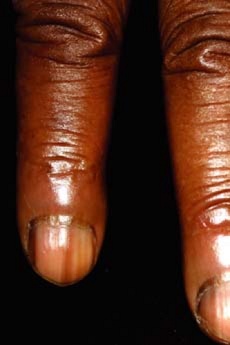 Figure 16-2 Longitudinal melanonychia. |
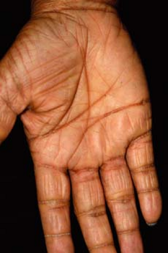 Figure 16-3 Palmar crease hyperpigmentation. |
Oral Hyperpigmentation
Pigmented areas range in color from brown to bluish black
Develops in childhood
Affects gingivae, but may include tongue and rest of oral mucosa as well
Nail Pigmentation (Melanonychia) (Fig. 16-2)
May be either diffuse or a longitudinal band, often bilateral
Affects at least 50% of African Americans, increases with age
Due to increased melanin deposition in the nail plate
Must be differentiated from a subungual melanoma (solitary lesion, occurs at a young age, very dark or varied in color, spills over the nail)
Palmar Hyperpigmentation (Fig. 16-3)
Creases in the palms are darker than the skin on the remainder of the palms
Dermatoses that Occur More Frequently in Ethnic Skin
 |
A 25-year-old Dominican female student presents to the office with large, well-defined, tuberous lesions on both of her ear lobes (Fig. 16-4). The lesions are 1 centimeter in diameter, firm, and smooth. The last time she was home, the areas on both ears were excised, but the large scars regrew. According to the patient, the lesions seem to have gotten bigger since she first noticed them. She reports no previous history of these lesions and no other medical problems.
Keloids
Background
Keloids are more common among darker-skinned patients such as African Americans, Asian Americans, and Latin Americans, who have more pigmented skin. Up to 15% of the population is at risk, although there appears to be genetic loci linked to increased susceptibility to keloid formation. Keloids occur most often in the second and third decade of life. Men and women are equally affected but women have a higher reported incidence.
Key Features
A keloid is excessive scar growth at areas of self-inflicted or surgical trauma.
The scar enlarges beyond the original border of injury compared to a hypertrophic scar, which does not.
Keloids occur more frequently in places with increased tension, such as the sternum, shoulder, earlobe, and cheek.
Patients may present with lesions that also cause pain, burning, itching, and restriction of movement.
Treatment is most effective with intralesional triamcinolone.
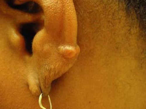 Figure 16-4 Keloid on the helix. |
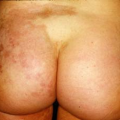
 Figure 16-5 Old hypertrophic surgical scar with active psoriasis surrounding. |
Pathogenesis
Keloids result from pathologic wound healing after injury to the skin. They may occur after local skin trauma such as ear piercing, surgery, injection, burn, or cut, or after local inflammatory skin problems, such as acne, bites, or infectious abscesses. Keloids occur more frequently in places with increased tension, such as the sternum, shoulder, earlobe, and cheek. Patients may present with lesions that also cause pain, burning, itching, and restriction of movement. It is unknown why certain individuals develop keloids. Current theories include intrinsically abnormal fibroblasts and increased growth factor receptors.
Clinical Presentation
Keloids are generally firm nodules, with a smooth but irregular border (Fig. 16-4). Initially, they may appear erythematous, but can become paler or red-brown with time. Alibert coined the term “cheloid” in 1806 from the Greek word for “crab’s claw” because the lesions push beyond the borders of the original wound. Keloids also do not regress, and usually recur after excision. In contrast, hypertrophic scars (Fig. 16-5) do not extend beyond the borders of the original wound, retain their shape over time, and can occasionally partially regress.
Diagnosis
The diagnosis is generally a clinical one; however, a skin biopsy may be helpful for confirming the diagnosis. Most often, the differential diagnosis is a hypertrophic scar. Keloids and hypertrophic scars are distinct entities. Clinically, hypertrophic scars grow intensely and then regress, whereas keloids may grow indefinitely. Histologically, collagen fibers in keloids are larger, thicker, and have a wavy random pattern, whereas collagen fibers in hypertrophic scars lie parallel to the epidermis. Biopsy is not recommended, as the procedure can worsen the keloid.
If the keloid fails to resolve after multiple intralesional steroid injections.
Treatment
Unfortunately, no single effective therapeutic exists for keloids. Thus, prevention is all-important, as patients with one keloid are predisposed to other keloids with future skin injury. These patients should be encouraged to minimize elective skin injury, such as ear piercings, tattoos, and elective mole
removals. Additionally, causes of inflammation, such as acne and infections, should be treated quickly and aggressively to minimize scarring.
removals. Additionally, causes of inflammation, such as acne and infections, should be treated quickly and aggressively to minimize scarring.
Differentiate between keloids and hypertrophic scars; the latter will involute on its own, while keloids may benefit from therapy.
Avoid further skin trauma/surgery/biopsy if possible in keloid patients as they likely will again form keloids.
Intralesional steroid injections are the mainstay of treatment. Triamcinolone acetonide (10 mg per linear centimeter of keloid) is injected every 2 to 6 weeks until the keloid involutes. Steroids inhibit the proliferation of fibroblasts and collagen synthesis, increase collagenase production, and reduce levels of collagenase inhibitors. Surgery can be attempted, but patients must be closely followed to prevent recurrence. Radiation therapy can reduce keloid recurrence, but the risk of inducing malignancy has dampened its use. Silicone gel sheeting can be used alone or as an adjuvant therapy, but requires patient compliance, as the sheets must be worn 12 to 24 hours/day for at least 2 to 3 months. Pressure therapy has been found to be effective for earlobe keloids. Patients can be instructed to wear compression earrings after keloid excision. Cryosurgery, interferon therapy, intralesional bleomycin or 5-FU, imiquimod, and laser therapy are helpful, at times using more than one treatment modality concurrently.
“At a Glance” Treatment
No clearly effective single therapy which is uniformly beneficial
The most effective treatment is intralesional triamcinolone acetonide (10 mg per linear centimeter of keloid) injected every 2 to 6 weeks until the keloid involutes
Other second-line treatments: silicone gel sheeting, excision, cryosurgery, interferon therapy, intralesional bleomycin or 5-FU, imiquimod, and laser therapy
Clinical Course and Complications
Keloids can have a variable clinical course. Most will continue to grow for weeks to months, though some may grow for years. Growth tends to be slow, and once stopped, keloids remain stable. At this stage, they usually do not cause symptoms. Intralesional steroid injections may be started as soon as possible after keloid formation, which can lead to symptomatic relief in 72% of patients and complete flattening in 64% of lesions.
ICD9 Code
| 701.4 | Keloid scar |
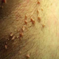
Pseudofolliculitis Barbae
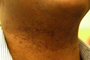 |
A 30-year-old black man presents to the office with painful red bumps on his lower face and neck (Fig. 16-6). When you examine him, you find nodular scars mixed in with the inflammatory pustules. He has always shaved regularly, but these “razor bumps” have gotten worse lately. Upon questioning, he recalls that he has recently switched to a “nicer, four blade” razor. He does not feel comfortable having a beard at his work place, and would like to know what alternative treatments are available.
Background
Pseudofolliculitis barbae (PFB), also known as barber’s itch or razor bumps, is a chronic inflammation of shaved skin. PFB is the most common dermatologic complaint of African American males. The U.S. Army, which requires clean shaven faces, found that 83% of their black recruits had PFB. Occasionally, white men and hirsute black women can also be affected.
Pathogenesis
PFB is mostly seen in people with tightly curled hair and occurs when the newly
cut, sharp tip of hair coils upon itself and penetrates the skin. Patients present with painful papules and pustules in the beard and neck area. Two alternatives for PFB caused by curving shaved hairs are presented in Figure 16-7.
cut, sharp tip of hair coils upon itself and penetrates the skin. Patients present with painful papules and pustules in the beard and neck area. Two alternatives for PFB caused by curving shaved hairs are presented in Figure 16-7.
Key Features
Pseudofolliculitis barbae (PFB) presents as inflammatory papules and pustules limited to the beard area.
PFB is the most common dermatologic complaint of African American males.
Caused by curled, newly shaved hair coiling upon itself and penetrating the skin.
PFB is best treated by prevention: not shaving and allowing hair growth. Beards can be neatly maintained with clippers.
 Figure 16-6 Pseudofolliculitis barbae papules on the neck. |
Clinical Presentation
Follicular-based papules and pustules are found in the beard area (Fig. 16-6). They may be pruritic, erythematous, and hyperpigmented. Generally, one finds more papules than pustules in PFB. However, intraepidermal micro-abscesses may form, accompanied by foreign-body giant cell immune reaction. Long term, these inflammatory sites may scar, forming small hard bumps all over the affected area.
Diagnosis
The diagnosis is generally clinical. If performed, biopsies will show inflammation and abscess formation. Cultures are usually sterile or have only normal skin flora. Unlike true folliculitis, a pathogenic microorganism is not involved in PFB unless secondary infections have occurred.
Treatment
PFB is best treated by prevention: not shaving and allowing hair growth. Beards can be neatly maintained with clippers. Hair-releasing techniques, such as washing the affected area with warm water and soap in a circular manner, may speed recovery.
 Figure 16-7 Pseudofolliculitis barbae: extrafollicular penetration. (A) A curly hair grows from a sharply curved follicle. When shaved, the hair is left with a sharp point. (B) As this hair grows, the sharp tip curves back and pierces the skin. Right: Transfollicular penetration. (C) Alternatively, when hairs are cut too closely, the hairs penetrate the side of the follicle. (D) Both types of follicular reentry cause a foreign body-like reaction (papule). Modified from Crutchfield CE 3rd. The causes and treatment of pseudofolliculitis barbae. Cutis 1998;61:351–356, with permission. |
If the patient chooses more invasive treatment techniques such as laser hair removal, referral to a dermatologist expert in laser surgery is indicated.
If hair or beard growth is not possible, other removal options are available. Chemical depilatories remove hair without leaving sharp ends, and may prevent PFB. However, depilatories cause irritation if used too frequently or left on the skin for too long. A hair removal product, eflornithine hydrochloride cream 13.9%, inhibits an enzyme required for hair growth. It needs to be applied twice a day and washed off 4 hours after application. Like depilatories, a side effect of this cream is local irritation. Electrolysis has been shown to be ineffective and may even cause hyperpigmentation. Laser hair removal is the definitive treatment, and its use in the treatment of PFB is increasing. Patients need to know that laser hair removal requires multiple visits, and is the only treatment that results in long-term diminution of hair growth. It is essential that long-wavelength lasers, such as the Nd:YAG or diode, be used for this therapy.
Related posts:

Stay updated, free articles. Join our Telegram channel

Full access? Get Clinical Tree


