Key Points
- ▪
The chest radiograph can assist with establishing “situs” by identifying the orientation of the apex, the side of the aortic arch, and the stomach (gastric air bubble).
- ▪
The chest radiographic findings of complex disorders reflect not only the underlying abnormality but also often the interventions and their successes and complications.
- ▪
The classic physiology of Eisenmenger syndrome is represented on the chest radiograph by enlargement and centralization (“pruning”) of the pulmonary arterial vasculature, diminished pulmonary venous vasculature, and right heart enlargement.
Situs
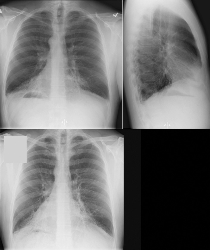
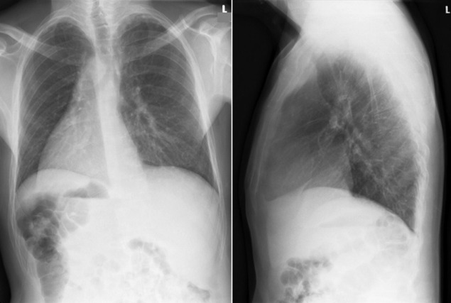
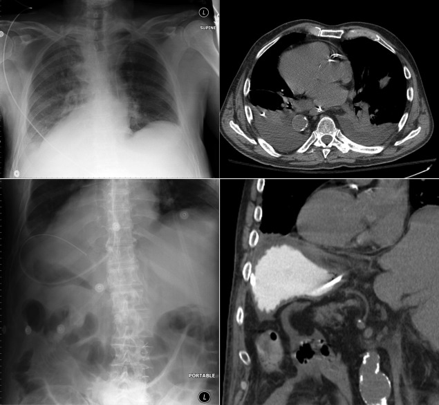
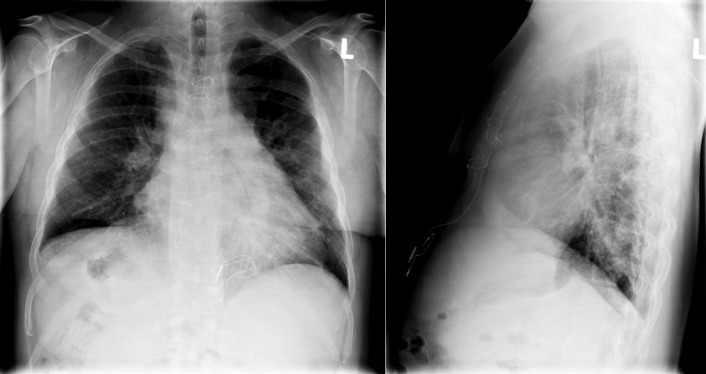
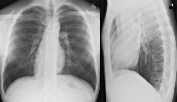
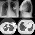
Situs refers to the position/arrangement of the thoracic and abdominal organs ( Figs 20-1 to 20-5 ). The posteroanterior/frontal chest radiograph is able to depict the location of the heart, the left ventricular apex, the aorta, and the gastric air bubble, and it may thereby determine situs.
There are three patterns of situs and one variable pattern:
- 1.
Situs solitus
- 2.
Dextrocardia
- 3.
Situs inversus
- 4.
Situs ambiguous/indeterminate situs/heterotaxy
Situs Solitus
Situs solitus is the term for the normal arrangement of thoracic and abdominal organs ( Table 20-1 ).
- □
Dextrocardia: (where the apex is) heart in the right side of the chest; refers to “reversal of the heart” position in the chest or of its structures. The incidence is less than 1:10,000
- □
DextroVERSION: ventricular loop
- □
Dextrocardia of embryonic arrest (situs solitus and a right-sided heart): heart located rightward in the chest, with the other organs in their normal positions (left gastric air bubble, left aortic arch). With dextrocardia of embryonic arrest/situs solitus with a right-sided heart, there is a 98% incidence of associated congenital heart disease, and 80% of affected individuals have anatomically corrected transposition of the great arteries. The next most frequent association of situs solitus and a right-sided heart is with a ventricular septal defect and pulmonary stenosis.
- □
Dextrocardia situs inversus: reversal/mirroring of the orientation of the heart chambers. The incidence is less than 1:30,000 (3.3% of cases of dextrocardia).
- □
Dextrocardia situs inversus totalis: reversal/mirroring of all visceral organs as well as the heart. Ninety to 95% of patients with situs inversus totalis do not have associated congenital heart disease, and they lead normal lives. Five to 10% of patients with situs inversus totalis have associated congenital heart disease, most commonly transposition of the great arteries. Primary ciliary dyskinesia/Kartagener syndrome is present in 25% cases of situs inversus totalis. The incidence is less than 1:15,000,000 (0.02% of cases of dextrocardia situs inversus).
- □
Situs inversus: left-right inversion/reversal of organ position
- □
Situs inversus with levocardia: 95% associated with congenital heart disease
| Right-Sided | Left-Sided |
|---|---|
| Thoracic Organ | |
| Right atrium | Left atrium |
| Trilobed lung | Bilobed lung |
| Aorta | |
| Abdominal Organ | |
| Liver | Stomach |
| Gallbladder | Spleen |
| Inferior vena cava | |
Situs Ambiguous/Heterotaxy/Indeterminate Situs
The gastric air bubble and the aortic “knob” are on different sides; therefore, the situs is not predictable (“ambiguous”). Manifestations of ambiguous situs include the following:
- □
Errors of cardiac looping
- •
Tetralogy of Fallot
- •
Transposition of the great arteries
- •
Pulmonic stenosis
- •
Atrial septal defects and ventricular septal defects
- •
- □
“Derangement” of abdominal organ symmetry
- •
Isolated stomach or splenic reversal
- •
Midline organs: stomach, liver, adrenal gland
- •
- □
Organ malformation
- •
Asplenia/polysplenia
- •
Horseshoe kidney or adrenal gland
- •
- □
Caval abnormalities
- •
Inferior vena caval interruption with azygous continuation (nearly always)
- •
Bilateral superior vena cava or inferior vena cava
- •
Situs Determination by Chest Radiography
Appearance on chest radiography gives clues to the type of situs.
- □
Situs solitus (normal): left apex and left-sided stomach ( Fig. 20-6 )
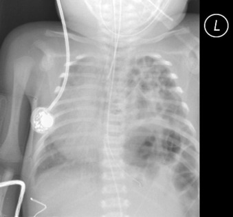
Figure 20-6
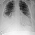
In this chest radiograph from an infant, the heart is displaced to the right side, not the apex. The position of the gastric air bubble is ambiguous. The responsible lesion is a large left-sided diaphragmatic hernia, with the stomach and bowel moved into the left chest, displacing the otherwise normal heart and rendering the gastric air bubble obscure.
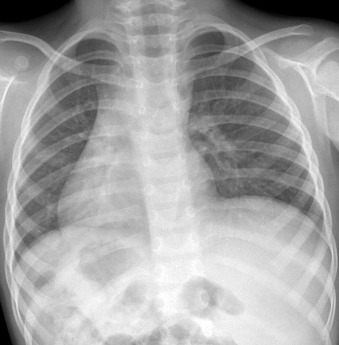
- □
Situs inversus: right apex and right-sided stomach ( Fig. 20-7 )
Related posts:
 Radiographic Findings by Diagnosis: Cardiomyopathies
 Radiographic Findings by Diagnosis: Coronary Artery Disease–Complications of Infarction
 Cardiac and Vascular Trauma
 Radiographic Findings by Diagnosis: Pericardial and Pleural Diseases
 Tubes and Drains
 Pacemakers and Implantable Cardioverter Defibrillators

Stay updated, free articles. Join our Telegram channel

Full access? Get Clinical Tree


