Radiographic Findings by Diagnosis: Congenital Heart Disease—Shunts and Closure Devices


Key Points


  • The chest radiograph is predominantly useful to detect/characterize the pulmonary vascularity as influenced by congenital shunt lesions, such as pulmonary vascular shunt plethora of larger shunts and signs of pulmonary hypertension complicating large shunts.


  • Small shunt lesions are not evident on chest radiography, other than pulmonary arteriovenous malformations.


  • A minority of shunt lesions are directly suggested by chest radiography, such as the vertical vein of left-sided anomalous pulmonary venous return, the “scimitar sign” of the scimitar variant of right-sided anomalous pulmonary venous return, and ductal calcification.


Left-to-right shunts initially result in volume overload of the chambers carrying the shunt volume, causing enlargement of the volume-overloaded chambers and an increase in pulmonary blood flow. If the amount of pulmonary blood flow is sufficiently high, obliterative disease of the pulmonary arterioles develops (“pulmonary vascular disease”), resulting in pulmonary hypertension. The chest radiographic appearance of pulmonary hypertension is one of centralization of pulmonary flow and accelerating enlargement of the right-sided chambers.


The size of a shunt is represented best by the shunt ratio (volume of blood through the pulmonary circuit vs. the systemic circuit, such as 2:1, 3:1, 4:1) and by the degree of elevation of the pulmonary pressures.


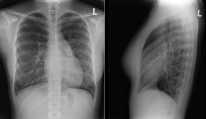
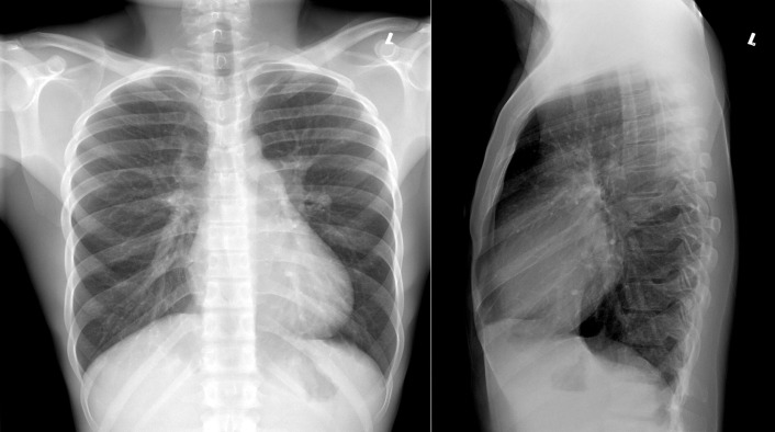
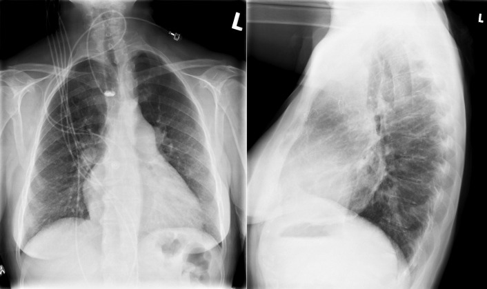
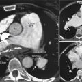
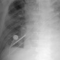
Patent Foramen Ovale


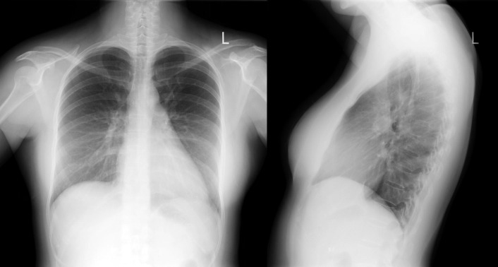
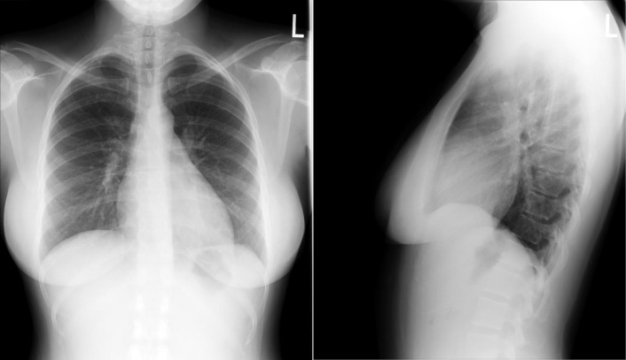
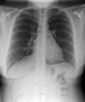
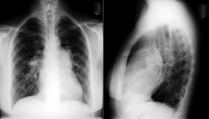
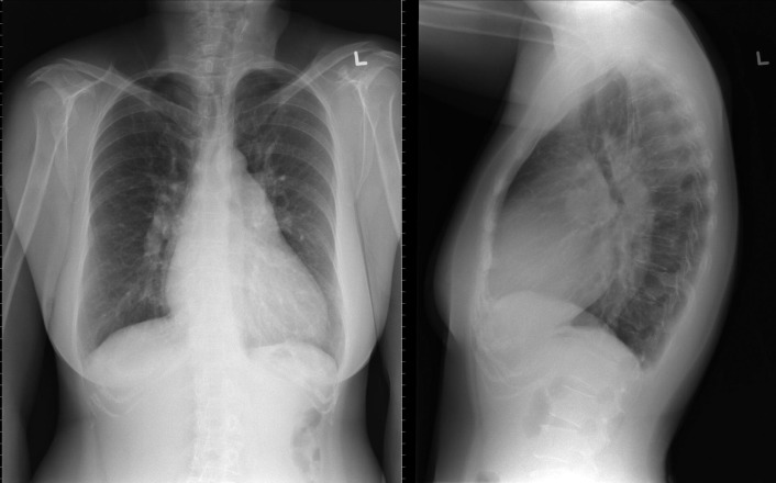
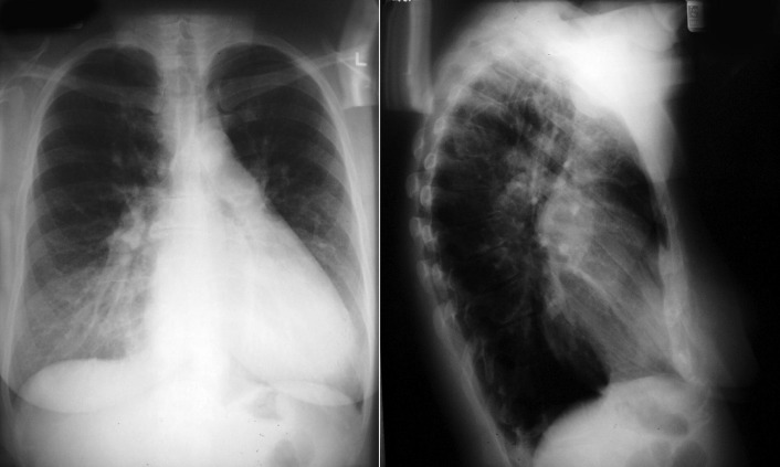
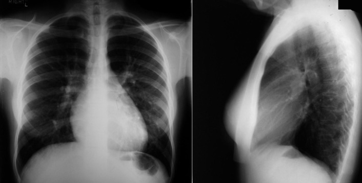
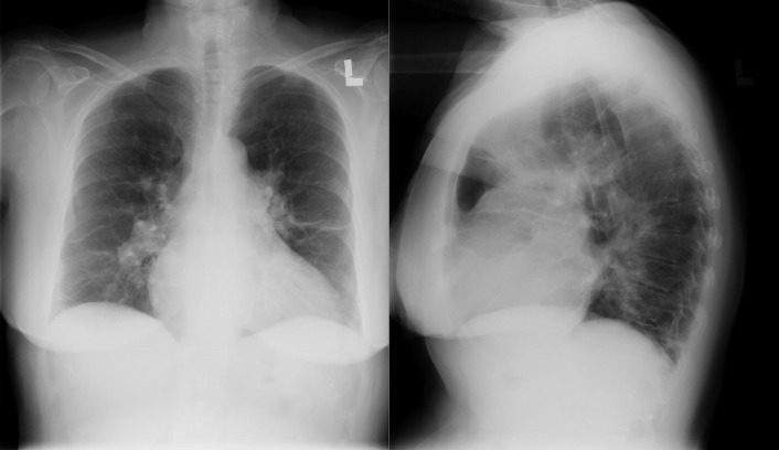
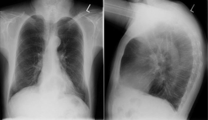
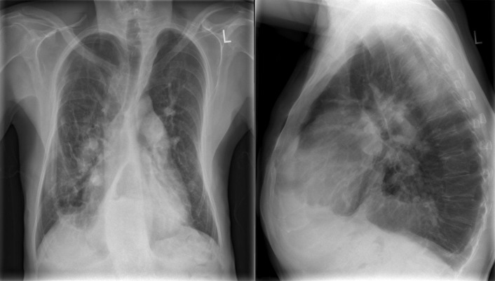
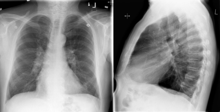
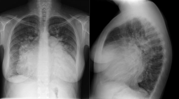
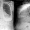
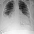
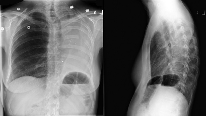
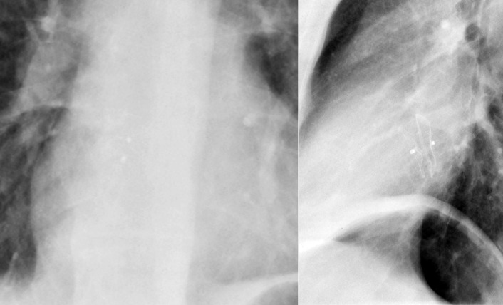
A patent foramen ovale is present in 25% to 30% of the adult population ( Figs. 18-1 to 18-4 ). Any magnitude of shunting across it in normal circumstances is undetectable radiographically. Percutaneous patent foramen ovale closure devices are fairly commonly inserted and are radiographically evident.


Figure 18-1


Chest radiographs of a patient who underwent percutaneous closure of both a large atrial septal defect and a patent foramen ovale with Amplatzer devices.


Figure 18-2


Following a transient ischemic attack–like event, a patent foramen ovale (PFO) and a left lower lobe pulmonary arteriovenous malformation (AVM) were identified ( upper radiograph ). To eliminate the possibility of right-to-left shunting at either the cardiac or pulmonary vascular level, both the PFO and AVM were occluded with Amplatzer closure devices ( lower radiographs ).


Figure 18-3


There has been a prior left pneumonectomy; there are surgical clips/staples at the former left hilum. An Amplatzer patent foramen ovale occluder device is seen “en-face” on the frontal radiograph and side-on on the lateral radiograph.


Figure 18-4


Following a transient ischemic attack–like event, a patent foramen ovale (PFO) was identified. To eliminate the possibility of right-to-left shunting at the cardiac level, the PFO was occluded with an Amplatzer closure device.


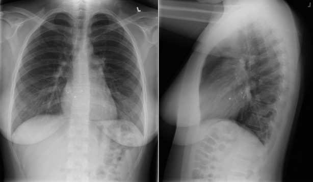
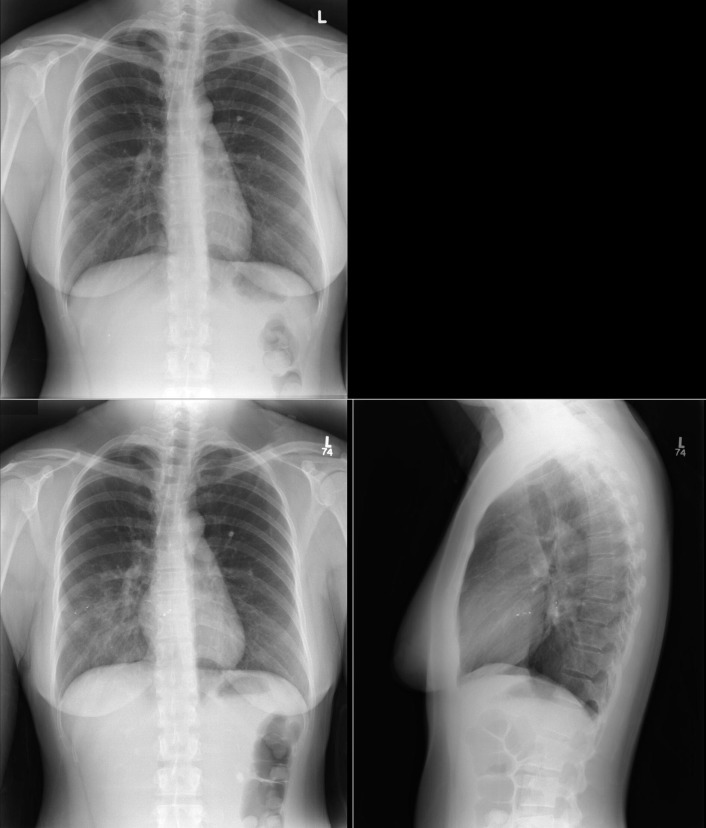
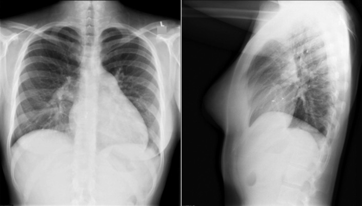
Atrial Septal Defect


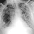
See Figures 18-5 to 18-31 . Possible findings are discussed in the following sections.


Figure 18-5


There is borderline cardiomegaly and some increase in pulmonary vascularity due to a small secundum-type atrial septal defect. The left superior cardiac silhouette is straightened, consistent with an enlarged left atrial appendage. There are no frank signs of left atrial enlargement.


Figure 18-6


There is no cardiomegaly, and there are no signs of left atrial or right heart enlargement (other than possibly of increased right ventricular apposition to the sternum), enlargement of the pulmonary arteries, or shunt vasculature in this case of a small secundum-type atrial septal defect.


Figure 18-7


There is no cardiomegaly, but there is straightening of the left heart border consistent with left atrial (appendage) enlargement in this moderate-sized secundum-type atrial septal defect. The right atrial contour is prominent, and the pulmonary vasculature has generalized accentuation–shunt vasculature.


Figure 18-8


There is borderline cardiomegaly, an ambiguous left upper heart border, increased apposition of the right ventricle to the sternum, gross dilation of the main and central pulmonary arteries, and generalized increase of the pulmonary vasculature–shunt vascularity in this case of a large secundum-type atrial septal defect with moderate pulmonary hypertension.


Figure 18-9


There is definite cardiomegaly, a straightened left upper heart border, increased apposition of the right ventricle to the sternum, gross dilation of the main and central pulmonary arteries, and generalized increase of the pulmonary vasculature–shunt vascularity in this case of a large secundum type atrial septal defect with moderate pulmonary hypertension.


Figure 18-10


There is gross cardiomegaly in this case of a sinus venosus atrial septal defect with a large (4:1) shunt. The left upper heart border is straightened, consistent with left atrial (appendage) enlargement. The main pulmonary artery is enlarged, and the pulmonary arteries and veins are both increased in size and in peripheral representation (shunt vascularity).


Figure 18-11


The heart size is not increased, but the contours are abnormal in this case of a medium-sized secundum atrial septal defect with a moderate-sized shunt. There is straightening of the left upper heart border consistent with left atrial (appendage) enlargement. The pulmonary vasculature is mildly accentuated.


Figure 18-12


There is cardiomegaly with straightening of the left upper heart border, prominence of the right atrial contour, and increased right ventricular apposition to the sternum in this case of a moderate-sized secundum atrial septal defect with moderate pulmonary hypertension. The main and central pulmonary arteries are enlarged and there is prominence of the pulmonary vasculature peripherally—shunt vascularity.


Figure 18-13


There is no cardiomegaly in this case of a moderate-sized secundum atrial septal defect with moderate pulmonary hypertension, but the lungs appear enlarged, and there has been obstructive lung disease to render plausible lung hyperinflation. The left upper heart border is straightened from left atrial (appendage) enlargement, and there is increased apposition of the right ventricle to the sternum. The central pulmonary arteries are enlarged and the peripheral vasculature is accentuated.


Figure 18-14


The cardiac silhouette contours are difficult to recognize on the posteroanterior radiograph, but the central pulmonary arteries and main pulmonary arteries are considerably dilated. The lateral chest radiograph is suggestive of right ventricular dilation. There is a large atrial septal defect in this patient, with extensive thoracic skeletal deformities and pulmonary disease associated with Marfan syndrome.


Figure 18-15


The frontal radiograph does not give the impression of cardiomegaly, but the lateral radiograph does in this patient with an atrial septal defect with moderate pulmonary hypertension. The left atrium in particular is dilated. The main and central pulmonary arteries are dilated, but there is only mild extenuation of the pulmonary vasculature.


Figure 18-16


Prominently enlarged pulmonary arteries and veins, as well as a dilated main pulmonary artery from advanced pulmonary hypertension from a large secundum atrial septal defect. In addition, there is severe cardiomegaly, mainly from right and left atrial and right ventricular chamber enlargement.


Figure 18-17


Mild cardiomegaly, straightening of the left upper heart border due to left atrial appendage enlargement, and mildly increased pulmonary vascularity due to a medium-sized secundum atrial septal defect.


Figure 18-18


Mild cardiomegaly with signs of right ventricular and left atrial enlargement in particular, with enlargement of the central and peripheral pulmonary arteries, as well as veins, due to a sinus venosus atrial septal defect and right-sided anomalous pulmonary venous return.


Figure 18-19


Posteroanterior and lateral chest radiographs of a patient with a previously repaired atrioventricular canal. Note the enlarged central pulmonary arteries, the right ventricular and right atrial prominence, and the left atrial and left ventricular enlargement.

Apr 10, 2019 | Posted by in General Surgery | Comments Off on Radiographic Findings by Diagnosis: Congenital Heart Disease—Shunts and Closure Devices

Full access? Get Clinical Tree

Get Clinical Tree app for offline access