Key Points
- ▪
Valvular pulmonic stenosis is often associated with dilation of the main and left pulmonary arteries.
- ▪
Secondary signs of right heart chamber enlargement occur in proportion to the severity of the stenosis.
- ▪
Coarctation of the aorta may be apparent radiographically—either by an abnormal contour of the proximal descending aorta or by rib-notching—a sign of collateral vessels.
Congenital Heart Disease: Obstructions
Pulmonary Valvular Stenosis
See Graphic 19-1 and Figures 19-1 to 19-13. Possible findings are discussed in the following sections.
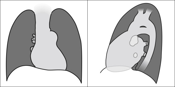
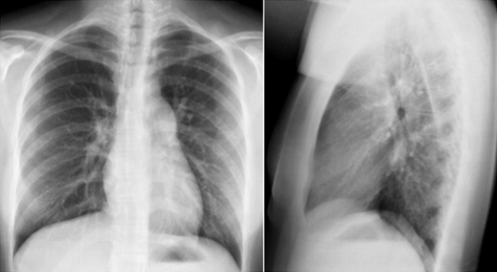
Posteroanterior and lateral projections: Valvular pulmonic stenosis. Note dilated main pulmonary artery seen on the frontal projection, and slight accentuation of the right atrial curvature. On the lateral projection, note the indirect sign of right ventricular dilation—increased apposition of the heart to the sternum, and dilation of the right main pulmonary artery, seen end-on.
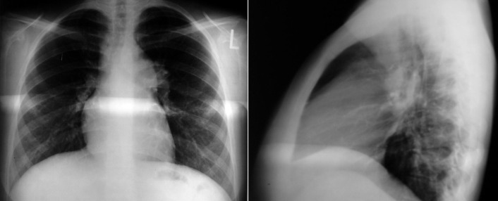
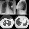
Pulmonic stenosis and a secundum atrial septal defect. There is dilation (poststenotic) of the main pulmonary artery. Right atrial enlargement is suggested by the right heart contour on the posteroanterior radiograph, and right ventricular enlargement is suggested by the sternal apposition on the lateral radiograph.
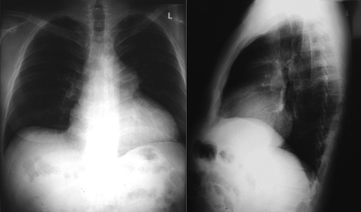
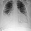
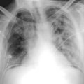
Pulmonic stenosis and dilated cardiomyopathy. The cardiothoracic ratio is increased, and the cardiac contour is globular with multichamber enlargement. There is dilation (poststenotic) of the main pulmonary artery.
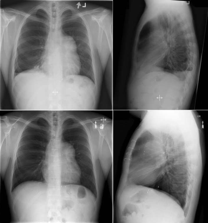
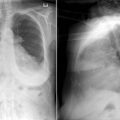
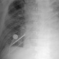
Upper (preoperatively) and lower (postoperatively) chest radiographs of a patient with severe valvular pulmonic stenosis who underwent insertion of a Hancock II bioprosthesis.
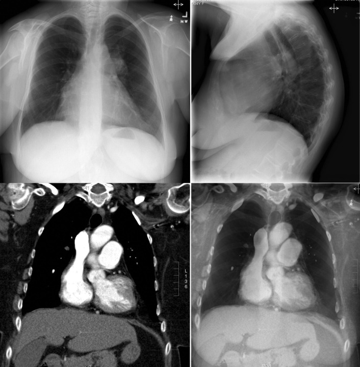
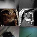
Chest radiographs depicting cardiomegaly. The anteroposterior radiograph shows some increase of the curvature of the right atrium and enlargement of the main pulmonary artery. The lateral chest radiograph shows a significant increase in the apposition of the right ventricle to the sternum, albeit in a patient with severe kyphosis. The contrast-enhanced computed tomography (CT) scan in the left lower image reveals the enlargement of the right atrium and the main pulmonary artery. The superimposition of the coronal CT scan on that of the anteroposterior chest radiograph is depicted in the right lower image, corroborating the impression of enlargement of the pulmonary artery appreciated on the chest radiograph.
Valvular pulmonary stenosis with poststenotic dilation of the main pulmonary artery and with right ventricular enlargement.
Related posts:
 Radiographic Findings by Diagnosis: Cardiomyopathies
 Radiographic Findings by Diagnosis: Coronary Artery Disease–Complications of Infarction
 Cardiac and Vascular Trauma
 Radiographic Findings by Diagnosis: Pericardial and Pleural Diseases
 Tubes and Drains
 Pacemakers and Implantable Cardioverter Defibrillators
Stay updated, free articles. Join our Telegram channel

Full access? Get Clinical Tree


