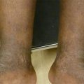
Figure 3.1
Pseudofolliculitis barbae. 2–4 mm skin-colored and hyperpigmented perifollicular papules, confined to the mandible and neck
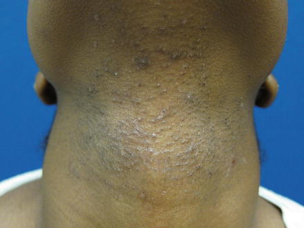
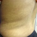
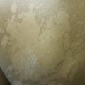
Figure 3.2
Pseudofolliculitis barbae in a woman. Scattered skin-colored and hyperpigmented perifollicular papules along the neck
Clinical Differential Diagnosis
The lesions were thought to be most consistent with pseudofolliculitis barbae. The differential included other conditions that can cause erythematous papules and pustules in the regions of terminal hair growth, such as acne vulgaris, folliculitis, tinea barbae, and acne keloidalis nuchae.
Acne vulgaris will include comedones and not is restricted to areas of terminal hair density. Primary features of bacterial folliculitis include pruritic erythematous papules and pustules, commonly observed in the beard region of men who shave. Bacterial folliculitis presents acutely, in contrast to the chronic nature of PFB. Cultures also help distinguish, as PFB is an inflammatory, sterile disorder. Tinea barbae may present as follicular pustules, inflammatory papules or scaly erythematous annular plaques. The lesions are confluent, while PFB lesions are isolated. Fungal forms can be detected with potassium hydroxide preparation. Acne keloidalis nuchae presents as perifollicular papules, pustules or keloidal plaques on the posterior scalp and, similar to PFB, disproportionally affects young men of African descent. The location on the scalp differentiates it from PFB.
Histopathology
Diagnosis can be made for this condition on clinical examination alone, and does not typically require skin biopsy. When biopsy is performed, histopathology reveals an invagination of the epidermis with intraepidermal neutrophils, granulomatous inflammation and foreign-body giant-cells surrounding an embedded hair tip. Other features include microabscess formation and fibrosis (Ramos-e-Silva and Pirmez 2014).
Diagnosis
Pseudofolliculitis barbae (PFB)
Case Treatment
The pathogenesis and chronic nature of PFB was reviewed with the patient, including the potential sequelae of post-inflammatory hyperpigmentation. Patient education is an important cornerstone of PFB management due to the multifactorial nature of treatment, which can require a combination of behavioral, procedural and pharmacologic interventions. The treatment options and reasonable expectations for each method were discussed, including cessation of shaving, alteration of shaving technique, concomitant medical treatments and permanent laser hair reduction. The patient denies personal, cultural, or occupational factors that limit his grooming practices, and therefore opted for the suggested first-line treatment of complete shaving cessation. He was advised to maintain a trimmed beard at a minimum length of 5–10 mm to prevent reactivation of the disease. He was counseled to expect his condition to persist for some time due to previously embedded hairs, and told that trapped hairs typically release from the skin naturally after 3–6 weeks of growth. The patient was instructed to contact the clinic for consideration of additional treatment options such as concomitant medical therapy if he did not experience improvement of his condition within 8 weeks.
Discussion
Pseudofolliculitis barbae is a common and chronic inflammatory condition that develops in areas of terminal hair growth as a result of hair epilation. Other names for PFB include folliculitis barbae tramatica, pili incarnate, ingrown hairs, or razor bumps. Though pseudofolliculitis can occur in all ethnicities, it is relatively unique to skin of color (Kundu and Patterson 2013). Men of African descent are disproportionally affected, with a reported prevalence between 45 and 83 %, compared to just 3 % of white men. Hispanic and Middle Eastern men, as well as women who tweeze or pluck facial hair are also frequently susceptible to PFB. The reported incidence of PFB in women of African or Hispanic descent with hirsutism or hypertrichosis closely approximates the incidence in men (Perry et al. 2002). In addition to the potential impact on self-esteem, PFB can cause significant problems for individuals, especially those with strict occupational grooming policies, such as servicemen in the US military who are required to maintain a clean-shaven face.
The predominance of PFB in individuals of African descent is considered attributable to unique biology of African hair, particularly the prevalence of tightly coiled hair and the natural tendency of the hair shaft to curl during growth and reenter the skin. This reentry occurs through extrafollicular and transfollicular penetration. Extrafollicular penetration describes reentry of a hair shaft that has already exited the follicle; as the hair grows along its curvature it pierces the surface of the skin 1–2 mm from the follicle. Transfollicular penetration occurs when the sharpened tip of the shaved hair retracts beneath the surface of the skin and pierces the follicular wall as it grows. This type of penetration is typically caused by close-shaving practices such as shaving against the grain and pulling the skin taut while shaving. When hair cut at an acute angle (from shaving or other means of epilation) penetrates the interfollicular dermis or epidermis, it creates an invagination of the skin and forms a “pseudofollicle.” This pseudofollicle can rupture, provoking an inflammatory response that manifests as firm, inflamed perifollicular papules or pustules. The ensuing inflammation can result in post-inflammatory hyperpigmentation (PIH) due to the stimulation of melanogenesis in the epidermis and an increased release of melanin in the dermis. Hypertrophic scarring or keloid formation can also develop in predisposed individuals. Often, the enduring sequelae are more cosmetically disturbing than the PFB itself.
In addition to hair removal practices, a genetic risk factor has been identified that may contribute to the pathophysiology and phenotypic expression of PFB. One study demonstrated that a single nucleotide polymorphism, which causes a disruptive Ala12 Thr substitution in the 1A α-helical segment of the hair-follicle-specific keratin 75 (previously K6hf), occurred in 36 % of individuals with PFB compared to only 9 % of unaffected individuals (P < 0.000006) (Winter et al. 2004). It is believed that this mutation structurally weakens the companion layer of the hair follicle and may increase the risk of PFB for some men and women, especially when combined with tightly coiled hair and shaving practices.
Related posts:

Stay updated, free articles. Join our Telegram channel

Full access? Get Clinical Tree


