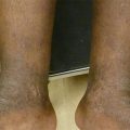
Figure 9.1
Pityriasis rosea. Numerous erythematous to violaceous and hyperpigmented ovoid scaly papules on the trunk
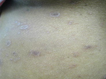
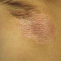
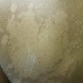
Figure 9.2
Pityriasis rosea. Close up. Erythematous to violaceous and hyperpigmented ovoid papules on the trunk with fine collarettes of scale
Clinical Differential Diagnosis
The rash was felt to be most consistent with pityriasis rosea. However, other diagnostic possibilities included nummular eczema and secondary syphilis. A complete differential diagnosis of pityriasis rosea also includes guttate psoriasis, pityriasis lichenoides chronica, tinea versicolor, tinea corporis, HIV acute seroconversion illness, Lyme disease, and drug eruption (Goldstein et al. 2015).
Histopathology
A biopsy was not undertaken in this case because the history and physical examination were felt to be diagnostic. When a biopsy is performed, histopathologic evaluation is often non-specific. Generally, there is subacute spongiotic dermatitis in the setting of a superficial perivascular lymphohistiocytic infiltrate. Other features include erythrocyte extravasation and subsequent transepidermal elimination. Non-herald patch lesions also frequently show a leading spongiosis with trailing parakeratosis, a feature also seen in erythema annulare centrifugum (Elston 2009). Notably, the finding of plasma cells favors a diagnosis of secondary syphilis.
Diagnosis
Pityriasis rosea (PR)
Case Treatment
The nature of the diagnosis of pityriasis rosea was reviewed with the patient, including the possible sequela of post-inflammatory hyperpigmentation (PIH). Treatment options were discussed, including observation, topical corticosteroids, acyclovir, erythromycin, and/or phototherapy. Given her pruritus, triamcinolone 0.1 % ointment BID was prescribed for a 4-week course, and the patient was instructed to contact the clinic for consideration of phototherapy if she did not experience a significant improvement in pruritus. In addition, after a discussion of the risks and benefits and relatively limited data for acyclovir, the patient opted for a 1-week course of acyclovir 800 mg five times daily. Finally, after a discussion of the clinical similarities between pityriasis rosea and secondary syphilis, the patient agreed to a serum rapid plasma reagin (RPR) with prozone, which was non-reactive.
Discussion
Pityriasis rosea is an acute, self-limited, papulosquamous eruption that involves the trunk and proximal extremities of healthy adolescents and young adults, generally between the ages of 10 and 35 (Chuang et al. 1982). The disease is more common in females than males, but individuals of all races and ethnicities are affected equally. The rash generally clears within 2 months but occasionally can last somewhat longer. Lesions that persist longer than 5 months should prompt consideration of pityriasis lichenoides chronica. A viral etiology for pityriasis rosea has long been postulated. A recent review article of the existing data concludes that there is strong evidence for an association between a “reactive response” to HHV-7 and to a lesser extent HHV-6 and development of pityriasis rosea (Drago et al. 2009). Less robust evidence exists to support an association with HHV-8 and influenza A (H1N1).
Clinically, the classic presentation of PR is a “herald patch” (a pink- to salmon-colored scaly patch or thin plaque) that gradually enlarges with central clearing and the development of a “collarette” of scale over several days. In people of color, this patch can appear brown or hyperpigmented. Of note, the presence of the “herald patch” exists in a majority of cases but is not required for diagnosis. Subsequently, there is a more diffuse eruption of smaller but morphologically similar papules and small plaques that involve the trunk and proximal extremities. Lesions tend to be distributed along skin cleavage lines (Langer’s lines), leading to the characteristic description of a “fir tree” or “Christmas tree” pattern on the trunk. Importantly, in skin of color, the subsequent lesions tend to be more papular, monomorphic, and folliculocentric, and larger lesions sometimes have central hyperpigmentation with a papular border; this is conceptually similar to the papular eczema variant of atopic dermatitis in ethnic skin. There is also more frequent involvement of the face and scalp (Amer et al. 2007) and rarely, the oral cavity (Jacyk 1980). Additionally, post-inflammatory hyperpigmentation may be significant and prolonged, especially in skin of color (Amer et al. 2007).
A prodrome of headache, malaise, and/or pharyngitis is present in a small minority of patients. Pruritus occurs in approximately 25 % of patients and ranges in severity (Hartley 1999; Allen et al. 1995). There is also a well-described inverse variant of pityriasis rosea, which affects the axillae, groin, distal extremities, and rarely the face. This subtype of PR is thought to be more common in younger children (Trager 2007). Additional less common variants that have been described include pustular, purpuric, vesicular, urticarial, and erythema multiforme-like.
Biopsy is typically not necessary for the diagnosis of pityriasis rosea. However, if undertaken, the clinician should consider the risk of hypertrophic scarring and keloid formation. Keloids are known to occur at high rates in patients of Hispanic and African ancestry (Robles and Berg 2007). He or she should discuss this risk with patients, especially in those with a history of keloids, and the clinician should be sensitive to cosmesis while choosing a site if biopsy is indicated. Furthermore, the physician could consider intralesional corticosteroid injection or other peri-procedural therapies to minimize the risk of keloid formation in patients with a known keloidal diathesis.
Dermoscopy can also be a useful tool in the diagnosis of pityriasis rosea and in discriminating among inflammatory dermatoses in general. In one large study investigating the dermoscopic features of plaque psoriasis, dermatitis, lichen planus, and pityriasis rosea, dermoscopy was found to be both sensitive and specific for detecting pityriasis rosea. PR was found to be significantly associated with the following features: yellowish background color, dotted vessels, patchy vascular distribution, and white peripheral scale (the “collarette” of scale). Of note, the dotted vessels seen in PR are also seen in psoriasis and dermatitis, although they are much more abundant in the latter conditions (Lallas et al. 2012). Unfortunately, the population studied largely consisted of white Europeans, so further investigations are needed to validate these dermoscopic findings in ethnic skin.
Related posts:

Stay updated, free articles. Join our Telegram channel

Full access? Get Clinical Tree


