33 Procedures to Treat Benign Conditions
Many skin tumors and growths are benign and can be diagnosed based on their clinical appearance and history. These lesions can arise from the epidermis, the dermis, or the subcutaneous tissues. This chapter provides a detailed discussion of the most common benign skin lesions and their treatment options. Chapter 12 covered epidermal cysts, lipomas, digital mucous cysts, and hydrocystomas so these will not be covered here.
Acne Surgery
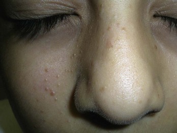
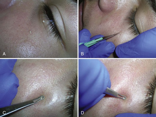
Acne surgery is the name given to the removal of open comedones (blackheads) with a comedone extractor in the clinician’s office. It can also be performed on actinic comedones or senile comedones. It is often performed for cosmetic reasons, but can also decrease pain around a comedone that is under pressure. Large inflammatory nodules and cysts are best treated with intralesional steroids rather than acne surgery (see Chapter 16, Intralesional Injections).
After informed consent, the comedones are cleaned with alcohol. No anesthesia is needed. The comedone is nicked with a No. 11 blade, a sterile needle, or the sharp end of a comedone extractor. Sebum, cells, and other debris are expressed out using pressure from a comedone extractor (Figure 33-1). If a comedone extractor is not available, one can be fashioned from a small paperclip bent to produce a homemade device. Clean the paperclip with an alcohol wipe before using it. Bill using acne surgery CPT code = 10040.
Acrochordons (Skin Tags)
Diagnosis
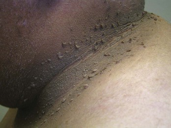
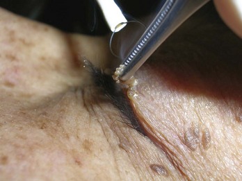
Treatment
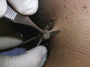
Snip with Iris Scissors (Figure 33-3)
Angiomas/Angiokeratomas/ANGIOFIBROMAS
Diagnosis
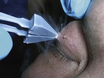
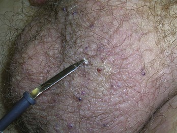
Electrodesiccation for Small Lesions
Shave Excision with Electrodesiccation of the Base for Larger Lesions
Chondrodermatitis Nodularis Helicis
Diagnosis
Treatment
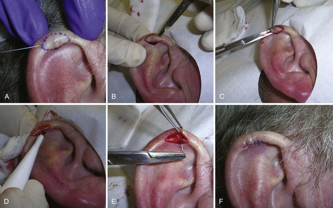
Elliptical Excision
Electrodesiccation and Curettage
Cutaneous Horn
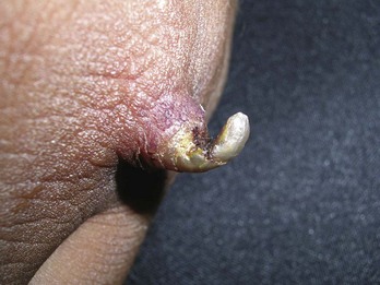
Treatment
Dermatofibromas
Diagnosis
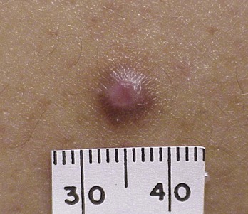
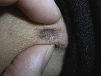
FIGURE 33-10 Typical dermatofibroma with a hyperpigmented halo and a white and pink scar at the center.
(Copyright Daniel L. Stulberg, MD.)
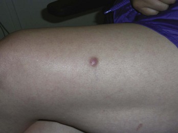
FIGURE 33-11 Large dermatofibroma on the leg that was not dermatofibrosarcoma protuberans.
(Copyright Richard P. Usatine, MD.)
Treatment
Keloids and Hypertrophic Scars
Treatment
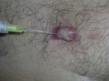
FIGURE 33-14 Injecting an acne keloid with triamcinolone using a 27-gauge needle.
(Copyright Richard P. Usatine, MD.)
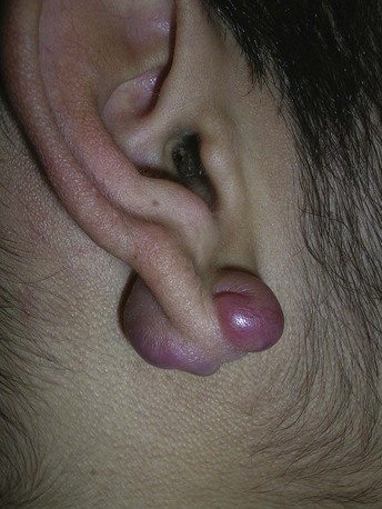
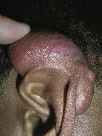
FIGURE 33-15 Keloids formed on both sides of the earlobe secondary to ear piercing.
(Copyright Richard P. Usatine, MD.)

