12 Cysts and Lipomas
Epidermal Inclusion Cysts and Variants
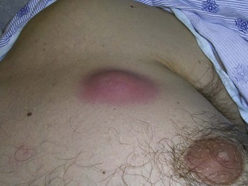
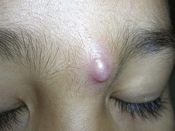
Epidermal inclusion cysts arise from a plugged follicular opening, resulting in a nodular cyst filled with cheesy malodorous keratin material. These are also commonly and incorrectly called sebaceous cysts but do not in fact contain any sebum. Unfortunately, the ICD code for these cysts is named “sebaceous cyst” so you must know this misnomer for billing purposes. Other names include infundibular cysts, keratinous cysts, and epidermoid cysts. Generally, these present as a compressible but nonfluctuant nodule, which can be diagnosed clinically due to a comedo-like central punctum that can be distinguished at the apex (Figure 12-1). Because the keratin material inside the cyst wall is highly inflammatory, any cyst that has ruptured can spark a vigorous inflammatory response that can be mistaken for cellulitis (Figure 12-2). Even if these do become superinfected, the treatment of choice is incision and drainage to remove the inflammatory and/or infected contents.
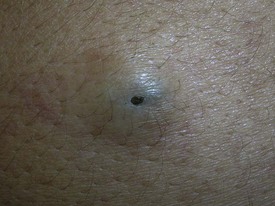
FIGURE 12-1 An epidermal inclusion cyst with a central punctum that looks like an open comedone.
(Copyright Richard P. Usatine, MD.)
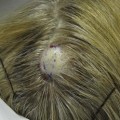
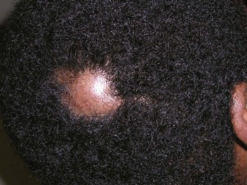
Pilar cysts (also known as trichilemmal cysts and commonly called wens) are very similar to epidermal inclusion cysts with the exception that the capsule is much thicker and the location generally on the scalp (Figures 12-3 and 12-4). Pilar cysts rarely have a punctum but the thickness of the capsule makes removal somewhat easier. These can also become very large but in most cases they are 5 to 20 mm in size. It is not unusual for the hair to become thinned or actually stop growing over the cyst (Figures 12-3 and 12-4).
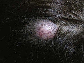
FIGURE 12-3 A typical presentation of a pilar cyst (trichilemmal cyst or wen) on the scalp.
(Copyright Richard P. Usatine, MD.)
One variant of the epidermal cyst is the milium cyst. Milia are histologically identical to classic epidermal cysts but only grow to 1 to 2 mm. They are generally located around the eyes and central portion of the face (See Figure 33-20 in Chapter 33, Procedures to Treat Benign Conditions).
Cysts can be located almost anywhere but are common on the trunk, neck, face, and behind the ears. They range in size from millimeters to centimeters. Epidermal inclusion cysts are benign; however, there are rare proliferating epidermal inclusion cysts that can become carcinogenic. Any cyst that does not look 100% typical on excision should be sent to the pathologist. A full differential diagnosis is given in Box 12-1.
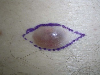
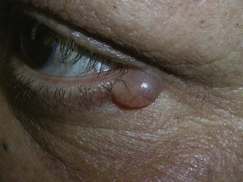
FIGURE 12-8 Hydrocystoma around the eye. It is benign and has a very thin wall that is easy to cut.
(Copyright Richard P. Usatine, MD.)
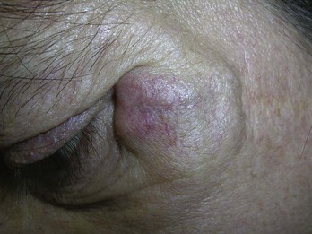
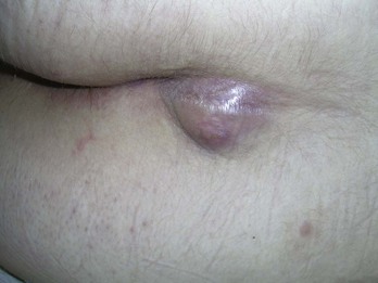
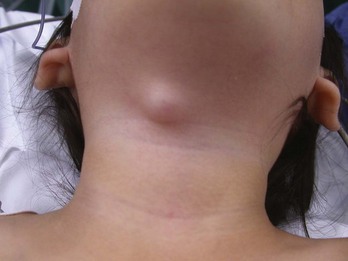
FIGURE 12-12 A thyroglossal duct cyst in a young girl. Note how it is midline. These will move up with swallowing.
(Courtesy of Frank Miller, MD.)
The occurrence of multiple true sebaceous cysts at a younger age is termed steatocystoma multiplex, which is a genetically inherited problem. These capsules are extremely thin. Small fluid-filled very thin-walled cysts around the eyes are called hidrocystomas (Figure 12-10).
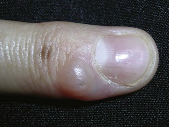
FIGURE 12-10 Digital mucous (myxoid) cyst on the finger. They are most commonly found distal to the DIP joint.
(Copyright Richard P. Usatine, MD.)
Contraindications for Cyst Removal
Minimal Excision Technique
Advantages
Advantages of the minimal excision technique for the clinician include the following:
Advantages for the patient include the following:
Disadvantages
Disadvantages of the minimal excision technique for the clinician include:
Disadvantages for the patient include:
Be aware that this procedure is more difficult where the skin is very thick such as on the back.
Minimal Excision Technique: Steps and Principles
Preoperative Measures