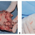
Pressure Injuries (Sacral and Pelvic Region, Columella—From CPAP)
David A. Billmire
Kim A. Bjorklund
DEFINITION
A pressure injury is a localized injury of skin and underlying tissue secondary to prolonged pressure, frequently in combination with shear forces.
Severity ranges from nonblanchable erythema to fullthickness tissue loss.
Common locations are over bony prominences such as the sacrum, coccyx, ischium, and heels.
In the pediatric population, children with special needs and those who are critically ill are at particularly high risk for pressure injuries.1
PATHOGENESIS
Pressure injury formation is a complex process that involves numerous factors.2
Extrinsic contributing factors include pressure, friction, and shear.
As muscle is more sensitive to pressure than is skin, areas with a significant amount of muscle may be susceptible to an inverted cone pattern of injury, in which most of the necrosis is deep to the skin.5
Paraplegia, sensory impairment
Cognitive impairment
Kyphoscoliosis or kyphosis
Chronic fecal or urinary soiling
Trauma
Immobility
Poor nutrition
NATURAL HISTORY7
Nonblanchable erythema may be observed within 30 minutes of unrelieved pressure and typically disappears within 1 hour after pressure is eliminated.
Ischemia develops after pressure is present for 2 to 6 hours.
Necrosis may occur if pressure is not relieved within 6 hours.
Ulceration tends to occur over bony prominences within 2 weeks after development of necrosis.
PATIENT HISTORY AND PHYSICAL FINDINGS
Determine factors contributing to the pressure injury (extrinsic and intrinsic) and whether they can be eliminated postoperatively.
Note any chronic medical conditions contributing to the pressure injury, such as myelodysplasia, cerebral palsy, paraplegia, and scoliosis.
Note any temporary conditions contributing to the pressure injury (prolonged immobility, repetitive trauma) that require resolution.
Ambulatory status
Previous pressure injuries
Compliance and motivation with treatment
Assess risk factors using Braden Scale, including mobility, activity, sensation, moisture, friction/shear, and nutrition.
Assess sensation and lower extremity motor function, including spasticity.
Palpate for any fluctuance or bony prominences.
Note any signs of infection such as warmth, erythema, tenderness, purulent drainage, and systemic signs of infection.
Assess ongoing factors that may be contributing to wound breakdown, such as items causing pressure, spasticity, and fecal or urinary incontinence.
Staging of pressure injuries based on the updated guidelines of National Pressure Ulcer Advisory Panel8:
Stage 1: Nonblanchable erythema of intact skin
Stage 2: Partial-thickness skin loss with exposed dermis
Stage 3: Full-thickness skin loss
Stage 4: Full-thickness skin and tissue loss
Unstageable pressure injury: Obscured full-thickness skin and tissue loss
Deep tissue pressure injury: Persistent nonblanchable deep red, maroon, or purple discoloration
IMAGING AND DIAGNOSTIC STUDIES
Nutritional assessment (including albumin, prealbumin, electrolytes)
Wound cultures if concern for infection
WBC and ESR if concern for infection/osteomyelitis
MRI or CT scan may help in identification of osteomyelitis and communicating sinus tracts.
Bone biopsy and culture if concern for osteomyelitis prior to treatment with antibiotics
NONOPERATIVE MANAGEMENT
Generally indicated for stage 1 and 2 pressure injuries
Correct nutritional deficiencies and medical comorbidities.
Control spasticity.
Treat infection with local wound care and culture-directed antibiotics.
Cleaning, debridement (mechanical, autolytic, enzymatic, sharp), and local wound care of pressure injuries
Local wound care should be adjusted based on the nature of the wound (exudative, granulating, fibrinous).
Pressure relief through frequent repositioning and protective padding; support surfaces that minimize pressure and reduce shear.
Prevent contamination from urinary and fecal soiling.
Routine inspection by caregivers for early pressure changes and timely referrals during growth spurts for assessment of orthotics, prosthetics, or wheelchairs are critical.Related posts:

Stay updated, free articles. Join our Telegram channel

Full access? Get Clinical Tree