Sunlight emits a wide spectrum of radiation energy, extending from radio waves through infrared, visible, and ultraviolet (UV) light, to X-rays. The wavelength range of visible light is 400 to 800 nm and is relatively harmless, except for individuals with photosensitivity disorders, such as porphyria, solar urticaria, and polymorphous light eruption (PMLE). The infrared range is 800 to 1800 nm. It is the UVA and UVB wavelengths (290 to 400 nm) that cause most cutaneous reactions, including in normal individuals who are exposed to sunlight, tanning booths, and an ever-expanding number of photosensitizers in the environment. Wavelengths less than 220 nm are absorbed by atmospheric gases, including oxygen and nitrogen, and those less than 290 nm are absorbed by the atmospheric ozone layer. The remaining middle wavelength (UVB, 290 to 320 nm) and long wavelength (UVA1, 340 to 400 nm; UVA2, 320 to 340 nm) UV radiation can reach the earth and be absorbed by biologic molecules. The skin is quite effective at protection from UV penetration, but the depth of penetration depends upon the wavelength. UVA easily reaches the deeper dermis, whereas UVB is absorbed in the epidermis and little reaches the upper dermis.
UV light also reaches the skin through reflection from snow (80% to 85%); sand (17% to 25%); water (5%, but up to 100% when the sun is directly overhead); sidewalks, and turf. UV light exposure also increases by 4% for every 1000-foot elevation above sea level. On a bright, cloudy day with thin cloud cover, it is possible to receive 60% to 85% of the amount of UV radiation present on a bright clear day. Hats and parasols provide only a moderate degree of protection, and surfaces with reflectivity greatly increase sunlight exposure.
Tanning and Sunburn Reactions
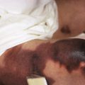
The visible short-term effects of UV light exposure are sunburn ( Fig. 19-1 ; see Fig. 26-15 ) and tanning. The ability to cause sunburn markedly declines with increasing wavelength. UVA light at 360 nm is 1000-fold less effective in causing skin erythema (sunburn) than UVA light at 300 nm. Thus UVB light is largely responsible for sunburn, with peak induction 6 to 24 hours after exposure. Sunburns gradually fade during the next 3 to 5 days, as the skin starts to desquamate. Reactivity to UVB light may range in severity from a mild asymptomatic erythema to a more intense reaction, with redness accompanied by tenderness, pain, edema, and, at times, vesiculation and bulla formation, particularly the day after the sunburn first appears. If the sunburned area is extensive, constitutional symptoms may include nausea, malaise, headache, fever, chills, and even delirium. Sunburn during childhood correlates with a higher risk of developing melanocytic nevi, as well as UV light-induced skin cancers.
Tanning is also wavelength-dependent and is biphasic. Immediate pigment darkening results primarily from exposure to UVA light, is caused by alteration and redistribution of melanin, and fades in 6 to 8 hours. Delayed tanning usually results from exposure to UVB and peaks at about 3 days after exposure. Fair skin is only able to tan with UVB dosages about the erythema threshold (i.e., a sunburn is required on type II skin; type I skin cannot tan; Table 19-1 ). In contrast, darker skin types (i.e., type III and higher) can tan significantly without burning (at suberythemogenic doses). Sunburn causes apoptosis (cell death) of keratinocytes (“sunburn cells”) or, if the dosage is high enough, induces cell cycle arrest, allowing the cell to undergo repair of their deoxyribonucleic acid (DNA) template before proliferating. Sunburn also depletes the protective Langerhans cells, causes epidermal thickening (which reduces exposure of the basal keratinocytes to UV radiation) as a protective mechanism, stimulates release of inflammatory cytokines, and induces the formation of antioxidative enzymes (which reduce oxidative DNA damage). The tan induced by UVB involves increased melanin synthesis, increased numbers of melanocytes, and increased transfer of melanosomes to keratinocytes.
| Skin Type | Reactivity to Sun | Examples |
|---|---|---|
| I | Very sensitive: always burns easily and severely, tans little or not at all | Individuals with fair skin, blond or red hair, blue or brown eyes, and freckles |
| II | Very sensitive: usually burns easily, tans minimally or lightly | Individuals with fair skin; red, blond, or brown hair; and blue, hazel, or brown eyes |
| III | Moderately sensitive: burns moderately, tans gradually and uniformly | Average white individuals |
| IV | Moderately sensitive: burns minimally, tans easily | Individuals with dark brown hair, dark eyes, and white or light brown skin |
| V | Minimally sensitive: rarely burns, tans well and easily | Brown-skinned (Middle Eastern and Hispanic) individuals |
| VI | Deeply pigmented: almost never burns, tans profusely | Blacks and others with heavy pigmentation |
The long-term effects of chronic sun exposure include photoaging, photocarcinogenesis, and immunosuppression. Given its potential to penetrate more deeply into the dermis, UVA light is thought to play a particularly important role in photoaging. UVA light, but not UVB light, is able to penetrate through window glass, even if tinted. Thus individuals who sit in offices exposed to UVA light through windows or drive cars extensively can show significant asymmetry in UVA damage on the face. An exception is the laminated glass of windshields (as opposed to the nonlaminated glass on the sides of cars), which blocks most UVA light (up to 380 nm). Window films can be applied to block UVA radiation but allow vision.
UVA light is also the light used in indoor tanning, a practice prevalent particularly among older female adolescents. This practice promotes skin damage, leading to an increased risk of both melanoma and nonmelanoma skin cancer and photodamage, but no increased protection against sun exposure. The danger of using tanning facilities has led to legislation to control use by minors and limitations of exposure in several states. Nevertheless, the prevalence of use of a tanning salon by college-aged students was 60.4% in 2009, with 33.1% using tanning salons more than five times a year. Many facilities now offer “safe” tanning, and these self-tanners are also available commercially. With all of these self-tanners, an artificial tan is produced by dihydroxyacetone (DHA), a sugar that interacts with the stratum corneum proteins to produce a brown pigment made of polymers called melanoidins, which resist washing. Approximately 10.8% of adolescents have used a self-tanner, especially older Caucasian adolescents with a high to moderate level of sun sensitivity. Self-tanners, unfortunately, provide minimal and transient sun protection, and their use is often in conjunction with tanning salon use rather than as a substitute.
Prevention of sunburn depends primarily on the utilization of measures that reduce exposure to strong sunlight. This is especially important for fair-skinned individuals, particularly blue-eyed persons, redheads, blonds, and those with freckles who withstand actinic exposure poorly, burn easily, and, over the years, tend to suffer chronic effects of light exposure. Prophylactic measures to reduce the impact of harmful UV rays include timing of outdoor activities to avoid peak UV light exposure between 10:00 am and 3:00 pm in the warmer seasons of the year; wearing broad-rimmed hats, sun protective clothing, and sunglasses; and staying in the shade. Light-textured materials such as T-shirts (especially when wet) give only partial protection. Clothes with a tighter weave are commercially available (e.g., www.coolibar.com ; www.solumbra.com ), or clothes can be laundered with a chemical (Tinosorb FD), which provides sun protection (e.g., Sun Guard).
Sunscreens occupy an important position in the management of UV light exposure. The lifetime use of sunscreen and sun avoidance has been calculated to reduce the lifetime risk of developing UV light-induced skin cancer by 78%, although whether use of sunscreens reduce the development of nevi remains controversial. Promoting routine sunscreen use in the pediatric population is important, including among adolescents whose behaviors escape parental influence. In order to be effective, adequate amounts of sunscreen must be applied to all areas exposed to UV light, and the sunscreen must be reapplied every few hours. It is critical that individuals continue sensible sun protection by means other than sunscreens and not increase their exposure because of sunscreen availability.
The most common sunscreen components and their absorbance capacity are listed in Table 19-2 . Inorganic sunscreens (titanium dioxide and zinc oxide) protect skin by reflecting and scattering UV and visible light (290 to 700 nm); often, both of these agents are usually included in an inorganic sunscreen. Organic sunscreens absorb light at particular wavelengths into specific chemical UV filters and reemit the energy as insignificant quantities of heat. Newer organic sunscreen components now absorb in both the UVA and UVB spectrum, thus providing protection against the damaging effects of the broad spectrum of UV light. These newer sunscreens are photostable (in contrast to avobenzone, an earlier generation component), and include bis-ethylhexyloxyphenol methoxyphenol triazine (anisotriazine [Tinosorb S]), drometrizole trisiloxane (silatriazole [Mexoryl XL]), methylene-bis-benzotriazolyl tetramethylbutylphenol (Tinosorb M), and terephthalylidene dicamphor sulfonic acid (Mexoryl SX). Unfortunately only avobenzone and Mexoryl SX are FDA-approved, so other newer sunscreen ingredients are not available in the US. Ideally, organic sunscreens should be applied 20 to 30 minutes before the onset of sun exposure, so that there is adequate time to bind to the stratum corneum and show effectiveness. The inorganic sunscreens can be applied immediately before sun exposure. Sunscreens should be reapplied after swimming, periods of excessive perspiration, and washing or showering; water resistance for 40 minutes is specified on labeling. Oral sunscreens containing antioxidants (e.g., lycopene, vitamins C and E) and botanicals (e.g., polyphenols, such as green tea and flavonoids such as genistein) are now commercially available. They provide some protection against acute sun damage, but they are not as effective as topical sunscreens in preventing sunburns, their long-term protective effects are not clear, and they should not replace other forms of photoprotection for children.
| Sunscreen Filter | Wavelength Protection * |
|---|---|
| ORGANIC SUNSCREENS | |
| PABA derivatives | UVB |
| PABA and padimate O (octyl dimethyl PABA) | |
| Salicylates | UVB |
| Homosalate (homomethyl salicylate), octisalate (octyl salicylate), trolamine salicylate | |
| Cinnamates | UVB |
| Cinoxate (2-ethyoxyethyl p-methoxycinnamate), octinoxate (octyl methoxycinnamate, Parsol MCX) | |
| Benzophenones | |
| Dioxybenzone (benzophenone-8) | UVB, some UVA2 |
| Oxybenzone (benzophenone-3) | UVB, UVA2 |
| Sulisobenzone (benzophenone-4) | UVA2, some UVA1 |
| Others | |
| Avobenzone (butyl methoxydibenzoylmethane, Parsol 1789) † | UVA2 and UVA1 |
| Ensulizole (phenylbenzimidazole sulfonic acid) | UVB |
| Meradimate (menthyl anthranilate) | UVA2 |
| Drometrizole trisiloxane (Mexoryl XL) | UVB, UVA2, UVA1 |
| Ecamsule (terephthalylidene dicamphor sulfonic acid, Mexoryl SX) † | UVA2, UVA1 |
| Octocrylene | UVB |
| Bisoctrizole (methylene bis-benzotriazolyl tetramethylbutylphenol. Tinosorb M) | UVB, UVA2, UVA1 |
| Bemotrizinol (bis-ethylhexyloxyphenol methoxyphenyl triazine, Tinosorb S) | UVA2, UVA1 |
| INORGANIC SUNSCREENS | |
| Titanium dioxide | UVA and UVB; if large enough particle, visible light |
| Zinc oxide | UVA and UVB; if large enough particle, visible light |
* UVB = 290 to 320 nm, UVA2 = 320 to 340 nm, UVA1 = 340 to 400 nm, visible light = 400 to 800 nm.
Sunscreen labels in the United States are now required to reveal both the sun protection factor (SPF) and the capability of broad spectrum UVA protection. The SPF rating can be determined by dividing the least amount of time it takes to produce erythema on sunscreen-protected skin by the time it takes to produce the same erythema without sunscreen protection. Thus individuals using a sunscreen with an SPF of 15 who normally burn after unprotected sun exposure, can theoretically stay out 15 times longer before getting the same degree of erythema, and thus only informs about protection against sunburn from UVB light. The additional benefit from use of a sunscreen with an SPF above 30 is small compared with the incremental benefit at lower SPF numbers, but higher SPF may be important for individuals with high photosensitivity or in intense sunlight. Furthermore, it should be recognized that the SPF is tested with a concentration of 2 mg/cm 2 , which is more than the majority of individuals apply. The designation “broad spectrum” provides information about the ability of the sunscreen to protect again nonerythema effects, especially from UVA light, including immune suppression, photoaging, and skin cancer.
The degree to which a person sunburns or tans depends on genetic factors and the natural protection of the skin. Skin types, accordingly, are ranked from skin type I, the most sensitive, to skin type VI, the least sensitive to sun damage (see Table 19-1 ). Because sun damage begins in children and is cumulative, it is strongly recommended that everyone adopt a program of sun protection and daily sunscreen use, preferably with an SPF of 15 or greater, from infancy on. Because increased melanin is not totally protective, even darker skinned individuals can burn and should use sunscreen.
Considerable media attention has focused on the need for UV light-induced vitamin D synthesis in the skin as a rationale for sun exposure. Indeed, UVB light induces the synthesis of vitamin D 3 in epidermal cells, and this vitamin D 3 is then hydroxylated in the liver (to 25-OH-vitamin D 3 ) and kidney (to 1,25-OH-vitamin D 3 ). However, the UV light exposure that stimulates vitamin D 3 production in the skin is inseparable from UV light exposure that is carcinogenic. Although vigorous sunscreen use may reduce the capacity of skin to produce vitamin D (and greater vitamin D deficiency has been linked to darker skin color), sunscreen use has not been linked with deficiency and oral administration of vitamin D likely suffices. The American Academy of Pediatrics recommends a minimum daily intake of vitamin D for infants, children, and adolescents to 400 IU/day, beginning shortly after birth. Patients who require strict photoprotection as treatment should be monitored for possible vitamin D deficiency and provided dietary supplementation.
Treatment of sunburn consists of cool compresses or cool tub baths in colloidal oatmeal (such as Aveeno), baking soda, or cornstarch; topical formulations with pramoxine or menthol; mild topical corticosteroid formulations; an emollient cream; and systemic preparations with analgesic and anti-inflammatory properties, such as nonsteroidal anti-inflammatory drugs (NSAIDs). When symptoms are severe, a short course of systemic corticosteroids (oral prednisone, or its equivalent, in dosages of 1 mg/kg per day, with tapering after a period of 4 to 8 days) will abort severe reactions and afford added relief.
Certain disorders predispose individuals to the adverse effects of UV light. For example, children with alopecia totalis (see Chapter 7 ), just as adults with androgenetic alopecia and balding at the vertex, have a higher risk of developing skin cancer at the exposed site if not protected. Patients with diminished or absent melanin, as in oculocutaneous albinism (see Chapter 11 ), or with defective DNA-repair mechanisms, as in xeroderma pigmentosum (XP), have an increased tendency to develop UV light-induced DNA damage and cutaneous malignancy. Individuals with nevoid basal cell carcinoma syndrome (see Chapter 9 ), which predisposes to the early onset of numerous basal cell carcinomas, have mutations in the PTCH gene, a gene that can also be mutated by UV light exposure in sporadic basal cell carcinomas. Sunlight can also exacerbate or trigger certain dermatoses, among them acne (see Chapter 8 ); herpes simplex infection (see Chapter 15 ); lupus systematosus, neonatal lupus, and dermatomyositis (see Chapter 22 ); Darier disease (see Chapter 5 ); pemphigus and bullous pemphigoid (see Chapter 13 ); and lichen planus and psoriasis (see Chapter 4 ).
Photodermatoses
Photosensitivity is a broad term used to describe abnormal or adverse reactions to sunlight energy in the skin. Photodermatoses must be distinguished from reactions to sunlight from exaggerated exposure, which is a normal response. Photodermatoses have been classified into four groups: 1) immunologically mediated; 2) drug- or chemical-induced; 3) with defective DNA repair; and 4) photoaggravation of existing conditions.
Photosensitivity in a child should be suspected if the child develops a sunburn reaction, swelling, or intense pruritus after limited exposure to sunlight or shows a rash or scarring predominantly in sun-exposed areas (face, V of the neck, and dorsal surface of the arms and hands). The history in a patient with a photosensitivity disorder is of great importance in determining the cause ( Table 19-3 ). Examination should focus on the distribution of lesions, including the areas of sparing. In a photosensitivity disorder, the upper eyelids, postauricular and submental areas, nasolabial and neck folds, volar aspect of the wrist, and the antecubital fossae generally tend to be spared. The morphology of the lesions may be helpful as well (urticarial versus papular versus vesicular, and the presence of lichenification, which suggests chronicity).
| History | Age of onset; exposure to potential photosensitizers; season of the eruption; time of onset after exposure to the sun; duration of eruption; effect of window glass and exposure to other light sources, including tanning booths; history of atopy and other medical problems; response to medications and use of sunscreens; family history |
| Examination | Distribution and morphology |
| Phototesting | Action spectrum, time to onset, minimal urticarial dose, inhibition and augmentation spectra |
| Photopatch testing | If photoallergy is suspected |
| Laboratory | Complete blood count, metabolic panel, erythrocyte sedimentation rate, autoantibodies, esp. antinuclear antibody, porphyrins |
| Biopsy | Useful for lupus and possibly porphyrias, but not for other photodermatoses |
| Fibroblast cultures for XP testing |
Phototesting can be helpful in determining the cause of an acquired photodermatosis. Exposure to UV light of different wavelengths may replicate the lesions, offering the opportunity to see morphology that may not be present at the time of the examination, confirming the suspicion of photosensitivity, and determining the UV range that triggers the disorder. Further laboratory investigations, such as antibody testing for suspected collagen vascular disease (such as antinuclear antibodies [ANAs], anti-ds DNA, anti-Ro, and anti-La antibodies), blood and 24-hour urine porphyrin levels, and photopatch testing in patients with suspected photoallergy, may be necessary. Performing a biopsy is rarely useful, except for suspected lupus erythematosus.
Immunologically Mediated Photodermatoses
Solar Urticaria
Solar urticaria accounts for less than 1% of type I immunoglobulin (Ig) E-mediated type of sensitivity reactions and is characterized by a sensation of pruritus or burning and erythema. Typically affected areas are the arms, legs, and upper chest; areas with regular sun exposure, such as the hands and face, are less commonly involved. It usually appears within 5 to 10 minutes of sunlight exposure and is followed almost immediately by a localized urticarial reaction confined to the exposed areas and an irregular flare reaction that may extend onto unexposed skin. Within 24 hours, lesions tend to resolve (usually in 1 to 3 hours); new lesions will not develop for 12 to 24 hours, even if subsequent exposure to sunlight occurs. Fixed solar urticaria, characterized by recurrent eruptions on the same body parts, is rare and less severe than typical solar urticaria. Delayed fixed solar urticaria, occurring 6 hours after exposure, has been described. Although the reaction is generally transient, scratching and rubbing may lead to secondary eczematization with persistent cutaneous changes. The disorder usually does not manifest until the third or fourth decade of life, and females are affected three times more often than males. It has been reported, however, as early as 1 week of age. The condition persists for more than a decade in the majority of affected individuals and often for a lifetime. Systemic signs have occasionally been reported in association, including headache, nausea, wheezing, dizziness, syncope and rarely shock.
The cause of solar urticaria is unclear, but the pathogenesis involves interaction of a cutaneous photoallergen with IgE-specific mast cells, leading to mast cell degranulation. Often phototesting can confirm the diagnosis and determine the wavelength range that causes the photosensitivity. If positive, whealing generally occurs within minutes. In some patients, the sensitivity involves the UVB range through the visible light range; the majority of patients react within the range of 290 to 480 nm. A negative phototest occurs in many patients and does not exclude the diagnosis. In patients with mild forms of solar urticaria (those in whom the threshold is high), the disorder may be controlled simply by appropriate sunscreens and avoidance of prolonged unprotected sun exposure. Those individuals highly sensitive to sunlight, however, must completely avoid daytime exposure.
Nonsedating antihistamines, antimalarials, corticosteroids, intravenous Ig (IVIG), and plasmapheresis have been beneficial. Omalizumab may be an option for patients with high levels of IgE who fail antihistamine therapy and has been successful in several (but not all) adult patients. In some individuals, sun tolerance may be established by carefully metered, increasing exposures (“hardening”) to natural or artificial light or the oral administration of a psoralen followed by UVA (PUVA) light.
Polymorphous Light Eruption
Polymorphous light eruption (PMLE) is the most common of the immune-mediated disorders associated with photosensitivity. It is predominantly a disorder of females in the second and third decades of life, and is estimated to occur in 10% to 15% of the US population. It occurs more often in individuals with lighter skin types. Although its etiology remains only partially understood, PMLE has been hypothesized to involve resistance to UV light-induced immune suppression and subsequently cell-mediated immune reactivity to a cutaneous photoantigen. An autosomal dominant inherited form of PMLE has been described in Native Americans of both North and South America. Onset of this hereditary form is in childhood, and female patients outnumber male patients 2:1.
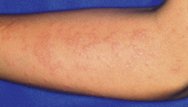
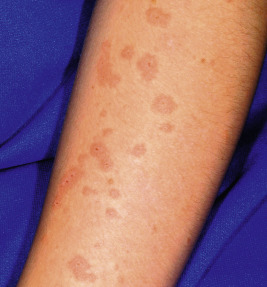
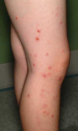
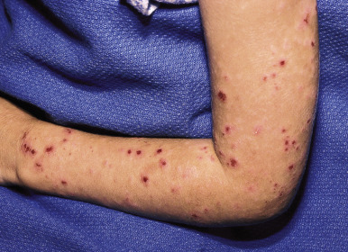
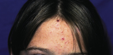
Often referred to as sun allergy or sun poisoning, the clinical eruption consists of a group of polymorphic lesions that usually occur 1 to 2 days after intense sunlight exposure, often while on vacation. In some individuals, the eruption is first seen in the spring and persists with continued sun exposure, but may improve later in the summer as the skin “hardens” from UV light exposure. The lesions may range from small papular ( Fig. 19-2 ), urticarial ( Fig. 19-3 ), vesicular, or eczematous reactions to large papules, plaques, or patterns resembling erythema multiforme. A “pinpoint” variant that resembles lichen nitidus clinically and histologically occurs more often in individuals with darker skin colors. The areas of the body most commonly involved include the face, the sides of the neck, and sun-exposed areas of the arms and hands. In children, it most commonly begins on the face as an acute, erythematous, eczematous eruption with small papules. Pruritus may be severe. Lesions usually involute spontaneously in 1 to 2 weeks, provided no additional exposure to sunlight occurs.
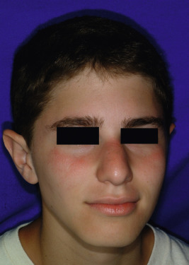
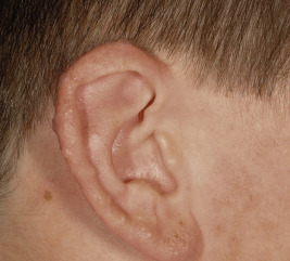
Juvenile spring eruption is considered a subset of PMLE, and is characterized by photo-induced, dull-red edematous papules that are largely confined to the helix of the ears ( Fig. 19-4 ). Lesions may become vesicular and crusted, and occasionally appear on the dorsal aspects of the hands and on the trunk. Juvenile spring eruption occurs more commonly in boys than in girls, and particularly between the ages of 5 and 12 years. Protuberant ears and lack of hair cover have been strongly associated, but skin color and use of sunscreen have not. Lesions heal within a week, without scarring, unless secondary infection develops. Lesions are more difficult than PMLE to reproduce by exposure to UV light.
The diagnosis of PMLE is suggested by the character of the lesions, their distribution, and their relationship to sun exposure. Although often unnecessary, PMLE can be confirmed by provocative light testing, which involves exposing the same site for about 4 consecutive days; it is important to use a site that has previously reacted but to perform the test early in the spring before hardening has occurred. The reaction, in contrast to that of solar urticaria, occurs within hours rather than minutes, lasts for days rather than hours, and is not urticarial. Some patients with systemic lupus erythematosus (SLE) acquire sun-induced lesions indistinguishable from those of PMLE, and serologic testing should be performed (see Chapter 22 ). In addition, erythema multiforme may be photodistributed and thus confused with PMLE ; in contrast with erythema multiforme, PMLE skin samples have shown no evidence of herpes simplex virus.
Prevention of PMLE consists of sunscreens with good coverage of the UVA spectrum (when applied adequately with 2 mg/cm 2 ), sun-protective clothing, and the avoidance of midday sun exposure. Hardening or desensitization of skin by gradually increasing exposure 2 to 3 times weekly to narrowband UVB or PUVA light for 4 to 6 weeks before exposure helps the majority of treated patients. The addition of oral flavonoids (especially polypodium leucotomos given for 12 weeks) or antioxidants in sunscreens has shown benefit in preventing PMLE. If patients severely affected by this disorder anticipate temporary intense or prolonged sun exposure, a short course of systemic corticosteroids can be administered. Topical steroids may provide relief in mild cases. Recent evidence suggests that topical vitamin D 3 or topical liposomal DNA-repair enzymes may be of value. Antimalarials, β-carotene, and nicotinamide are of limited effectiveness.
Actinic Prurigo
This photosensitivity disorder, also called hydroa aestivale and Hutchinson summer prurigo, is most commonly seen in the Indian and mestizo (mixed ancestry) populations of Mexico and other regions of Central and South America, although it has been described in the Caucasian and Asian populations. It has been seen in many children in the United States and Canada but is rare in Europe. Most cases begin in childhood before puberty. The condition is characterized by intensely itchy papules, plaques, and nodules, along with excoriations and scars ( Figs. 19-5 and 19-6 ). Some patients can be very uncomfortable and show secondary eczematization and lichenification. Actinic prurigo predominantly affects exposed sites on the face and distal extremities, but may involve covered areas as well, particularly the sacrum and buttocks. Healed facial lesions leave minute linear or pitted scars. Seasonal exacerbation at the beginning of spring with improvement in the fall is common, although the lesions commonly do not clear during the winter; greater seasonal change occurs with higher latitudes. The oral or ocular mucosae are involved in 30% to 50% of cases. Cheilitis alone is seen in 28% of patients, but 83% of patients experience pruritus, tingling, and pain of the vermilion. Ocular findings most commonly include photophobia and conjunctivitis.
The diagnosis is generally based on the clinical appearance. Biopsy of skin is generally not useful, although histologic evaluation of lip and conjunctival biopsies shows the characteristic well-formed lymphoid follicles. The presence of the eruption on both exposed and covered sites, its occurrence in winter, mucosal and conjunctival involvement, persistence beyond 4 weeks, and residual scarring of skin distinguish actinic prurigo from PMLE. Photosensitivity testing for minimal erythema dose is abnormal in up to two-thirds of patients. Human leukocyte antigen (HLA) DRB1*0407 is found in 60% to 70% of patients with actinic prurigo, but only in 4% to 8% of DR4+ controls) ; HLA DRB1*0401 is present in up to 20% of affected individuals with actinic prurigo. Although individuals with PMLE do not show these associations, 35% of patients with typical actinic prurigo have a history that suggests coexistence of actinic prurigo and PMLE or transition from one to the other. Although the etiology of actinic prurigo is unknown, the strong association with HLA markers suggests a role for major histocompatibility complex (MHC)-restricted antigen presentation in the pathomechanism of the condition.
The disorder often has a chronic course that persists into adulthood; however, spontaneous resolution may occur during late adolescence. Vigorous sun protection and use of topical anti-inflammatory agents for the pruritus lead to improvement in the majority of patients. Short courses of systemic steroids can be helpful, but antimalarials and other immunosuppressive medications have not had much effect. Complete resolution may require the addition of thalidomide (usually 50 to 100 mg/day), which results in rapid clearing. The gradual introduction of exposure to narrow band UVB light has also helped some patients. Oral cyclosporine and azathioprine have had some success in a few refractory cases.
Hydroa Vacciniforme
Hydroa vacciniforme (HV) is a rare disorder that is now known to result from chronic Epstein–Barr virus infection. The classic lesions tend to appear each summer in children on uncovered parts of the body after exposure to sunlight. Boys are more often affected than girls. Rare familial cases have been described. The mean age of onset is 8 years. Most patients show sensitivity to UVA light in monochromator phototesting, which may induce papulovesicular lesions.
The primary lesion is a pruritic edematous papule, vesicle, or bulla that occurs within hours or days on uncovered surfaces exposed to sunlight. Itching and burning, as well as mild constitutional symptoms may occur a few hours before the outbreak of the cutaneous lesions. Lesions tend to appear on the face, the sides of the neck, and extensor surfaces of the extremities, and are arranged symmetrically over the nose, cheeks, ears, and dorsal surfaces of the hands. The vesicles or bullae usually develop on an erythematous base and initially are rather tense. These are followed by central necrosis and umbilication that leads to healing with individual or confluent varioliform scarring within 1 to 2 weeks ( Fig. 19-7 ). Mild conjunctivitis or keratitis may be associated. The disease usually flares after sun exposure, and, in most instances, involutes spontaneously by the late teenage years with a mean duration of 9 years.
A more severe and persistent vesiculonecrotic HV-like skin eruption is likely to be lymphoma (HV-like), which is associated with higher titer latent Epstein–Barr virus infection and monoclonal T-cell receptor gene rearrangements (see Chapter 10 ). Patients have marked facial edema, hemorrhagic bullae, atrophic scarring, and severe disfigurement in both sun-exposed and sun-protected sites. Fever, hepatosplenomegaly, abnormal liver function testing, lymphadenopathy, hemophagocytosis, and hypersensitivity to mosquito bites may be associated. HV-like T-cell lymphoma occurs primarily in pediatric patients from Central and South America and Asia.
The diagnosis can be confirmed histologically. Treatment of HV consists of strict sun protection, including broad-spectrum sunscreens (SPF at least 30), protective clothing, and avoidance of midday sun exposure. No intervention has been uniformly successful. Antimalarial drugs, β-carotene, oral fish oils, and “hardening” by treatment with narrowband UVB light have been used with some therapeutic success. Low-dose prophylactic narrowband UVB light during spring months may also reduce disease severity. The psychosocial and emotional impairment of HV on quality of life is significant, given the extensive disfigurement from scarring and the effect on daily life of strict sun avoidance. Because of the potential risk of HV progression to HV-like lymphoma, patients should be closely monitored for at least 10 years after diagnosis.
Photosensitivity Induced by Exogenous Sources
Photosensitivity reactions may be phototoxic (photoirritant) or photoallergic ( Table 19-4 ). Exogenous photosensitizers may reach the skin by topical or systemic routes. The clinical course is brief, and elimination of the offending drug or sunlight exposure usually results in improvement. In rare cases, however, the photosensitivity may persist for months after the last known exposure to the offending chemical. Such individuals are known as persistent light reactors and the eruption (chronic actinic dermatitis) ranges from a chronic dermatitis initially restricted to sun-exposed surfaces to thickened hyperpigmented plaques. This rare disorder generally affects men and not pediatric patients.
| Phototoxic Medications | Photoallergic Medications |
|---|---|
|
|
|
|
|
|
| |
| |
| |
|
Phototoxicity
Phototoxic reactions are common and can be likened to a primary irritant reaction. A phototoxic reaction refers to a nonimmunologic exaggerated sunburn or sunburn-like reaction characterized by erythema (and at times swelling and blistering), occurring within a few minutes to several hours (usually within a period of 2 to 6 hours) after exposure to UVA light and followed by hyperpigmentation and desquamation confined to the exposed areas. This type of sensitivity usually occurs with the first exposure to the photosensitizing substance, when the systemic or percutaneous absorption of the sensitizing substances is in high enough concentration to result in a photo-induced cutaneous reaction.
Plant-induced photosensitivity (phytophotodermatitis) is the most common phototoxic reaction of children. The large majority are phototoxic reactions caused by the presence of furocoumarin compounds (psoralens) found widely in such plants as Rutaceae (e.g., limes and lemons); Umbelliferae (e.g., parsnips, carrots, dill, parsley, meadow grass, common rue, giant hogweed, and celery, most often celery infected with a fungus that causes pink rot disease); and Moraceae (e.g., fig). Psoralens can also reach the skin after ingestion, as has been noted after ingestion of contaminated celery and subsequent outdoor and tanning salon UV light exposure. With systemic ingestion, all sun-exposed areas are susceptible to reaction. Psoralens have also been used therapeutically in topical or oral formulations in combination with UVA light (PUVA therapy) as treatment of psoriasis and vitiligo; given the safety issues with PUVA, this intervention is rarely used in children. Furocoumarins can also be components in Chinese herbal medications. Inhalation of traces of giant hogweed have been reported to cause obstructive pulmonary symptoms, and contact with the eye can lead to blindness.
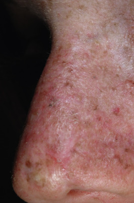
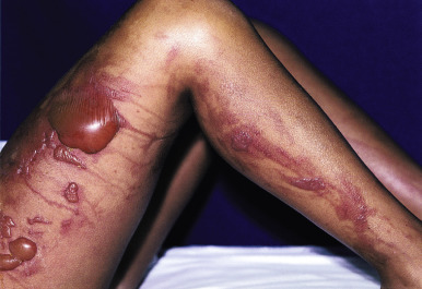
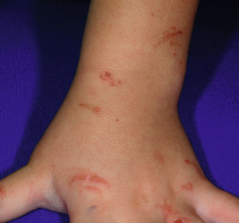
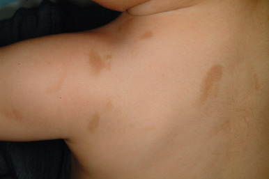
Phytophotodermatitis usually begins within a day after exposure to the furocoumarin and sunlight, ranges in severity from mild erythema with or without erosion to severe blistering ( Fig. 19-8 ), and eventuates in a characteristic dense inflammatory hyperpigmentation. A bizarre linear streaking configuration of the dermatitis ( Fig. 19-9 ), with subsequent hyperpigmentation, especially on the face, chest, hands, and lower legs of children, is characteristic. At times, only the hyperpigmented streak appears without prior erythema ( Fig. 19-10 ). The purple coloration of skin and bizarre patterning can be mistaken for child abuse and the blistering for herpes simplex infection. Streaks on the trunk have been noted after dripping of lime juice (sometimes used as a hair rinse or in drinks), and thumbprint-shaped macules on the lateral aspects of the trunk may be described after a parent with furocoumarins on the fingers picks up a child. Usually no treatment is necessary once the diagnosis is made, and the often-intense hyperpigmentation fades spontaneously over several weeks to months. However, severe burns have been induced in children by contact with phototoxins, especially giant hogweed and prolonged sunlight exposure, and have required debridement and surgical wound closure.
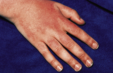
Phototoxicity may present with a variety of manifestations. For example, sunburn reactions can occur with thiazide diuretics, tetracyclines ( Fig. 19-11 ), retinoids, voriconazole, ciprofloxacin, and others, whereas skin fragility and blisters (pseudoporphyria) are the typical manifestation of exposure to NSAIDs, and telangiectasia results from from calcium channel antagonists. Photoonycholysis most commonly occurs after administration of doxycycline (see Chapter 8 , Fig. 8-16 ). The phototoxicity from voriconazole, used to prevent fungal infections in transplant patients, occurs in 20% of treated children overall and in 47% of children treated for more at least 6 months ; a dosage of at least 6 mg/kg twice daily has also been associated with a higher risk of developing voriconazole-induced phototoxicity. The eruption can be can be confused with graft-versus-host disease in these immunocompromised patients and has been associated with a 4.6% risk of developing nonmelanoma skin cancer in areas that had a phototoxic reaction at a mean age of 15.5 years.
Pseudoporphyria is a non-immunologically mediated phototoxicity reaction characterized clinically by increased cutaneous fragility, vesiculobullae, and histopathologic features similar to those of patients with porphyria, but levels of porphyrins are normal. It has been described in approximately 11% of children taking NSAIDs, especially naproxen sodium ( Fig. 19-12 , for juvenile idiopathic arthritis ) and is especially common in patients with blue/gray eye color and fair skin. The disorder has also been described in individuals receiving other NSAIDs, dapsone, ciprofloxacin, furosemide, imatinib, metformin, nalidixic acid, tetracyclines, retinoids, amiodarone, and voriconazole. A similar, if not identical, disorder has also been described in patients receiving hemodialysis ( Fig. 19-13 ).
A careful history and the presence of normal porphyrin levels in serum, erythrocytes, urine, and feces will generally establish the diagnosis. The cutaneous fragility tends to reverse rapidly upon withdrawal of the causative medication, but may only gradually reverse or even persist with new lesions for up to 1 month after discontinuation of the medication. Pseudoporphyria associated with hemodialysis has responded to treatment with N-acetylcysteine or glutathione. Disfiguring facial scarring is a residua in some affected children.
Photoallergy
Photoallergy is relatively uncommon and presumably is a form of cell-mediated delayed hypersensitivity. The individual must first be sensitized to the allergen, which can require 7 to 10 days. When exposed to UV light, the light is absorbed by the photoantigen, which is thought to cause a change in the molecule. Instead of sunburn-type reactions, photoallergic responses are generally characterized by immediate urticarial or delayed papular or eczematous lesions that are not followed by hyperpigmentation. After the first sensitization, subsequent photoallergic reactions generally appear within 24 hours, even after very brief periods of exposure. Sunscreens with chemical components (especially benzophenones) are the leading cause because of their extensive use, although the risk of reaction with sunscreens containing these agents is less than 1% of that for allergic contact dermatitis. In the past, children showed photoallergic reactions to salicylanilides (antimicrobial agents formerly in soaps but now only in industrial cleaners) and fragrances (musk ambrette, oil of bergamot). These chemicals are no longer used in personal products, but children or adolescents have been known to find a cologne with oil of bergamot, apply it to the skin, and develop berloque (berlock) dermatitis after UV exposure. Promethazine hydrochloride cream and topical nonsteroidal anti-inflammatory agents have caused photoreactions.
Suspected photoallergic contact dermatitis may be confirmed by photopatch testing. Photopatch testing is similar to traditional testing for contact dermatitis (see Chapter 3 ), except that two sets of patch tests are placed on the back. Twenty-four hours later, one set is uncovered and irradiated with UVA light. The following day a comparison is made of the covered and irradiated sites. A positive test reproduces the clinical eczematous lesion at the phototest site. Therapy is treatment with topical anti-inflammatory agents and avoidance of exposure to the offending antigen.
Genetic Disorders Associated with Photosensitivity
Photosensitivity is a prominent feature of several genetic disorders, many of which are associated with defective DNA repair. Some of these are described elsewhere in this text, such as Kindler syndrome (see Chapter 13 ), trichothiodystrophy (see Chapter 7 ), and ataxia-telangiectasia (see Chapter 12 ).
Xeroderma Pigmentosum
Xeroderma pigmentosum (XP) is a rare autosomal recessive disease characterized by cutaneous photosensitivity, a decreased ability to repair DNA damaged by UV radiation, and the early development of cutaneous and ocular malignancies. The disorder is estimated to occur in one in a million individuals in the United States and Europe, and with an incidence in Japan as high as one in 40,000 persons. The risk of developing cutaneous malignancy does not diminish in patients with darker skin types. Historically, seven complementation groups (XPA through XPG) have been described based on in vitro cell fusion studies ( Table 19-5 ), which are now recognized to correlated with mutations in specific genes whose products affect DNA repair.
| Gene/Protein | Protein Function | Incidence | Skin Disease | Neoplasia | Neurologic Change | Comments |
|---|---|---|---|---|---|---|
| XPA | Confirms damage | Especially in Japan | +++ | +++ | + to +++ | Lowest repair activity |
| XPB/ERCC3 | Helicase | Very rare | ++ to +++ | +++ | +++ |
|
| XPC | Detects damage | Most common | ++ to +++ | ++; melanoma | Rare | |
| XPD/ERCC2 | Helicase | 20% of cases | ++ | + | Late onset or none | Tremendous variability in phenotype; TTD; XP/CS |
| XPE/DDB2 | Detects damage | Rare | + | Rare | None or mild | Mild phenotype |
| XPF/ERCC4 | Nuclease | Fairly rare | ++ | Few to none | Usually none | |
| XPG/ERCC5 | Nuclease | Very rare | +++ | Few or none | Cockayne type | CS has been associated |
| XPV/POLH | Polymerase | 30% of cases | ++ to +++ | Later onset | ++ in a few |
The basic abnormality is an absence of a component of the nucleotide excision repair complex, a multistep mechanism that includes recognition of the UV light-induced DNA lesion, unwinding of DNA, and then resynthesis and ligation of the DNA. Complementation groups XPC and XPE recognize and bind to damaged DNA. XPA may help to assemble the DNA machinery around the damaged DNA site. This binding signals the XPB and XPD proteins to unwind the DNA in the damaged region and to allow DNA transcription. The XPF and XPG nucleases cut the DNA and allow excision of the UV light-induced pyrimidine dimers. DNA repair rates may range from 0% to 50% of normal levels. A variant form has been described (XPV). In the variant group, postreplication repair is defective, but excision repair is normal. More than half of all cases in the United States are caused by mutations in the genes encoding XPC ( XPC ), XPD ( ERCC2 ), or XPV ( POLH ), although mutations in the genes encoding XPA and XPF are more common in Japan than in the United States and Europe.
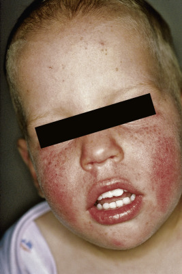
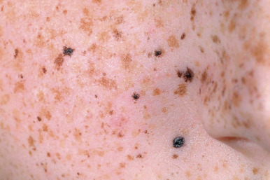
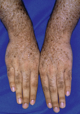
In 75% of cases the first signs appear between 6 months and 3 years of age. The presenting feature of this disorder is photosensitivity to UV light, primarily at wavelengths 290 to 340 nm, but this sunburn after minimal UV light exposure only occurs in 50% to 64% of patients and is not a feature of XP patients with mutations in XPC, XPE, or XPV. The sunburn may be severe with vesicles and bullae ( Fig. 19-14 ) and is often accompanied by photophobia with chronic conjunctivitis during the first months of life. The other 40% of cases do not show this sunburn reaction and instead tend to manifest initially with the lentigines (freckle-like pigmentation) that otherwise follow the early sunburns ( Figs. 19-15 and 19-16 ). The skin ages prematurely with atrophy, telangiectasia, mottled hyper- and hypopigmentation, keratoses, and ulcerations, but not wrinkling; these observations likely reflect the sensitivity primarily to UVB rather than to UVA light, which is the primary cause of solar elastosis. With continued exposure, the skin becomes xerotic, leading to the name XP. Areas of skin ordinarily protected by clothing remain relatively normal or may eventually show similar features, but to a lesser degree. Affected children resemble adults with severe actinic damage by early childhood.
Other issues are epithelial neoplasms, severe eye involvement, progressive neurologic degeneration in some patients, and malignancy. Several tumors occur with increased incidence in XP (basal cell and squamous cell carcinomas, angiosarcoma, fibrosarcoma, atypical fibroxanthoma, keratoacanthoma, and melanoma) ( Fig. 19-17 ). In patients younger than 20 years of age with XP, the overall risk of developing basal cell and squamous cell carcinomas is 10,000 times that of the normal population (median age 9 years), and of melanoma 2000-fold greater (median age 22 years). Interestingly, the melanomas in XP show a high rate of PTEN mutations (mammalian target of rapamycin [mTOR] pathway activation) rather than the typical RAS pathway activation, which may be important in considering choice of small molecule inhibitors. The tendency to develop these neoplasms is a function of exposure to UV light and the subtype of XP (see Table 19-5 ). The lower risk of developing skin cancer in patients who show early sunburns may reflect the earlier recognition of XP and intense early photoprotection. These tumors and their therapy cause considerable facial distortion. Patients with XP also have an approximately 10- to 20-fold increase in internal malignancies, including of the brain, lungs, hematopoietic system, kidney, and gastrointestinal tract, which may reflect concomitant sensitivity to physical and chemical carcinogens, such as cigarette smoke. Although skin cancer is the most common cause of death (34%), internal cancer leads to death of 17% of patients.