Papulosquamous Diseases
Susan J. Huang
Arturo Saavedra-Lauzon
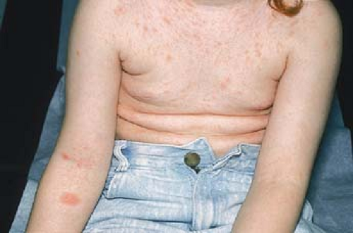
Papulosquamous disorders are a varied group of conditions, all sharing the common morphologic feature of scaling papules and plaques. Psoriasis is one of the most common papulosquamous disorders, seen in almost 2% of the general population. Seborrheic dermatitis, pityriasis rosea, and keratosis pilaris are dissimilar but common conditions, which are frequently seen in primary care. Lichen planus is an uncommon condition that can be idiopathic, caused by common oral medications or even by systemic viral infections such as hepatitis B and C. Lastly, pityriasis rubra pilaris is very rare, idiopathic condition that is challenging to treat. All of these conditions will be reviewed in detail in this section.
Psoriasis
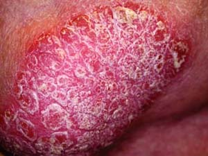 |
Ms. K is a 25-year-old white student who complains of a 2-month history of an itchy rash. She states that the rash first started as raised bumps, which then coalesced into plaques. She also states that her mother is being treated for a longstanding skin condition. On examination, well-demarcated papules and plaques with overlying thick, white scale (Fig. 12-1) over an erythematous base involve her elbows, knees, and her lower back. The patient also shows you areas where the lesions have appeared after scratching. When gently curetting the scale off a lesion, the scale lifts off in intact sheets. Removal of the scale reveals pinpoint bleeding, consistent with the Auspitz sign.
Background/Epidemiology
Psoriasis is a fairly common skin condition in the United States, affecting up to 2% of individuals. In its common form, it is marked by thick white or silvery lamellated scale (often called micaceous) with or without accompanying pruritus, often in visible areas. Such visibility, accompanying symptoms, and chronic nature can severely decrease quality of life. Individuals with psoriasis have a higher incidence of depression. Furthermore, patients with psoriasis are at increased risk of having lymphoma, metabolic syndrome, and cardiovascular disease. Psoriasis primarily affects adults with bimodal age peaks in the second and fifth decades of life, though any age may be affected.
Key Features
Psoriasis affects ∼2% of individuals.
The most common presentation is with thick white or silvery lamellated scale on typical areas: elbows, knees, scalp, umbilicus.
It primarily affects adults, with bimodal age peaks in the second and fifth decades of life, though any age may be affected.
Psoriasis patients are at increased risk of having lymphoma, metabolic syndrome, and cardiovascular disease.
Treatment of psoriasis is complicated, and individualized regimens are needed depending on the extent and severity of disease. No cure exists, so treatment is aimed at controlling the disease.
Pathogenesis
A positive family history predisposes an individual to psoriasis. Studies have shown that between 36% and 91% of patients with psoriasis have positive family histories. Furthermore, certain HLA types including HLA-B13, B17, B37, Bw16, and DR7 are more common among those with psoriasis. Clinically, there
are several triggers and exacerbating factors. Streptococcal infection often leads to guttate psoriasis, and viral exanthems are known to trigger episodes. Life stressors; direct cutaneous trauma; medications such as lithium, beta blockers, nonsteroidal anti-inflammatory drugs (NSAIDs); and steroid tapers have all been reported to trigger psoriasis. Human immunodeficiency virus (HIV) infection does not increase the incidence of psoriasis but is linked to more aggressive psoriasis.
are several triggers and exacerbating factors. Streptococcal infection often leads to guttate psoriasis, and viral exanthems are known to trigger episodes. Life stressors; direct cutaneous trauma; medications such as lithium, beta blockers, nonsteroidal anti-inflammatory drugs (NSAIDs); and steroid tapers have all been reported to trigger psoriasis. Human immunodeficiency virus (HIV) infection does not increase the incidence of psoriasis but is linked to more aggressive psoriasis.
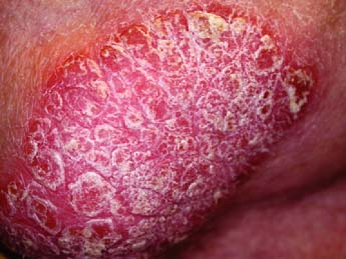 Figure 12-1 Micaceous scale overlying an erythematous plaque consistent with psoriasis on the elbow. |
Clinical Presentation
The most frequent type of psoriasis accounting for 90% of cases is plaque psoriasis. Other variants include pustular, guttate, erythrodermic, inverse, and nail psoriasis.
The most common form of psoriasis is plaque psoriasis. Lesions are well-demarcated circular, oval, or polycyclic papules or plaques marked by erythema and overlying silvery micaceous scale (Fig. 12-1). They are generally distributed symmetrically and often affect the scalp, elbows, knees, trunk, presacral region, hands, and feet. On hands and feet, painful fissuring can occur. Genitals are affected in 30% of cases. Lesions may be accompanied by pruritus or pain. In developing lesions, the active edge appears more erythematous and small papules may surround the lesion. Arrangement of lesions may be linear, perhaps reflecting the Koebner phenomenon, where lesions develop in sites of cutaneous trauma such as scratching (Fig. 12-2). The scales of psoriatic lesions can be lifted in coherent sheets. Upon removing the scales and then the overlying membrane, pinpoint bleeding can be observed. This is known as the Auspitz sign. In some cases, an inverse pattern of psoriasis may be observed. In these cases, thin erythematous plaques without scale are present in intertriginous or flexural areas such as the axilla, groin, inguinal crease, or under breasts.
Pustular psoriasis is a less common form of cutaneous psoriasis and can manifest as localized or generalized sterile pustules (Fig. 12-3). In many cases,
these patients are systemically ill, requiring hospitalization. It is a diagnosis of exclusion and as such, other causes of pustules such as infection must be ruled out.
these patients are systemically ill, requiring hospitalization. It is a diagnosis of exclusion and as such, other causes of pustules such as infection must be ruled out.
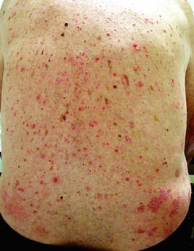
Guttate psoriasis presents as hundreds of small ∼1- to 10-mm plaques of psoriasis on the trunk and extremities (Fig. 12-4), often in an explosive manner. This is the most common type of psoriasis outbreak in those under the age of 30 years. In many cases, group A beta-hemolytic streptococcal infections (S. pyogenes), often pharyngitis, precede outbreak and are the cause of this type of psoriasis. This may be the first presentation of psoriasis. Treatment with appropriate oral antibiotics will often lead to completely clearance, though not all cases are infection-related. In some, this will be the only eruption of psoriasis, though many eventually develop plaque type psoriasis.
Erythrodermic psoriasis is another form of cutaneous psoriasis and is a medical emergency. Often it is in the setting of worsening or uncontrolled psoriasis, although it is sometimes the first presentation of psoriasis. Erythema and exfoliation affect large areas of skin. The condition can lead to infection and significant fluid, electrolyte, and protein imbalances, which can be fatal. Of note, hypocalcemia is a complication of erythrodermic psoriasis. These patients should be managed as inpatients.
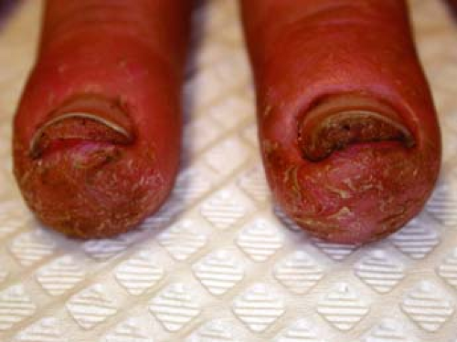
Psoriasis affecting the nails occurs in almost 80% of psoriasis patients. Nails may exhibit pitting, keratotic collections, “oil spots” (yellow-brown discoloration), and onycholysis (Fig. 12-5) (lifting of the distal nail).
Psoriatic arthritis occurs in about 10% of psoriasis cases. The majority of cases present as asymmetric arthritis of finger and toe joints, which in time can lead to a “sausage digit.” In patients with joint disease, tendons may also be affected. For patients with psoriatic arthritis, consider referring to dermatology or rheumatology for consideration of a systemic therapy, which will not only treat the psoriasis but also reduce joint inflammation and destruction.
Diagnosis
Diagnosis of cutaneous lesions is usually clinical but a biopsy may aid in diagnosis in questionable cases. To evaluate the
differential diagnosis of fungal infection, a potassium hydroxide (KOH) test of a skin scraping can be performed. This test has high sensitivity and low specificity, so a fungal culture should be obtained if you suspect a false negative KOH test (see Chapter 9).
differential diagnosis of fungal infection, a potassium hydroxide (KOH) test of a skin scraping can be performed. This test has high sensitivity and low specificity, so a fungal culture should be obtained if you suspect a false negative KOH test (see Chapter 9).
Joint disease typically coincides or follows cutaneous lesions, but could also precede cutaneous lesions. To evaluate joint disease, erythrocyte sedimentation rate (ESR), rheumatoid factor (RF), and radiologic studies may help determine the cause of disease. Contrary to rheumatoid arthritis, psoriatic arthritis is a seronegative arthritis. ESR is usually normal, serologies are negative, and radiographic erosive changes are present.
Differential diagnoses for psoriasis includes atopic dermatitis, lichen simplex chronicus, seborrheic dermatitis, pityriasis rosea, fungal infection, and syphilis. Psoriatic lesions may be secondarily infected. Consider the possibility of allergic contact dermatitis for psoriasis that is worsening despite adequate topical therapy. There are many preservatives that are potential causes of allergy and will worsen psoriasis if the patient becomes allergic.
Treatments
Treatment of psoriasis is complicated, and individualized regimens are needed depending on the extent and severity of disease. No cure exists so treatment is aimed at controlling the disease. Patients should be referred to a dermatologist and/or rheumatologist for long-term care. However, it is appropriate to start initial therapy for plaque psoriasis.
For involvement of less than 5% of the body surface area (BSA), topical medication should first be used. Topical steroids, coal tar, vitamin D analogs (e.g., calcipotriol, calcipotriene), or a topical retinoid (e.g., tazarotene) may be useful. Potent steroids should be avoided for the face and genital areas. For lesions of the palms and soles, topical therapy may be insufficient. Patients may benefit from local phototherapy and/or systemic treatment such as oral methotrexate, acitretin, or cyclosporine.
A typical regimen for a newly diagnosed patient with plaque psoriasis on elbows, knees, and scalp (<5% BSA) could consist of the following topicals: a vitamin D analog (calcipotriene) twice daily to affected areas on weekdays and a superpotent topical steroid (clobetasol propionate) ointment twice daily on weekends. For scalp involvement, a topical solution or foam such as fluocinonide 0.05% is usually helpful. An over-the-counter coal tar shampoo used 3 to 4 times weekly is helpful for many patients. It is important that the patient leave the shampoo in for 5 to 10 minutes before rinsing to receive full benefit. If palms and/or soles are involved, consider narrow-band UVB therapy (if available), a super-potent topical steroid, and a keratolytic such as salicylic acid 6% cream.
If lesions involve a larger percentage of the body surface area, systemic therapy, phototherapy with or without topical therapy should be considered. Oral methotrexate, acitretin, or cyclosporine can be used. If treatment is refractory, consider the use of biologics. These biologics include alefacept, efalizumab, adalimumab, etanercept, infliximab, and ustekinumab. Patients needing these medications should be referred to a dermatologist.
“At a Glance” Treatment
Treatment of psoriasis is complicated and individualized regimens are needed depending on the extent and severity of disease. No cure exists.
Less than 5% BSA:
Primary: topical medications: vitamin D analog (calcipotriene) BID to affected areas on weekdays and a superpotent topical steroid (clobetasol propionate) ointment twice daily on weekends.
Scalp: a topical solution/foam such as fluocinonide 0.05%. An over-the-counter coal tar shampoo used 3 to 4 times weekly. Leave in for 5 minutes.
Palmoplantar psoriasis:
Primary: super-potent topical steroid and a keratolytic such as salicylic acid 6% cream
Secondary: may require local phototherapy and/or systemic treatment such as oral methotrexate, acitretin, or cyclosporine
More than 5% BSA:
Topical therapy as appropriate as above
Systemic therapy, phototherapy with or without topical therapy should be considered. Oral methotrexate, acitretin or cyclosporine can be used.
If treatment is refractory, consider the use of biologics including alefacept, adalimumab, etanercept, infliximab, and ustekinumab. Patients needing these medications should be referred to a dermatologist.
Psoriasis is a chronic condition requiring follow-up and tailored treatment regimens. As such, the patient should be referred to a dermatologist and/or rheumatologist for advanced or recalcitrant disease.
Erythrodermic or pustular psoriasis patients should receive emergent care.
Erythrodermic psoriasis requires emergent treatment.
Course and Complications
In plaque psoriasis, individual lesions start as papules, which may then coalesce into plaques with variable amount of scaling. Lesions may last months to years in the same location. With treatment, lesions first clear centrally and clearance then proceeds outward. Upon resolution, postinflammatory hypo- or hyperpigmentation may be present. Improvement with therapy may be evaluated based on the Psoriasis Area and Severity Index (PASI) score or on quality-of-life scores. The PASI score considers the surface area affected, redness, thickness, and amount of scaling.
Psoriatic arthritis develops in about 10% of patients with cutaneous psoriasis. Usually joint disease coincides with or follows cutaneous disease, although this is not always the case. Psoriatic arthritis is usually progressive and may lead to long-term destruction of affected joints.
ICD9 Code
| 696.1 | Other psoriasis and similar disorders |
Pityriasis Rosea
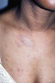 |
Ms. K is a 26-year-old waitress who complains of pruritic pink patches on her chest, back, and upper arms (Fig. 12-6). One week prior, she noticed a solitary pink ovoid patch on her abdomen, which then enlarged over the next few days. Starting 2 days ago, she noticed similar patches developing on her trunk. Ms. K recently had an upper respiratory infection before the onset of her rash, but otherwise has had no other medical issues. She is particularly concerned about the appearance of her lesions and wants to know if her lesions are contagious and when they will subside.
Background/Epidemiology
Pityriasis rosea (PR) is a common mild and self-resolving skin condition which most often affects healthy children and young adults between the ages of 10 and 35. In the typical scenario, a single salmon-colored patch called a “herald patch” appears first, frequently on the trunk. Multiple other similar lesions then appear, often on the trunk and proximal extremities and following skin cleavage lines, although other patterns may occur. Thus, on the back, these lesions may create the classical “Christmas tree” distribution. Patients often complain of pruritus, which can be severe. Variants of PR exist and include localized and inverse pityriasis rosea, and papular, urticarial erythema multiforme-like, vesicular, pustular, and purpuric variants.
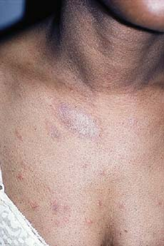 Figure 12-6 Pityriasis rosea. From Goodheart HP. Goodheart’s Photoguide of Common Skin Disorders, 3rd ed. Philadelphia: Lippincott Williams & Wilkins, 2009. |
Pathogenesis
An exact etiology is unknown, although viral etiologies—specifically, HHV-6 or HHV-7 infection—have been implicated. Some cases are preceded by a viral prodrome marked by headache, fever, malaise, abdominal discomfort, and/or arthralgias. PR has also been reported to occur more frequently in the spring and fall seasons. Infectivity is low to none.
Clinical Presentation
The herald patch in 80% of the cases precedes the wave of lesions and is frequently located on the trunk or neck. This patch is a single discrete pink or salmon-colored ovoid patch or plaque, which enlarges over the course of a few days. It is marked by a collarette of scale as well as fine central scale which may clear with time. After days or weeks, crops of other lesions in different phases arise, most often on the trunk and proximal extremities and neck, although other distributions are also possible. For instance, the palms, soles, and face are usually spared but may be affected in variant forms of PR. The lesions are similar to the herald patch in morphology but are often smaller in size (Figs. 12-6, 12-7). They are often arranged with their long axis along skin cleavage lines, thus resulting in the classic “Christmas tree” distribution on the back. Most of patients report either no symptoms or mild pruritus. In individuals with dark skin, lesions are more often hyperpigmented and papular and more widespread.
Key Features
Pityriasis rosea is a self-limited condition presenting initially as a single salmon-colored, thin scaling plaque on the trunk, then many smaller scaling plaques develop.
It is likely caused by a viral infection, though pathogenesis has not been proven.
Be aware that secondary syphilis is a mimic; screen appropriate cases.
Treatment is symptomatic: topical corticosteroids and antipruritics.
Diagnosis
Several other conditions can mimic PR (Table 12-1). Of primary importance, secondary syphilis should be ruled out (Fig. 12-8). Clinical suspicion increases with the presence of condyloma lata, a chancre, split papules (fissured papules seen at the angle of the mouth), and/or peripheral lymphadenopathy. It is important to be aware that, while unlikely to spread with minimal contact, the skin lesions of secondary syphilis may be infectious. Mucosal involvement is
also more common in secondary syphilis. A rapid plasma reagin (RPR), Venereal Disease Research Laboratory (VDRL) and fluorescent treponemal antibody-absorption (FTA-ABS) along with a biopsy can help distinguish between pityriasis rosea and secondary syphilis.
also more common in secondary syphilis. A rapid plasma reagin (RPR), Venereal Disease Research Laboratory (VDRL) and fluorescent treponemal antibody-absorption (FTA-ABS) along with a biopsy can help distinguish between pityriasis rosea and secondary syphilis.
Table 12-1 Differential Diagnosis for Pityriasis Rosacea | |
|---|---|
|
Drug eruptions may mimic PR (Table 12-2). In these cases, lesions tend to be fewer in number and larger in size and often lack the preliminary herald patch. Tinea corporis can also resemble pityriasis rosea and can be ruled out by a KOH preparation of a scraping. Viral exanthems can mimic pityriasis rosea although lesions are usually nonscaling. Nummular eczema and pityriasis lichenoides should also be excluded. In acquired immunodeficiency syndrome (AIDS) patients, a more chronic pityriasis rosea-like condition has been described.
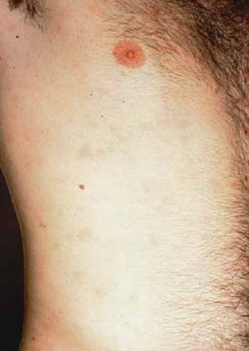 Figure 12-8 Secondary syphilis: body. From Goodheart HP. Goodheart’s Photoguide of Common Skin Disorders, 3rd ed. Philadelphia: Lippincott Williams & Wilkins, 2009.
Stay updated, free articles. Join our Telegram channel
Full access? Get Clinical Tree
 Get Clinical Tree app for offline access

|
