Nutritional Diseases


Abstract


Proper nutrition is fundamental to human life. From fetal development to adulthood, proper nutrition is essential for survival, physical growth, mental development, productivity, and overall health. The skin, hair, nails, and mucous membranes may all provide clues to the presence of a nutritional deficiency, and often a patient will have multiple deficiencies. In addition to inadequate oral intake, there are secondary causes of nutritional deficiencies, e.g. intestinal malabsorption. Two classic protein–energy malnutrition syndromes have been described, kwashiorkor and marasmus, and each has characteristic features. Deficiencies of vitamins and trace elements can lead to a wide range of mucocutaneous findings, from follicular keratoses to photodistributed erythema to periorificial erosions and atrophic glossitis.


Keywords

protein–energy malnutrition, vitamin deficiencies, obesity, kwashiorkor, marasmus, vitamins, trace elements, zinc deficiency, pellagra, acrodermatitis enteropathica, scurvy, phrynoderma, atrophic glossitis, vitamin D deficiency


Key features


  • Nutritional deficiencies may affect any organ system, including the skin, which may provide clues to the diagnosis


  • The two classic protein–energy malnutrition syndromes are kwashiorkor and marasmus


  • In addition to inadequate oral intake, there are secondary causes of nutritional deficiencies, e.g. intestinal malabsorption due to medical conditions or surgical procedures


  • Vitamin and trace element deficiencies can lead to a wide range of mucocutaneous findings, from follicular keratoses to periorificial erosions and glossitis


  • Insufficient and deficient levels of vitamin D have been noted worldwide, in both low-income and high-income countries


  • Obesity can be associated with high-calorie malnutrition and a number of skin changes, from acanthosis nigricans to lipodermatosclerosis


Introduction


Proper nutrition is fundamental to human life. From fetal development to adulthood, proper nutrition is essential for survival, physical growth, mental development, productivity, and overall health . Deficiency of one isolated nutrient is rare, as individuals are usually deficient in multiple nutrients at the same time. In general, nutrients are categorized as macronutrients (carbohydrates, proteins and fats) or micronutrients (vitamins and minerals). Most people who suffer from nutritional deficiencies live in low-income countries. However, there are certain conditions that predispose individuals from high-income countries to develop nutritional deficiencies, such as alcoholism, intestinal malabsorption, anorexia nervosa or bulimia, and restrictive diets.


Nutrients require a process of ingestion, digestion, absorption and circulation plus subsequent metabolism in order to be beneficial. When any one of these steps is disrupted, clinical manifestations may appear.


Epidemiology and Pathogenesis


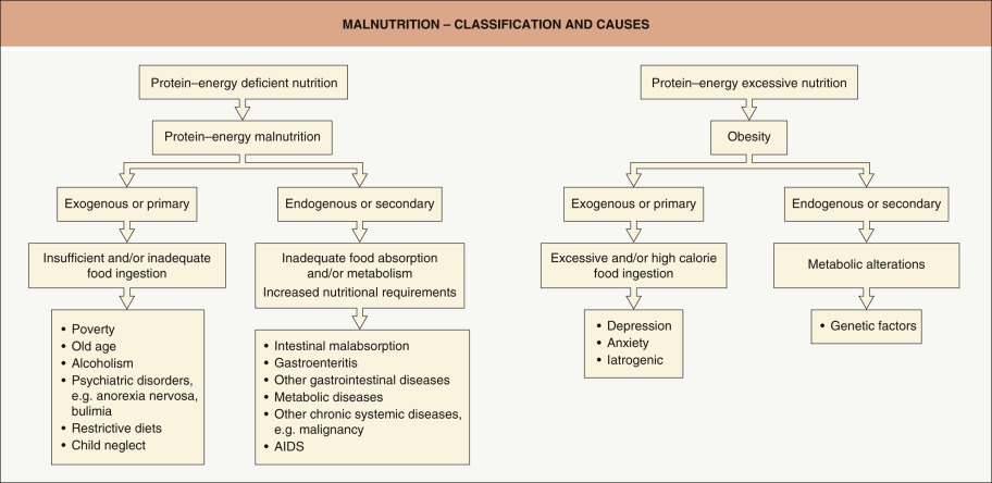
In low-income countries, malnutrition stems primarily from inadequate ingestion of all macronutrients as well as many micronutrients ( Fig. 51.1 ). The World Health Organization (WHO) has estimated that worldwide, ~50% of deaths in children <5 years of age are due directly or indirectly to the multisystem ramifications of malnutrition. In industrialized regions of the world, malnutrition is directly linked to the per capita gross national product; if a country has a per capita income that is less than US$300 per year, endemic malnutrition is likely, especially in infants and young children.


Fig. 51.1


Malnutrition – classification and causes.

Courtesy, Ramón Ruiz-Maldonado, MD.


At the same time, a global epidemic of obesity has emerged within industrialized countries, with increasing rates of death due to cardiovascular disease and diabetes mellitus . Excessive consumption of foods high in calories but low in nutrients can lead to obese patients who lack essential vitamins and minerals. This is supported by a study in which 57% of morbidly obese patients undergoing preoperative assessment for bariatric surgery were found to have a deficiency in at least one micronutrient .


Restrictive diets, including those based upon real or perceived need (e.g. food allergies), psychiatric illness (e.g. anorexia nervosa) or mental impairment (e.g. illicit drug abuse), also disrupt the intake of appropriate nutrients and can result in malnutrition.


In addition to a diet inadequate in the quantity and/or quality of macro- and micronutrients (i.e. primary causes), there are medical conditions that can lead to a functional malnutrition, i.e. secondary causes. The latter include an increase in metabolic requirements (e.g. severe infections) and/or a decrease in the transport and utilization of nutrients (e.g. intestinal malabsorption, bariatric surgery). Malnutrition is also seen in individuals with late-stage internal malignancies and advanced AIDS ( Table 51.1 ).


Table 51.1

Characteristics of protein–energy malnutrition and essential fatty acid deficiency.

AIDS, acquired immunodeficiency syndrome; HIV, human immunodeficiency virus.


CHARACTERISTICS OF PROTEIN–ENERGY MALNUTRITION AND ESSENTIAL FATTY ACID DEFICIENCY
Protein–energy malnutrition Essential fatty acid deficiency
Marasmus Kwashiorkor
Causes


  • Decreased energy intake


  • Develops over months to years


  • Primary causes: poverty, alcoholism, drug abuse, psychiatric disorders, restrictive diets, child or elderly neglect/abuse


  • Secondary causes: intestinal malabsorption, chronic diarrhea, malignancies, chronic systemic diseases (e.g. hepatic failure, end-stage renal disease), AIDS * , metabolic disorders


  • Decreased protein intake during periods of stress lasting a few weeks or more


  • Diets consisting primarily of rice or rice-based beverages (e.g. “rice milk”) for perceived milk intolerance


  • Other causes: protein-losing enteropathies, extensive gastrointestinal surgeries, HIV disease *


  • Protein–energy malnutrition


  • Gastrointestinal disorders/surgeries causing severe fat malabsorption


  • Long-term parenteral nutrition without lipid supplementation


  • Diets extremely low in fat


  • Nephrotic syndrome


  • Inborn errors of metabolism, e.g. deficiency of fatty acid desaturase 2 (FADS2; also referred to as delta (6) desaturase)

Cutaneous features


  • Dry, thin, pale, lax and wrinkled skin


  • Occasionally, fine scaling and hyperpigmentation ( Fig. 51.3 )


  • Follicular hyperkeratosis and folliculitis in adults


  • Ulcerations


  • Excess of lanugo-like hair


  • Thin hair that grows slowly and falls out readily


  • Impaired growth of nails and fissured nails


  • Purpura ( Fig. 51.4 )


  • Dyschromia is a common finding


  • Pallor due to distention of the skin and loss of pigment


  • Hypopigmentation following abrasions, and ulcerations and hyperpigmentation in areas subject to trauma


  • Superficial desquamation in mild cases (“enamel paint spots”), but in severe cases large areas of erosion (“flaky paint”) ( Fig. 51.6 )


  • Other cutaneous signs include erythema, thinning, petechiae, ecchymoses, and purpura


  • Hair is sparse, dry, lusterless and brittle with a reddish tinge; bands of light and dark coloration (“flag sign”) reflect intermittent periods of malnutrition; positive gentle hair pull test


  • Nails are soft and thin


  • Mucosal lesions: cheilitis, xerophthalmia and vulvovaginitis


  • The skin is dry, scaly, and leathery with underlying erythema


  • Intertriginous erosions


  • Alopecia and more lightly pigmented hair


  • Increase in transepidermal water loss


  • Petechiae

Systemic features


  • Starved appearance but alert


  • Reasonably preserved responsiveness to short-term stress


  • Bradycardia, hypotension and hypothermia


  • Loss of subcutaneous fat and muscle ( Fig. 51.3 )


  • “Monkey facies” or an aged appearance (due to loss of buccal fat pads)


  • Suppression of growth


  • Relatively well-nourished appearance


  • Edema or even anasarca ( Fig. 51.5 )


  • Apathy, anorexia, irritability


  • Failure to thrive (retardation of growth and mental development)


  • Superimposed bacterial and fungal (e.g. candidal) infections


  • Bilateral parotitis, hepatomegaly, diarrhea, loss of muscle mass


  • Growth failure


  • Poor wound healing


  • Impaired reproduction


  • Abnormal liver and kidney function tests


  • Capillary fragility


  • Increased susceptibility to infections


  • Neurologic damage

Lab findings and diagnostic criteria


  • Triceps skin-fold <3 mm


  • Mid-arm muscle circumference <15 cm


  • Creatinine–height index <60% of standard


  • Low plasma zinc levels


  • Epidermal acanthosis, confluent hyperkeratosis, pallor of upper layers of the epidermis (missing in later lesions)


  • Most hair bulbs are in the telogen phase


  • Abundant broken hairs


  • At least one of the following: poor wound healing, decubitus ulcers, or skin breakdown


  • Anergy


  • Hypoalbuminemia (<2.5 g/dl)


  • Total iron-binding capacity <200 mcg/dl


  • Peripheral lymphocyte count <1500/mcl


  • Steatohepatitis


  • Decreased number of anagen hair follicles and increased number of telogen follicles


  • Structural abnormalities in anagen follicles – severe atrophy, shaft constriction, depletion of pigment


  • Exclude nephrotic syndrome


  • Low plasma levels of linoleic and linolenic acids


  • Accumulation of 5,8,11-eicosatrienoic acid


  • Triene (eicosatrienoic acid) : tetraene (arachidonic acid) ratio >0.4


  • Elevated levels of palmitoleic and oleic acids


  • Anemia, thrombocytopenia


  • Orthokeratotic hyperkeratosis, acanthosis with hypergranulosis, atrophic sebaceous glands, and papillary dermal vasodilation with a mixed cell infiltrate (in rat models)


  • Steatohepatitis

Treatment


  • Treat or prevent hypothermia, dehydration, hypoglycemia, and electrolyte imbalances


  • Treat infections


  • Slowly replace proteins and calories, allowing for readaptation of metabolic and intestinal functions


  • Supplementation with linoleic acid and zinc


  • Monitor for hypophosphatemia and cardiorespiratory failure (associated with overly aggressive nutritional replacement)


  • Address underlying diseases, social issues


  • Aggressive nutritional support is indicated to rapidly restore metabolic balance; correction of any electrolyte disturbances or hypoglycemia


  • Institute diet with adequate protein and caloric intake; supplementation with minerals and vitamins is required as patients often have multiple nutritional deficiencies


  • Identify and attempt to treat underlying cause(s), including bacterial and parasitic diseases


  • Topical moisturizers as needed


  • Essential fatty acid replacement, depending upon severity


  • Correction of coexisting nutritional deficiencies

Prognosis


  • 10% mortality, often secondary to diarrhea or pneumonia


  • Mortality rate related to underlying disease(s)


  • Prognosis in adult patients is poor, even with aggressive nutritional support


  • In children, a lesser degree of stress is required to precipitate the disorder, so it is less foreboding


  • Severe, relapsing cases and those with coexistent HIV disease have a high mortality rate


  • Death usually occurs due to overwhelming infection despite antibiotic treatment


  • Prognosis depends on the severity and coexistence of other nutritional deficiencies


  • Depends on awareness of this entity


* HIV infection is more often associated with marasmus than with kwashiorkor.


Clinical Features


Weight-for-height scores and height-for-age scores are utilized to evaluate malnutrition; scores are compared to the standard deviations (SD) from the median value for the reference population. An individual is considered to have moderate malnutrition if the scores are between the 2 nd to 3 rd (lower) SD and severe malnutrition if below the 3 rd SD, with or without symmetrical edema . Cutaneous manifestations in part reflect the fact that epidermal maturation (from basal cell layer to stratum corneum) occurs over a period of 10–14 days. Deficiencies, especially of macronutrients, can interfere with this process, resulting in skin that is dry and thin in appearance with associated epidermal atrophy. Prolonged deficiency can reduce both protein production, including that of dermal collagen and muscle, and the amount of subcutaneous fat. As a result, patients may develop dermal atrophy, muscle wasting, and/or lax skin.


Additional clinical findings of marasmus and kwashiorkor, such as dyschromia (hypo- and hyperpigmentation), desquamation and erosions, are reviewed in Table 51.1 . Patients may also have cutaneous manifestations from a deficiency in one or more micronutrients, and these are outlined in Table 51.2 . Mucocutaneous clues to the possibility of a nutritional deficiency are summarized in Fig. 51.2 .


Table 51.2

Vitamin deficiencies in adults and children.

Adapted in part from refs . IM, intramuscularly; INR, international normalized ratio; LC–MS/MS, liquid chromatography–tandem mass spectrometry; mos, months; PO, orally; sc, subcutaneously; yrs, years.


VITAMIN DEFICIENCIES IN ADULTS AND CHILDREN
Vitamin Clinical manifestations Reference values Replacement therapy
Mucocutaneous Systemic Other
Fat-soluble vitamins – can easily accumulate and lead to toxicity if replaced in high doses for prolonged periods
A (retinol)


  • Phrynoderma – follicular papules with central keratotic plug; favors extensor surfaces of extremities ( Fig. 51.7 ) & buttocks


  • Generalized xerosis


  • Sparse, fragile hair


  • Night blindness


  • Xerophthalmia (dryness of the cornea & conjunctivae)


  • Bitot spots (gray–white patches on the conjunctivae)


  • Keratomalacia (softness of the cornea, sometimes with ulceration)


  • Stunted growth


  • Increase in measles-related mortality (see Ch. 81 )


  • Increased susceptibility to and mortality from respiratory and diarrheal illnesses


  • Serum: LC–MS/MS for vitamin A levels


  • Reference values (fasting):


    • 0–6 yrs: 11–65 mcg/dl


    • 7–12 yrs: 13–80 mcg/dl


    • 13–17 yrs: 14–98 mcg/dl


    • ≥18 yrs: 32–78 mcg/dl


  • Supplementation recommended when <20 mcg/dl


  • Severe deficiency when <10 mcg/dl


  • Based on severity of ophthalmologic impairment


  • Infants (<6 mos): 50 000 IU PO × 3 on days 1, 2, and at least 14


  • Infants (6–12 mos): 100 000 IU PO × 3 doses (see above)


  • Children >1 yr, men, post-menopausal women: 200 000 IU PO × 3 doses (see above) *


  • Higher doses given if keratomalacia

D
D 2 : ergocalciferol
D 3 : cholecalciferol


  • None


  • Muscle weakness


  • Rickets in children


  • Osteomalacia in adults


  • Possible decrease in innate immunity (see Table 51.3 )


  • Possible increase in internal malignancies (e.g. colon, prostate)


  • Total serum or plasma 25(OH) vitamin D is measured (D 2 plus D 3 )


  • Deficiency: <20 ng/ml (IU: <50 nmol/l)


  • Insufficiency: 20–29 ng/ml (IU: 51–75 nmol/l)


  • For deficiency, 20 000 IU of vitamin D 3 (preferred) or 50 000 IU of vitamin D 2 PO once weekly × 8 weeks


  • Maintenance dose is age-dependent (see text)


  • Plus 1 g/day of oral calcium if deficient

E (tocopherol)


  • None


  • Ophthalmoplegia, ptosis


  • Muscle weakness, ataxia, areflexia


  • Pigmentary retinopathy


  • Serum: LC–MS/MS for vitamin E levels


  • Reference values (fasting):


    • 0–17 yrs: 3.8–18.4 mg/l


    • ≥18 yrs: 5.5–17.0 mg/l


  • Significant deficiency: <3.0 mg/l


  • Children 200–300 mg/day PO


  • Adults 800–1000 mg/day PO

K
K 1 : phytonadione (source = diet)
K 2 : menaquinone (source = gut bacteria)
Purpura, ecchymoses


  • If severe hypoprothrombinemia, extensive hemorrhage at any body site


  • Hemorrhagic disease of the newborn – infants not given vitamin K at birth plus inadequate diet or mother on medications which inhibit vitamin K function ,


  • Elevated prothrombin time & INR


  • Serum or plasma vitamin K 1 level is measured


  • ≥18 yrs: 0.10–2.20 ng/ml versus 0.2–3.2 ng/ml


  • Newborns: 0.5–1 mg sc or IM


  • Children: 2 mg


  • Adults: 5–10 mg


  • PO if asymptomatic


  • Intravenously in emergencies (risk of anaphylaxis)


  • Fresh frozen plasma


  • Doses for K 1 and K 2 similar

Water-soluble vitamins – less likely to accumulate & lead to toxicity, but in high doses can lead to side effects, e.g. nephrolithiasis (vitamin C), hepatitis (niacin)
C (ascorbic acid)


  • Spongy gingivae with bleeding & erosions


  • Petechiae, ecchymoses


  • Follicular hyperkeratosis, especially forearms, abdomen & legs


  • Corkscrew hairs (flattened & curled) with perifollicular erythema or hemorrhage ( Fig. 51.8 )


  • Subperiosteal hemorrhage with “pseudoparalysis”, especially in children


  • Arthralgias, joint swelling, edema


  • Loose teeth


  • Impaired wound healing


  • Weakness, malaise, depression


  • Vasomotor instability


  • In infants, hemorrhage into GI and GU tracts


  • Anemia


  • Impeded growth


  • Cerebral & femoral sheath hemorrhages


  • Many of the signs of scurvy are related to impaired collagen synthesis


  • Fractures of cartilaginous matrix


  • Positive Rumpel–Leede capillary fragility test (>20 petechiae/inch )


  • Plasma: HPLC for ascorbic acid levels


  • Conventional (fasting): 0.6–2.0 mg/dl


  • <0.3 mg/dl = significant deficiency


  • Absence of vitamin C in urine


  • Adults: 200–800 mg/day PO (in divided doses)


  • Children: 100–300 mg/day PO (in divided doses)


  • Higher dosage (1–2 g/day) may decrease symptom duration

B 1 (thiamin; thiamine)


  • Skin breakdown may be seen in “wet” beriberi due to edema


  • Glossitis and glossodynia


  • Early – fatigue, apathy, irritability, depression, drowsiness, anorexia, nausea, abdominal pain


  • Late – peripheral neuropathy, weakness, cardiac failure, mental alterations, growth retardation


  • Prolonged deprivation – meningismus, coma


  • Beriberi is divided into “wet” and “dry” forms §


  • Korsakoff syndrome – amnesia, confabulation & peripheral neuropathy


  • Wernicke encephalopathy – ophthalmoplegia, horizontal nystagmus, ataxia, confusion


  • Whole blood or erythrocytes: HPLC for thiamin diphosphate (active form of vitamin B 1 ) levels


  • Reference values (fasting): 70–180 nmol/l


  • Thiamin diphosphate level <70 nmol/l suggests deficiency


  • 100 mg TID IV × several days, followed by 100 mg/day PO until complete recovery


  • Infantile beriberi: 5–20 mg IV

B 2 (riboflavin, lactoflavin)


  • Angular cheilitis


  • Glossitis – pebbly appearance, then atrophic with irregular denudation of papillae


  • In periorificial sites (head & neck, anogenital region) – painless scaly papules, ulcers, indolent fissures & seborrheic dermatitis-like lesions


  • Conjunctivitis


  • Superficial keratitis


  • Photophobia


  • Changes in iris (pigment clumping)


  • Anemia due to bone marrow hypoplasia


  • Mental retardation in newborns


  • In adults, oro-oculo-genital syndrome can occur, especially if there are additional B vitamin deficiencies


  • Plasma: LC–MS/MS for riboflavin levels


  • Reference values (fasting): 1–19 mcg/l


  • Riboflavin level <1 mcg/l = deficiency; marginally low levels should also be corrected


  • Riboflavin 3–10 mg/day PO


  • In refractory cases, 2 mg IM TID

B 3 (niacin, nicotinic acid)


  • Photodistributed erythema that becomes scarlet or hyperpigmented, with scale-crust; surface has shellac-like appearance ( Fig. 51.9 )


  • “Casal necklace” – well-demarcated band around the neck


  • Painful fissures of the palms & soles


  • Peri-anal & oral inflammation & erosions


  • Cheilitis & glossitis (atrophic, red)


  • Vaginitis with ulceration


  • Peripheral neuropathy with dysesthesias, including burning


  • Esophagitis


  • Anorexia


  • Lassitude


  • Dizziness


  • Irritability, disorientation


  • Abdominal pain


  • Late – irritability, anxiety, apathy, diarrhea, nausea, dementia


  • The classic triad of pellagra is dermatitis, diarrhea & dementia, with the fourth “d” possibly death


  • Because of large tissue stores, symptoms develop after months of niacin or tryptophan deficiency


  • Other causes of pellagra include carcinoid syndrome, Hartnup disease, and medications, e.g. isoniazid ¶¶


  • Plasma; HPLC for niacin levels


  • Reference values (fasting; mcg/ml):


    • Children <10 yrs


      • 0.50–8.91 (normal)


      • <0.50 (low)


    • Adults & children >10 yrs:


      • 0.50–8.45 (normal)


      • <0.50 (low)


  • Nicotinamide 300 mg PO in divided doses × 3–4 weeks

B 5 (pantothenic acid)


  • None


  • Fatigue, headache & vomiting


  • Hypoglycemia


  • Paresthesias


  • Rare


  • Serum: microbiological assay


  • Reference values (mcg/l):


    • ≤1 yr: 3.45–845


    • 1–10 yrs: 3.45–229.2


    • >10 yrs: 37–147


Vitamin B 5 is ubiquitous in the food supply and deficiency rare
B 6 (pyridoxine, pyridoxamine, pyridoxal)


  • Periorificial seborrheic dermatitis-like lesions


  • Angular cheilitis, stomatitis


  • Glossitis – atrophic with ulceration


  • Conjunctivitis


  • Intertrigo


  • Anorexia, nausea, vomiting


  • Neurologic symptoms including peripheral neuropathy, somnolence, confusion & seizures


  • Sideroblastic anemia, eosinophilia, lymphopenia


  • LC–MS/MS for pyridoxal 5-phosphate levels


  • Reference value (fasting): 5–50 mcg/l


  • Pyridoxine 50–100 mg/day PO to prevent neuropathy


  • 100 mg/day IV in those with seizures

B 7 (biotin)


  • Alopecia


  • Seborrheic dermatitis of the scalp, face, flexures & perianal region


  • Nummular eczema


  • Blepharitis, conjunctivitis


  • Erythroderma in neonatal form


  • Juvenile form may resemble acrodermatitis enteropathica


  • Depression, lassitude


  • Anorexia, nausea & vomiting


  • Paresthesias & hyperesthesias


  • Hypotonia, muscle pain


  • Developmental delay, hearing loss


  • Seizures, ataxia


  • Both forms fatal if not treated


  • Neonatal form – holocarboxylase synthetase deficiency (see Fig. 51.13 )


  • Juvenile form – biotinidase deficiency


  • Two forms have overlap; both autosomal recessive


  • Metabolic acidosis, lactic acidosis, hyperammonemia


  • Serum: microbiological assay


  • Reference values (pg/ml):


    • <12 yrs: 57.0–2460.2


    • ≥12 yrs: 221–3004


  • Increased urinary excretion of organic acids (e.g. 3-hydroxy-isovaleric acid)


  • Additional diagnostic tests include enzyme activity and genetic analysis (see Fig. 51.13 )


  • Infants & children: 5–20 mg/day PO or IM


  • Adults: 10–40 mg/day PO or IM

B 9 (folic acid)


  • Cheilitis, glossitis, mucosal erosions


  • Diffuse hyperpigmentation, with accentuation in sun-exposed sites


  • Megaloblastic anemia (macrocytosis plus hypersegmented neutrophils is pathognomonic)


  • Weakness secondary to anemia


  • Neuropsychiatric symptoms due to impaired synthesis of myelin


  • While folate can reverse the megaloblastic anemia of B 12 deficiency, it cannot reverse the neurologic degeneration


  • Serum folate levels: <4 mcg/l suggests folate deficiency


  • RBC folate levels: 140–628 ng/ml in packed RBCs


  • IU: 317–1422 nmol/l in packed RBCs


  • Replace folate and exclude B 12 deficiency


  • Folate 1 mg TID PO, followed by 1 mg/day


  • Folate 1–5 mg/day IV if acutely ill

B 12 (cyanocobalamin [CNCbl])


  • Cutaneous findings are uncommon


  • Diffuse or patchy hyperpigmentation, including flexural areas, palms, soles, nails, oral cavity


  • Glossitis with fissures of the oral mucosa (early sign prior to anemia)


  • Painful, red, swollen tongue


  • Megaloblastic anemia due to altered folic acid metabolism


  • Peripheral neuropathy, ataxia


  • Pernicious anemia – decrease in gastric intrinsic factor


  • Juvenile form – normal gastric acid secretion (in contrast to adult form)


  • Use of Schilling test has been supplanted by serologic testing for anti-parietal cell and anti-intrinsic factor antibodies


  • As an autoimmune endocrinopathy, associated with vitiligo, alopecia areata


  • Megaloblastic anemia with decreased serum B 12 levels


  • Conventional: 200–835 pg/ml


  • IU: 148–616 pmol/l


  • More sensitive assay is serum levels of methylmalonic acid and homocysteine **


  • CNCbl 1 mg IM twice weekly × 2 weeks then weekly × 2 months then monthly


  • Sublingual route is felt to be appropriate, even in pernicious anemia or in states of abnormal absorption – 500 mcg/day of CNCbl

Only gold members can continue reading. Log In or Register to continue

Stay updated, free articles. Join our Telegram channel

Tags:
Sep 15, 2019 | Posted by in Dermatology | Comments Off on Nutritional Diseases

Full access? Get Clinical Tree

Get Clinical Tree app for offline access