Fig. 2.1
Plate VI Figure 2 Mammary ducts injected with red, yellow, black, green, and brown. (From Cooper A. On the anatomy of the breast. London: Longman; 1840 with permission)
There is general consensus that duct systems are anatomically distinct [16], though they may lie in overlapping planes and with interlocking borders. However, Ohtake et al. prepared three-dimensional reconstructions from 2 mm slices through 20 quadrantectomy specimens and identified ductal anastomoses in several cases. In one specimen, intraductal tumour extended widely from the primary invasive carcinoma through a branch connecting adjacent ductal-lobular systems [17].
Several authors have called for a more in depth understanding of the macroscopic anatomy of the whole breast [16]. To date, a full anatomical description has not been undertaken. The variation seen in the external appearance of the breast is likely to be paralleled by substantial variation in the number and arrangement of duct systems. Concerns that using large parts of surgical specimens for research might compromise clinical care, the laborious nature of the work to produce 3D reconstructions, and the difficulty of relating the ductal architecture to radiological or surgical intervention have meant that this area has not attracted much research activity.
Supporting Structures
The breast tissue is supported by fascia, fibrocollagenous septa and the suspensory ligaments of Cooper. The latter arise from the clavicle, clavipectoral fascia and the retromammary fascia and branch out through the breast tissue to the dermis of the skin. Wueringer and Tschabitscher have described an internal breast septum running transversely across the lower pole of the breast, enhancing our understanding of the inferior pedicle blood supply [18]. It is well-vascularised and anatomically consistent and has therefore been utilised in reduction mammoplasty [19]. The infra-mammary fold deserves particular attention. This is formed where the superficial and deep layers of superficial fascia unite at the inferior border of the breast. If disrupted during the course of skin- or nipple-sparing mastectomy it should be recreated during breast reconstruction to help produce a natural-looking breast.
Blood Supply
The blood supply to the breast is derived from six sources: perforating branches of the internal mammary artery, the highest thoracic artery, the anterior and posterior branches of the intercostal arteries, the thoracoacromial artery and the lateral thoracic artery either of which can give off the superficial thoracic artery which also supplies the breast [20]. Van Deventer et al. have subsequently questioned the nomenclature [21], but there is agreement that the breast is supplied by several sources and with variation between individuals. Venous drainage maps to the main branches of arterial supply but there is, in addition, an extensive superficial network of veins. Although there is variation between individuals, the two sides are usually symmetrical.
Amanti et al. [22] recently studied the blood supply of the breast using MRI and identified three perforating branches other than the named branches of the internal thoracic artery (pericardiophrenic, anterior mediastinal and sternal and anterior intercostal branches). The superior perforating branch emerges from the pectoralis major muscle at the second to third intercostal spaces, an intermediate one, known as “major”, at the level of the third to fourth intercostal spaces, while the lower branch emerges in correspondence with the fifth to sixth intercostal spaces. They report that these perforating vessels supply blood to the pectoralis major muscle, to the breast skin envelope, and to the mammary gland. Whilst care must be taken to avoid leaving breast tissue, these perforating vessels can often be seen at the medial border of the breast and preserved.
Nerve Supply
In a manner similar to the blood supply, the nerve supply to the breast comes from several sources, namely the lateral and anterior cutaneous branches of the second to the sixth intercostal nerves, and from the supraclavicular nerves, leading to discrepancies in the literature as to the exact supply of the nipple. This is likely to be variable between individuals. Laterally, branches from the third, fourth and/or fifth, and medially branches from the second to fifth intercostal nerves may be found passing along the superficial fascia to contribute to a plexus in the subdermal tissue of the areola. Branches from the sixth intercostal nerve supply the lower part of the breast. A deep branch from the anterior division of the fourth lateral cutaneous nerve passes through the inferolateral part of the breast to reach the subareolar neural plexus. Together, these convey sensation from the skin of the breast and the sympathetic nerves to the blood vessels and smooth muscle in the skin and nipple.
Lymphatics
In his paper of 1874, Sappey demonstrated that the lymphatics of the breast drain predominantly to the axillary lymph nodes [23]. He noted the appearance of a subareolar plexus of lymphatics which led to the notion that lymphatic spread from a tumour to the axilla is via the subareaolar plexus. While Suami et al. also identified a dense network of lymphatic capillaries and pre-collectors in the dermis of the areola region, they concluded that the flow of lymph from most of the breast (even the infero-medial periphery) was direct to the axilla through the superficial lymphatic system, rather than via the subareolar plexus [24]. Furthermore, they identified that the superficial lymphatics have a “wavy” path, sometimes lying in the subdermal plane, sometimes passing deeper, through the breast tissue. They found no evidence of direct anastomosis between the superficial collecting lymphatics and the collectors associated with perforating arterial branches, but stated that this does not exclude a connection of small-calibre lymphatic vessels which could not be demonstrated radiologically. The presence of two draining systems (the superficial, and the perforating) offers a mechanism for anatomical false negative sentinel lymph node biopsy, and for metastasis of breast cancer to internal mammary nodes.
Obstruction of the lymphatics by extensive lymphovascular or nodal involvement gives rise to the edema and erythema mimicking acute inflammation, hence the name “inflammatory carcinoma ”. Anchoring of the skin by Cooper’s ligaments gives rise to the characteristic dimpling known as peau d’orange. Inflammatory breast cancer has been seen as a contraindication to skin- and nipple-sparing mastectomy even after neoadjuvant systemic therapy, though this may be challenged as pathological complete response rates continue to rise.
Nipple and Areola
The nipple, like the breast, can vary in size, shape and position. The skin of the areola is usually more pigmented than the surrounding skin and Montgomery’s tubercles (a group of three to six blind-ending ducts always associated with large sebaceous glands are present [25]). Contraction of underlying smooth muscle results in rugae. The nipple is formed of ducts surrounded by a connective tissue scaffold, rich in smooth muscle, and the necessary blood and lymphatic vessels.
Embryologically the ducts arise from the nipple and grow into the supporting mesodermal scaffold more proximally, but functionally, the flow of milk is from within the breast distally to the nipple. The number of ducts in a cross section of the nipple varies dramatically, from 5 to 50 on histological cross-section. In one series [14] the median was 23 with an interquartile range of 19–28. The ducts varied in size, many having a crenelated appearance suggesting the potential for expansion to form lactiferous sinuses as reported by Cooper [26]. Most remained very narrow (0.06 mm diameter) at 1.5 mm from the tip, increasing to 0.7 mm at 3 mm below the tip and many shared common orifices. These findings may explain why studies requiring cannulation or ultrasound visualisation of ducts report far fewer ducts than are present in histological studies.
The dominant blood supply to the nipple is via the internal thoracic artery. Clinically, vessels are often seen at the areolar border in the upper inner quadrant, concordant with the findings of O’Dey [20], that the internal thoracic vessels had a curved course with superior convexity and arrive at the supero-medial border of the nipple–areola complex. O’Dey also reported that the lateral thoracic artery supplied up to three separate branches to the nipple–areola complex during its descending course, and that these passed through deep breast tissue before ascending towards the superolateral edge of the nipple–areola complex [20].
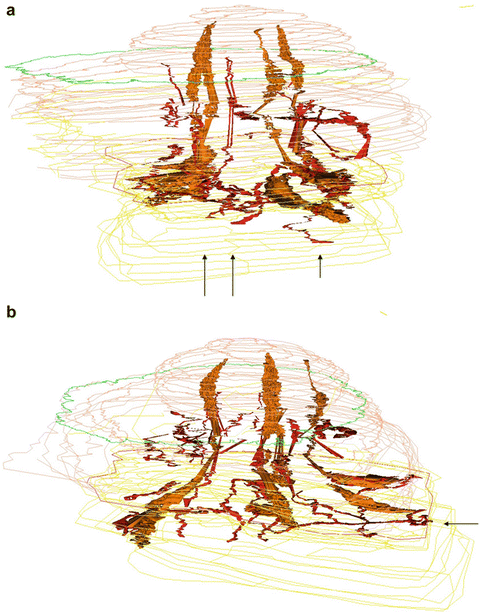
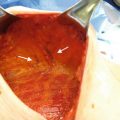
Microscopic studies also suggest a dual blood supply, part arising through the deep parenchyma, and part via the subdermal plexus (Fig. 2.2). Thus, as the deep blood supply is removed during a skin- and nipple-sparing mastectomy, careful attention must be paid to preserving the subdermal plexus of vessels to ensure the viability of skin flaps and nipple is maintained. Nakajima et al. showed parallel vessels ascending within the nipple before branching in the upper third [27]. This can be seen in figure 2.1 in that paper, and in the image from Cooper’s book reproduced below (Fig. 2.3).
 Patient Selection and Breast Imaging
 Plastic Surgery Considerations in Reconstruction After Nipple-Sparing Mastectomy
 The Inframammary Approach to Nipple-Sparing Mastectomy: The UCSF Experience
 Management of Complications Following Nipple-Sparing Mastectomy
 The Vertical Infra-Areolar Approach to Nipple Skin-Sparing or Total Skin-Sparing Mastectomy
 Techniques to Avoid Nipple and Flap Necrosis


Related posts:
Patient Selection and Breast Imaging
Plastic Surgery Considerations in Reconstruction After Nipple-Sparing Mastectomy
The Inframammary Approach to Nipple-Sparing Mastectomy: The UCSF Experience
Management of Complications Following Nipple-Sparing Mastectomy
The Vertical Infra-Areolar Approach to Nipple Skin-Sparing or Total Skin-Sparing Mastectomy
Techniques to Avoid Nipple and Flap Necrosis
Stay updated, free articles. Join our Telegram channel
Full access? Get Clinical Tree
