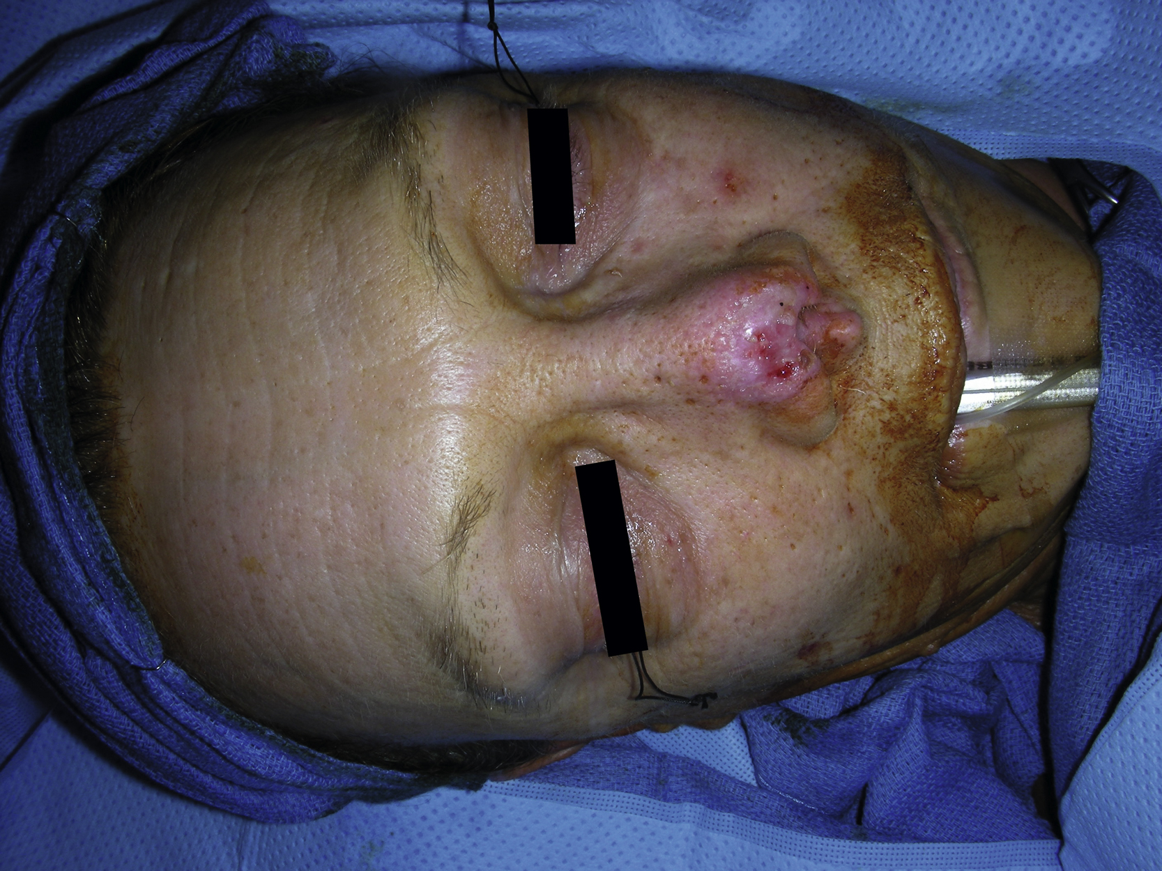
Clinical Presentation
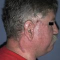
A 58-year-old White female sustained a dog bite to her nasal tip. Apparently, she lost a significant amount of nasal tip cartilage and also had a sizable nasal skin defect. After a period of local wound care, the nasal skin wound healed ( Figs. 8.1 and 8.2 ). She desired appropriate reconstruction for the partial composite defect of her nose and was brought to the operating room for definitive but delayed nasal reconstruction about 4 weeks after her initial injury.

Operative Plan and Special Considerations
This patient required nasal frame reconstruction with cartilage grafts and distal nasal skin defect reconstruction with a paramedial forehead flap. All necessary cartilage grafts could be harvested from the ear and a paramedial forehead flap from either side of the forehead based on the supratrochlear vessels could be used to provide adequate nasal skin coverage. The paramedial forehead is a good option for a partial nasal skin coverage when the size of the nasal skin defect is greater than 2 × 2 cm. The nasal reconstruction was done in staged procedures because a better outcome can be accomplished through several flap debulking procedures after initial reconstruction. The flap debulking procedure may be more reliable and effective while the pedicle of the flap is still intact. No internal nasal lining was needed for this patient.
Operative Procedures
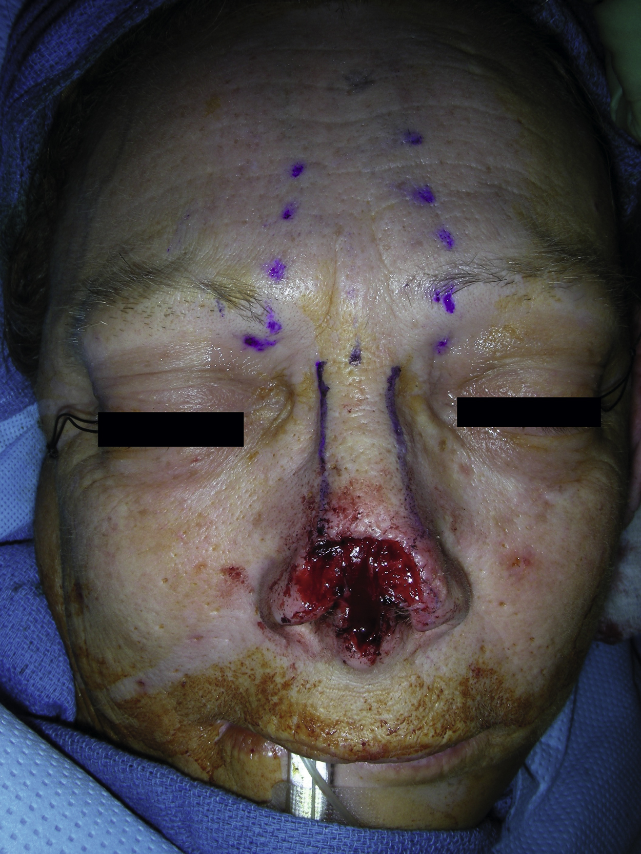
The first goal of the nasal reconstruction was to reopen healed nasal wounds and to recreate all composite defects of the nasal cartilages and nasal skin. Under general anesthesia, all scar tissue was excised and the wound was irrigated with normal saline solution ( Fig. 8.3 ).
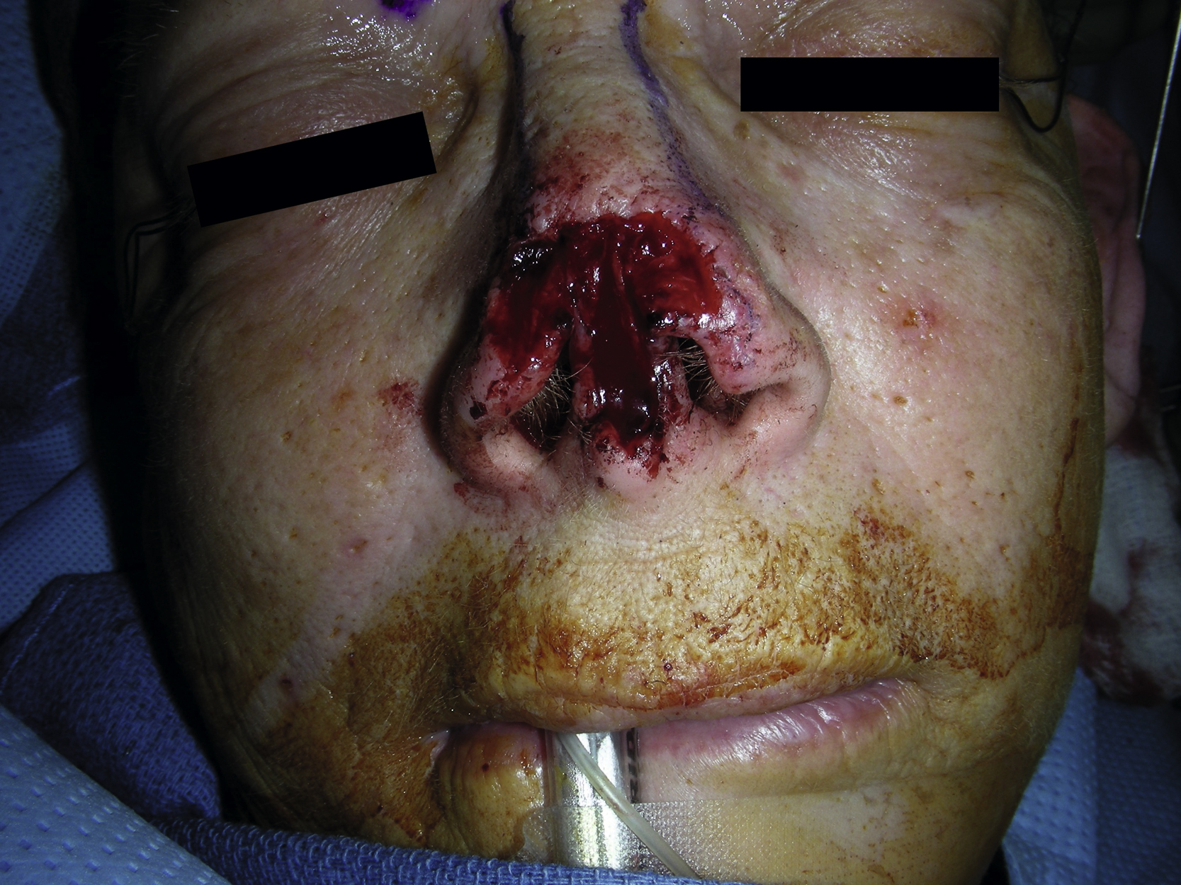
Contracture release surgery was performed for the nasal tip, alar septum, and columella. Under direct vision, the contracted wound was released with the scissors and the residual structures of the nasal skin and the cartilage-supporting structures were identified. The dissection was done to free the caudal septum, columella, nasal cartilage, and alar cartilage on each side. After these releases, it appeared that the patient would require extensive septal graft, the columellar strut, both right and left lower cartilage grafts for support and maintenance of the normal nasal shape ( Fig. 8.4 ). The decision was made to harvest cartilage grafts from each ear.
A posterior approach was used to harvest ear cartilage grafts. The proposed incision was infiltrated with 1% lidocaine with 1:100,000 epinephrine. Once the skin incision was made, the skin dissection was done to free ear cartilage. With preservation of the rim of the cartilage in the concha, a 2 × 1 cm of the cartilage was harvested from each ear and placed in the normal saline solution.
Based on the template, a septal cartilage extension graft was placed first. This was done with a cartilage graft and sutured to the caudal portion of the septum with several interrupted 5-0 PDS sutures. The columellar strut graft was placed to the nasal spine and secured to the adjacent soft tissue with 5-0 PDS sutures. It was also secured to the caudal portion of the septum.
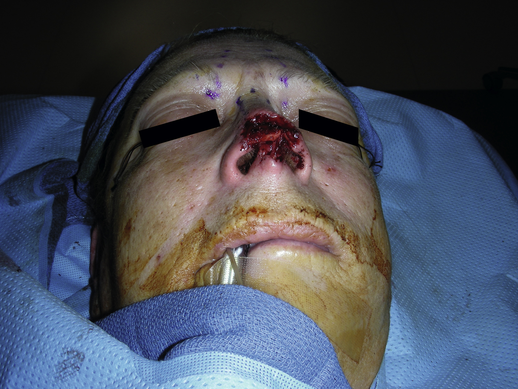
The right lower lateral cartilage graft was placed and sutured with several interrupted 5-0 PDS sutures. Both alar rim grafts were placed next to the columellar strut graft and secured with several interrupted 5-0 PDS sutures. At the end of procedure, the structure support of the nose was reestablished ( Figs. 8.5 and 8.6 ).
Related posts:


Stay updated, free articles. Join our Telegram channel

Full access? Get Clinical Tree


