Clinical Presentation
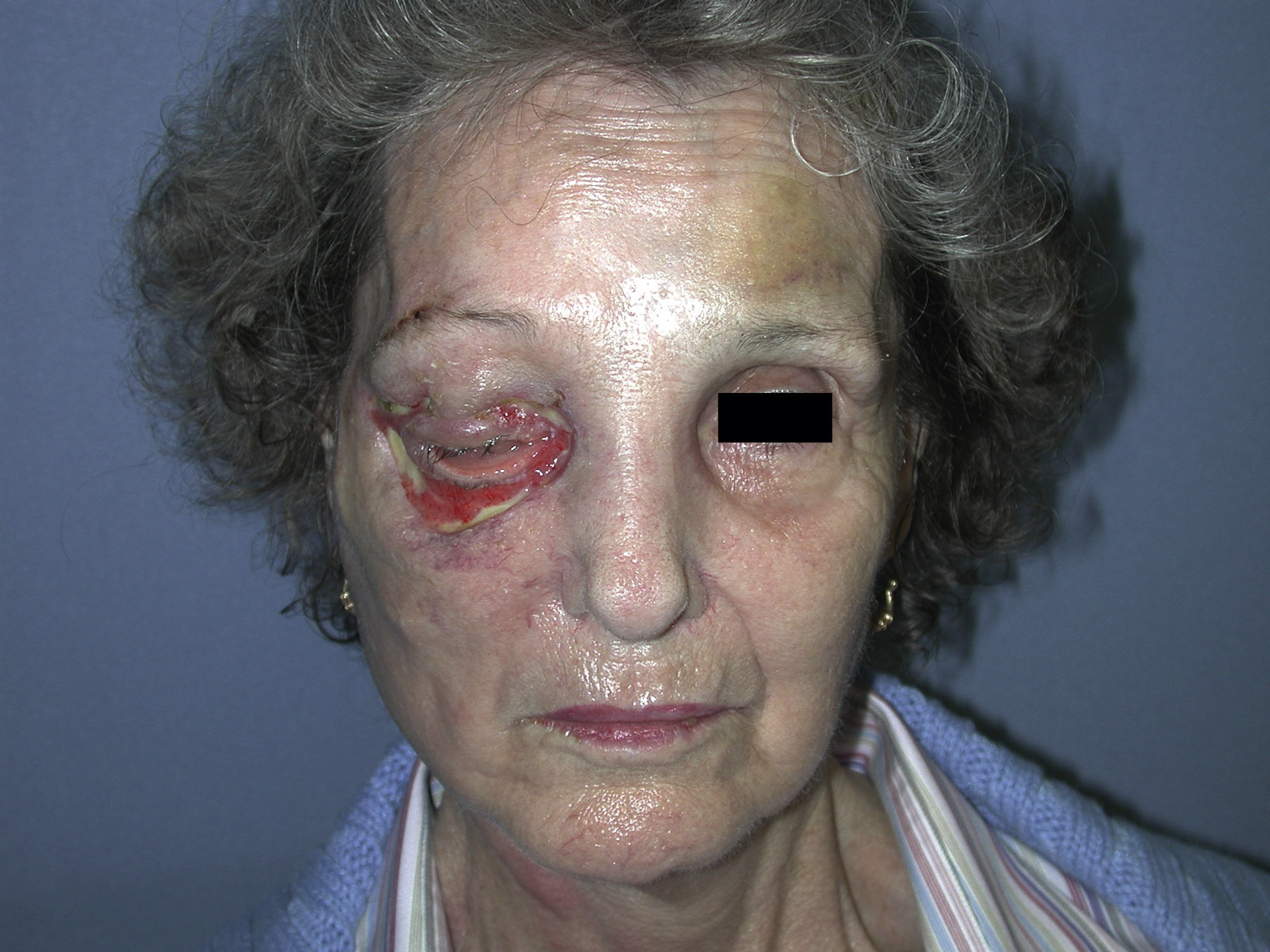
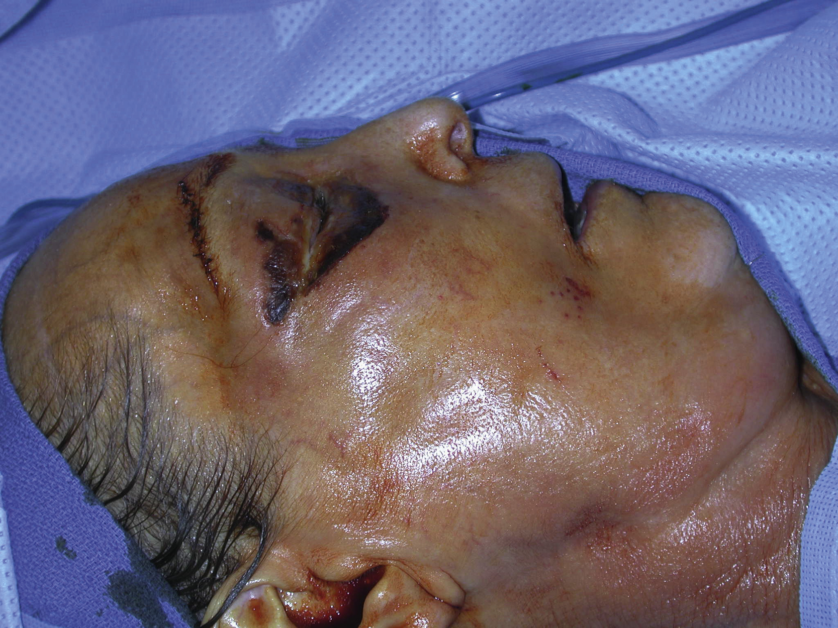
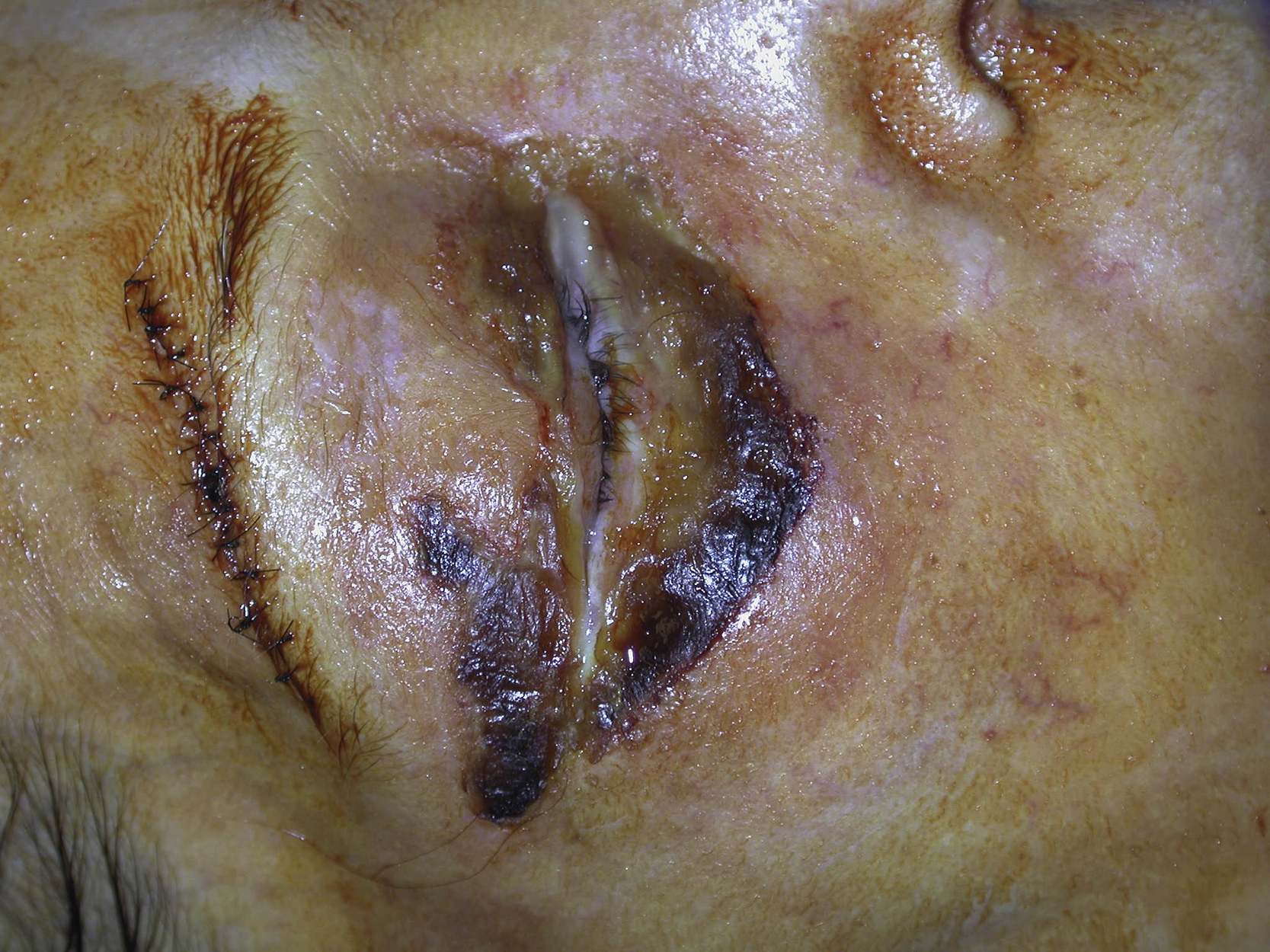
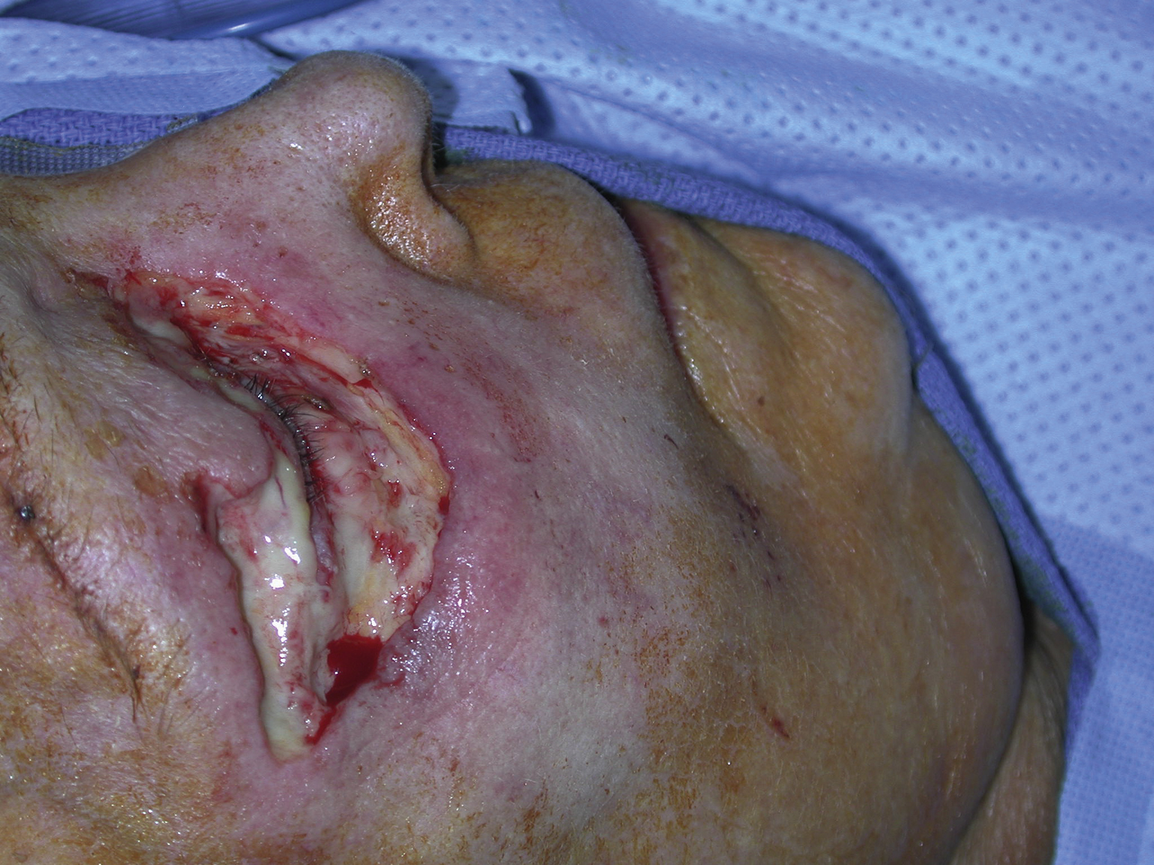
A 77-year-old White female sustained necrotizing fasciitis in her right periorbital area secondary to facial trauma. She was initially seen by an oculoplastic surgeon and urgently referred to the plastic surgery service for more definitive surgical management. On clinical examination, she had a periorbital soft tissue infection involving both upper and lower eyelids. She was urgently taken to the operating room for surgical debridement ( Figs. 6.1 and 6.2 ). The debridement was performed to preserve as much of the eyelid tissue as possible. After the debridement, open wounds remained in the entire lower eyelid and lateral portion of the upper eyelid ( Fig. 6.3 ).
Operative Plan and Special Considerations
After 4 weeks of proper local wound care, the upper and lower eyelid wounds were ready for definitive closure. Because the tissue at the base of the wounds was of good quality, the wounds could be closed with a full-thickness skin graft ( Fig. 6.4 ). The supraclavicular area was an excellent donor site because a skin graft taken from that area ensures the best color match to the lower eyelid. In addition, a lateral canthopexy or canthoplasty may be needed for correction of ectropion if indicated.