Orbital floor reconstruction in silent sinus syndrome


Table 68.1

Indications for surgery


Enophthalmos caused by orbital floor resorption in silent sinus syndrome
Secondary volume augmentation after enucleation/evisceration or orbital fracture repair


Table 68.2

Preoperative evaluation


History of orbital trauma, sinus disease, breast cancer or other malignancies
Exophthalmometry
Ocular ductions, cover/uncover testing
Photographs documenting eye movements in nine fields and worm’s eye views
Examination by otolaryngology
CT scan of orbit and sinuses


Introduction


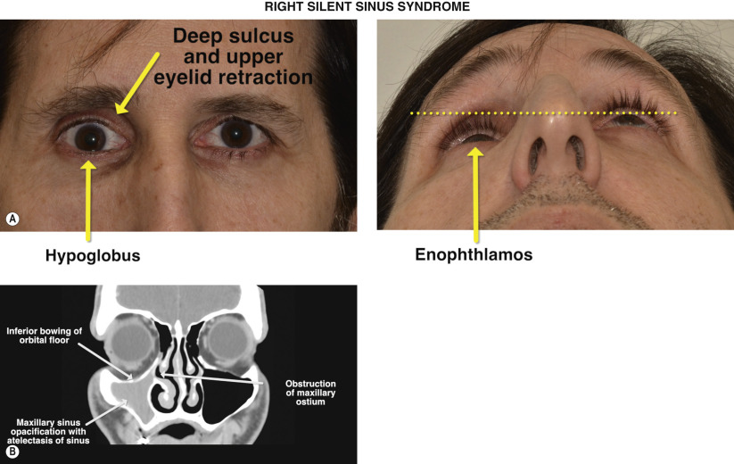
Silent sinus syndrome describes an indolent and chronic disease characterized by atelectasis of the maxillary sinus due to obstruction of the ostium. The resultant collapse of the maxillary sinus is associated with inferior bowing of the orbital floor that increases the effective orbital volume. Clinically, this is manifested by enophthalmos, hypoglobus, and a superior sulcus deformity. The condition is described as silent owing to little or no signs of sinusitis or nasal congestion.


Primary treatment is aimed towards aeration of the sinus by enlarging the native maxillary ostium with functional endoscopic sinus surgery. Microbial cultures of the sinus aspirate are typically sterile in nature and antimicrobial therapy has no role in the management of silent sinus syndrome. In a small percentage of patients who have underwent endoscopic sinus surgery alone, the orbital floor can re-expand, improving the enophthalmos but rarely to the pre-disease state. Treatment of the globe malposition is achieved by augmenting the orbital volume with alloplastic implants in a fashion similar to repair of an orbital fracture ( Chapter 67 ).


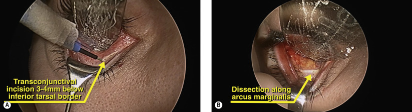
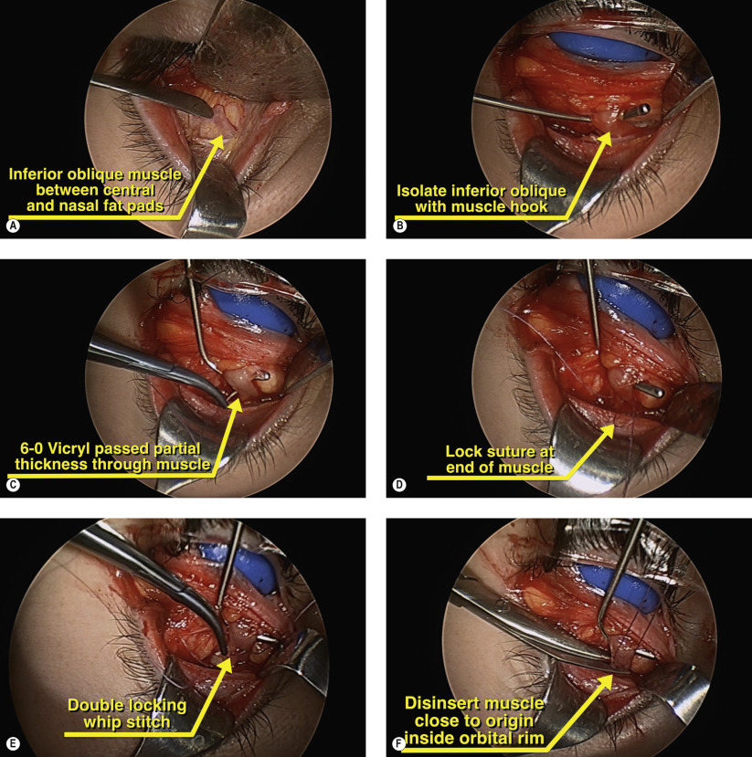
A variety of implants have been described, including autologous bone, porous polyethylene, hydroxyapatite, and titanium mesh. Our preference is to use a porous polyethylene enophthalmos wedge along the floor defect and covered by a thin barrier sheet of porous polyethylene with or without embedded titanium mesh. The surgery is carried out using a transconjunctival approach and can be performed simultaneously with endoscopic sinus surgery to address the maxillary sinus disease.
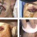
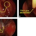


Figures 68.1A and 68.1B


Preoperative examination

Figure 68.1A shows a patient with the classic findings of silent sinus syndrome: hypoglobus, enophthalmos, superior sulcus deepening and upper eyelid retraction. Chronic obstruction of the maxillary ostium results in sinus opacification and atelectasis of the walls of the maxillary sinus on CT scanning ( Figure 68.1B ). These radiographic changes are virtually pathognomonic for silent sinus syndrome. With long-standing disease, maxillary hypoplasia can occur, resulting in significant midfacial hypoplasia as seen in Figure 68.1B . Restoration of sinus ventilation is achieved through endoscopic enlargement of the maxillary ostium and orbital reconstruction.


Surgical Technique


Figure 68.2


Lateral canthotomy and cantholysis

A lateral canthotomy and inferior cantholysis facilitates wide exposure of the inferior orbit when combined with a transconjunctival incision ( Figure 68.2 ). In patients with pre-existing lower eyelid laxity, a lateral canthotomy and inferior cantholysis may not be necessary.


Figures 68.3A and 68.3B


Transconjunctival incision

A transconjunctival incision is made 3–4 mm below the inferior tarsal border with cutting cautery ( Figure 68.3A ). Exposure and retraction are facilitated with the use of a Desmarres retractor on the lower eyelid and a malleable retractor to protect the globe and to isolate the inferior orbit. The dissection continues in a preseptal plane to expose the arcus marginalis along the inferior orbital rim ( Figure 68.3B ). Care is taken near the medial eyelid where inadvertent damage to the inferior punctum can occur during cauterization.


Figures 68.4A–F


Disinsertion of inferior oblique muscle

The inferior oblique muscle originates along the inner aspect of the orbital rim at the medial one-third of the orbit ( Figure 68.4A ). When dissecting along the arcus marginalis, care is taken near this origin to avoid transecting the muscle. Although optional, disinsertion of the inferior oblique muscle can provide a wide, panoramic view of the orbital floor and the medial wall of the orbit. This is particularly useful for repair of combined floor and medial wall fractures as well as three wall orbital decompression. In this case, disinsertion of the inferior oblique muscle allows for placement of the large floor implants needed to restore orbital volume. A Green or Von Graefe muscle hook is used to isolate the inferior oblique muscle and cotton-tipped applicators are used to strip off the orbital fat and fascia to expose the muscle fibers ( Figure 68.4B ). A double-armed 6-0 Vicryl suture is used to imbricate the muscle by a partial thickness pass through two-thirds of the muscle width ( Figure 68.4C ). At the terminal end, a full thickness locking bite is placed ( Figure 68.4D ). The other arm of the suture is passed in a similar fashion to complete the double-locking whip stitch ( Figure 68.4E ). Once the suture has been passed, the muscle is disinserted close to its origin, leaving a 2–3 mm stump ( Figure 68.4F ). The muscle is then tagged with a bulldog clamp for subsequent reinsertion.

Only gold members can continue reading. Log In or Register to continue

Stay updated, free articles. Join our Telegram channel

May 16, 2019 | Posted by in Reconstructive surgery | Comments Off on Orbital floor reconstruction in silent sinus syndrome

Full access? Get Clinical Tree

Get Clinical Tree app for offline access