Lower eyelid retraction repair with porcine acellular dermal collagen matrix


Table 34.1

Indications for surgery


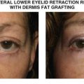
Lower eyelid retraction without need for significant volume augmentation
Posterior lamellar and/or middle lamellar shortening
No history of adverse reaction to porcine xenografting
Contraindication or aversion to hard palate or autologous grafting


Table 34.2

Preoperative evaluation


Quantify amount of eyelid retraction according to MRD2
Manual palpation and elevation of lower eyelid to feel for restriction within the middle lamella; if supple elevation is possible, spacer graft may not be required
Assess adequacy of anterior lamella; may need cheek lift or skin graft if severe shortening
Assess eyelid laxity
Assess globe and inferior orbital rim position for presence of negative vector
Assess orbicularis strength; weakness on closure may portend high risk of recurrence
Prior eyelid, facial surgery or trauma


Introduction


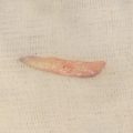
The use of acellular dermis as a spacer graft for lower eyelid retraction is similar to dermis fat ( Chapter 35 ) and autologous hard palate ( Chapter 36 ) with some distinguishing features. Dermis fat can restore lost orbital volume and provide a large surface area for posterior lamellar grafting with the benefit of being autologous, while hard palate supplies only surface area. Acellular dermis like hard palate provides posterior lamella but requires commercial preparation prior to use.


There are two sources of acellular dermis: the first is an allograft, taken from human cadaveric donors, and the second is a xenograft from porcine sources. Acellular dermis does not have a native epithelial layer but does provide a connective tissue framework for incorporation into host tissues. Conjunctival epithelialization, however, is required from the host.


Many decisions factor into the choice of posterior lamellar grafts. If patients are unwilling to have a second incision for an autologous graft, acellular dermis does provide a suitable option. Some potential downsides are prolonged inflammation and conjunctival injection during the epithelialization phase and the lack of volume that dermis fat provides. One benefit of acellular dermis and hard palate is no risk of ectopic hair transplantation that can be seen with dermis fat grafting. An additional benefit of acellular dermis over hard palate is the consistency of graft thickness and the virtually unlimited size compared to hard palate.


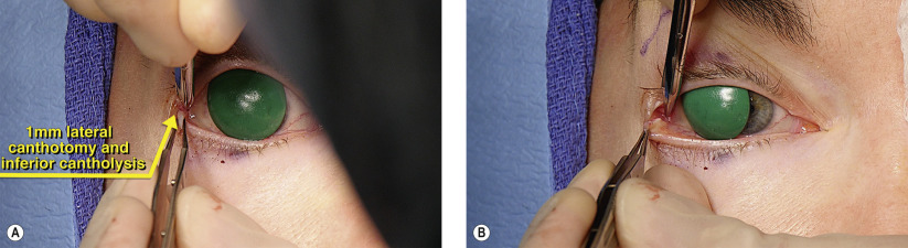
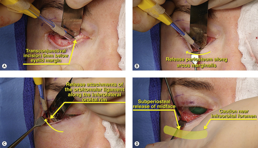
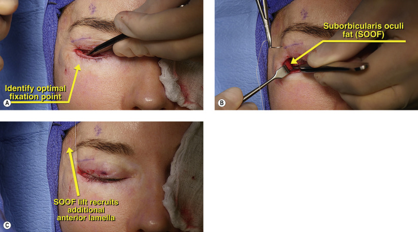
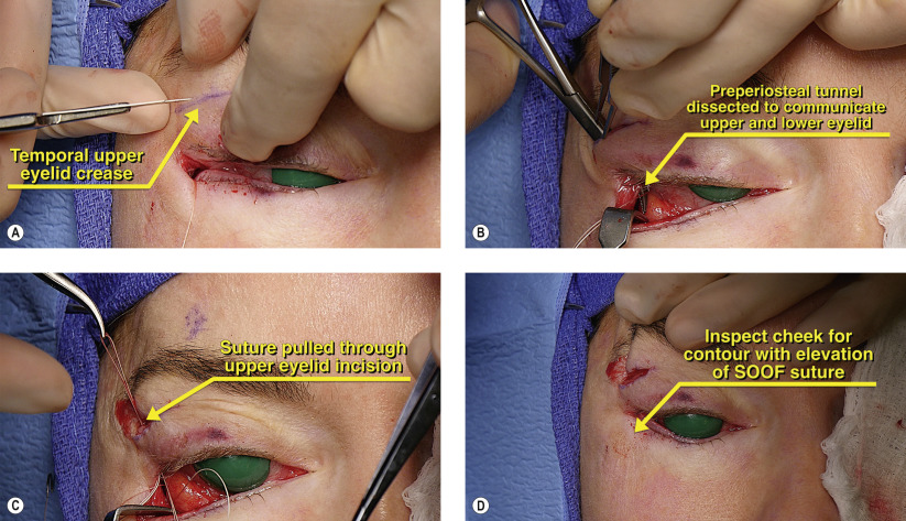
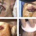
Conceptually, this chapter and the following two ( Chapters 35 and 36 ) are fundamentally similar. The surgery is performed through a transconjunctival approach. The lower eyelid cicatrix is released with an optional subperiosteal midface lift. Orbitomalar suspension can be performed to elevate the midface and to recruit additional anterior lamella. The spacer graft is secured posteriorly and the lower eyelid is tightened. The lower eyelid is then immobilized with Frost sutures.


For adequate surgical repair, slight oversizing of the graft is required as there will likely be some shrinkage postoperatively. Because the posterior lamellar graft can be irritating to the cornea, particularly if a Frost suture is placed, a collagen shield or a large-diameter contact lens can be used for comfort. Also helpful in reducing foreign body sensation are buried knots and fine, non-braided sutures to secure the graft.


Surgical Technique


Figures 34.1A and 34.1B


Lateral canthotomy and cantholysis

Our preference is to place posterior lamellar grafts from the transconjunctival approach. This prevents postoperative anterior lamellar contraction and preserves cosmesis. A small, 1 mm lateral canthotomy and inferior cantholysis is performed ( Figure 34.1A ). Complete release of the inferior crus of the lateral canthal tendon is performed to loosen any scarring and allow full access to the lower eyelid ( Figure 34.1B ).


Figures 34.2A–D


Inferior transconjunctival incision and cicatricial release along inferior orbital rim

Middle lamellar scarring is often a key factor in the development of lower eyelid retraction and fully releasing this cicatrix along the inferior orbital rim is essential for success. After the lateral canthotomy and inferior cantholysis, an inferior transconjunctival incision with monopolar cautery is performed 6 mm below the lower eyelid margin ( Figure 34.2A ). This incision is performed 2 mm below the inferior border of the tarsus so that this small flap of palpebral conjunctiva can be draped over the spacer graft and facilitate subsequent epithelialization. The dissection then continues in the preseptal plane using a malleable retractor to posteriorly displace orbital fat while a Senn retractor isolates the arcus marginalis at the inferior orbital rim. Cutting cautery is then used to release the periosteum along the arcus marginalis ( Figure 34.2B ). At the inferolateral orbital rim, cutting cautery is used to dissect along the inferolateral orbital rim to release the orbitomalar ligament which has its stoutest attachment to the orbital rim ( Figure 34.2C ). The zygomaticofacial foramen is located approximately 5 mm below the inferolateral orbital rim and if bleeding occurs, gentle bipolar cautery should be applied. If significant lower eyelid retraction (MRD2 >8 mm) and/or severe middle lamellar tethering is present, a subperiosteal midfacial dissection is performed ( Figure 34.2D ). Care is taken along the inferomedial orbit where the infraorbital nerve emerges 5 mm below the orbital rim. After full release of the cicatrix, the midface and lower eyelid should freely elevate. If any bands of traction still remain, these are lysed. A cotton-tipped “peanut” sponge on a hemostat can also be used to release residual attachments to the maxilla and zygoma.


Figures 34.3A–C


Placement of orbitomalar suspension suture

A suture is then passed through the soft tissues of the cheek for fixation later in the operation. The location of the suture placement is critical. If too superficial, dimpling may occur. If too deep, bunching may occur. Toothed Adson forceps are used to select an optimal fixation point through the suborbicularis oculi fat (SOOF) ( Figure 34.3A ). Once the desired location is selected, 4-0 Vicryl is passed though the SOOF ( Figure 34.3B ). With superolateral traction of the suture, the midface should freely elevate without dimpling or bunching of the SOOF and overlying skin. This SOOF lift recruits additional anterior lamella, which obviates the need for a cosmetically displeasing skin graft ( Figure 34.3C ).

May 16, 2019 | Posted by in Reconstructive surgery | Comments Off on Lower eyelid retraction repair with porcine acellular dermal collagen matrix

Full access? Get Clinical Tree

Get Clinical Tree app for offline access