Lower eyelid retraction repair with dermis fat


Table 35.1

Indications for surgery


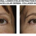
Lower eyelid retraction with volume loss
Posterior lamellar and/or middle lamellar shortening
Desire for use of autologous grafts
HIV-associated lipoatrophy
Prostaglandin-associated lipoatrophy
Lower eyelid retraction seen in craniofacial syndromes (ie Treacher Collins syndrome)


Table 35.2

Preoperative evaluation


Assess for volume loss/hollowing of lower eyelid
Quantify amount of eyelid retraction according to MRD2
Manual palpation and elevation of lower eyelid to feel for restriction; if supple elevation is possible, spacer may not be required
Assess adequacy of anterior lamella; may need cheek lift or skin graft if severe shortening
Assess eyelid laxity
Assess inferior orbital rim and presence of negative vector
Assess orbicularis strength; weakness on closure may portend high risk of recurrence
Prior eyelid, facial surgery or trauma
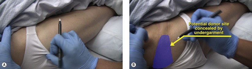
Suitability for donor site of dermis fat (retro/preauricular, hip/gluteal, flank/periumbilical)


Introduction


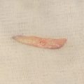
The use of dermis fat as a posterior lamellar graft for lower eyelid reconstruction has several advantages over acellular dermis and hard palate. Dermis fat can provide a virtually limitless amount of volume augmentation and surface area compared to acellular dermis ( Chapter 34 ) and hard palate ( Chapter 36 ). Dermis fat is autologous, which may appeal to patients who are adverse to use of xenografts. Hard palate, although autologous, has more associated donor site morbidity, such as postoperative pain, difficulty eating, and potential for fistula formation compared to dermis fat. The use of dermis fat, acellular dermis, and hard palate is technically similar and review of each of these chapters ( Chapter 34 , Chapter 36 ) should be performed to appreciate the subtle nuances of each graft.
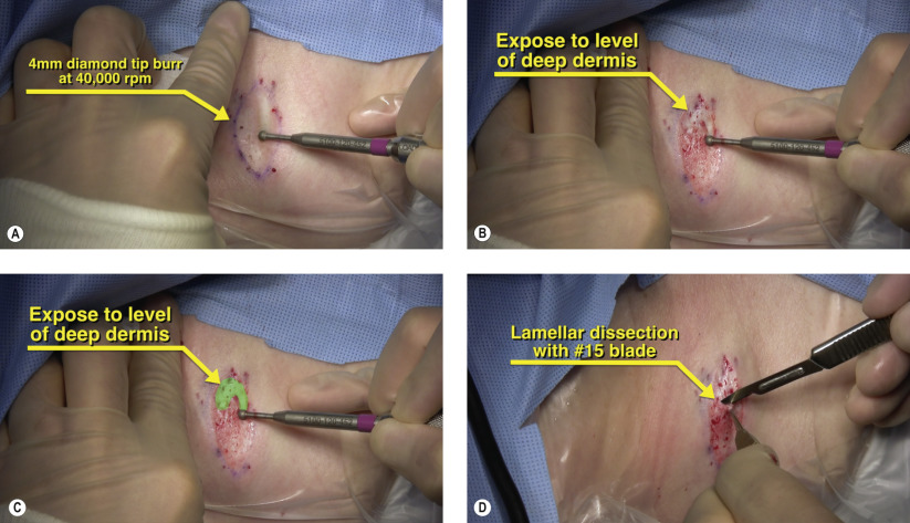
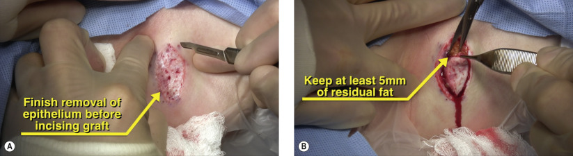
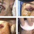


Surgical Technique


Figures 35.1A and 35.1B


Potential donor site for dermis fat graft

The gluteal and hip region provides excellent surface area for dermis and volume with respect to subcutaneous fat ( Figure 35.1A ). The donor site should be hidden under the patient’s undergarment ( Figure 35.1B ). Even in patients with HIV-associated lipodystrophy, there is often adequate subcutaneous fat present. When choosing a donor site, a hair-free area should be utilized. The size of the potential donor site should be increased by at least 1.5–2 fold to account for contraction after harvesting. Other suitable areas for harvesting dermis fat include the periumbilical area and flanks, but these carry the potential risk of peritoneal perforation. Preauricular or retroauricular grafts may be obtained but these donor sites contain less subcutaneous fat.


Figures 35.2A–D


Removal of epithelium from graft

Complete removal of the epithelium from a dermis fat graft is essential to minimize postoperative complications. Vellus hair growth, keratin production, and mucoid discharge may be seen with incomplete removal of the epithelium. A 4 mm diamond burr rotating at 40,000 rpm can be used for epithelial removal ( Figure 35.2A ). As the epidermis is removed, fine pinpoint bleeders can be noted. The dermis is reached when a pale appearance is noted after the debridement ( Figures 35.2B and 35.2C ). Lamellar dissection can also be performed with a #15 or #10 blade ( Figure 35.2D ). One theoretical advantage of the burr over the blade is the thermal damage that may be directed to the hair follicle base to minimize postoperative hair growth.


Figures 35.3A and 35.3B


Removal of dermis fat graft

The epithelium should be completely removed from the donor site prior to skin incision. Keeping the surrounding skin intact provides countertraction when debriding or excising the epithelium. Once the donor site has been prepared, a #15 blade is used to incise the skin slightly inside of the denuded epithelium to ensure no surface epithelium is transplanted to the eyelid ( Figure 35.3A ). A residual bed of at least 5 mm thickness of the fat side should be removed from the donor site ( Figure 35.3B ). This slight oversizing allows for subsequent trimming when placed into the lower eyelid. The donor site is closed with a deep 4-0 Vicryl suture followed by a running subcuticular 5-0 Vicryl suture. Alternatively a Monocryl suture may be used for less inflammatory response. The skin should be closed with Steri-Strips for reinforcement and the patient must be instructed to minimize squatting or bending over to prevent wound dehiscence.

Only gold members can continue reading. Log In or Register to continue

Stay updated, free articles. Join our Telegram channel

May 16, 2019 | Posted by in Reconstructive surgery | Comments Off on Lower eyelid retraction repair with dermis fat

Full access? Get Clinical Tree

Get Clinical Tree app for offline access