Abstract
Examination of the oral cavity can provide important diagnostic information for dermatologic diagnosis and therefore should be included in every skin examination. A variety of skin disorders can be accompanied by mucous membrane involvement. For example, erythema multiforme and systemic lupus erythematosus can cause erosions in the mouth, nose, or eyes. However, this chapter focuses on disorders either exclusively or predominantly confined to mucous membranes, usually of the oral cavity.
Mucous membrane disorders are divided into two broad categories: (1) erosions and ulcerations and (2) white lesions ( Table 22.1 ). Erosions are lesions in which the mucosal epithelium is partly denuded. Ulcerations extend through the epidermis into the underlying tissue, which in mucous membranes is called lamina propria rather than dermis. Erosive and ulcerative diseases range in frequency from common (aphthous stomatitis) to rare (pemphigus), and their causes are idiopathic, immunologic, infectious, and malignant.
Chapter Contents
Uncommon Causes of Oral Ulcers
Categorization of mucous membrand disorders:
- 1.
Erosions and ulcerations
- 2.
White lesions
| Etiology | History | Physical Examination | Differential Diagnosis | Laboratory Test | |
|---|---|---|---|---|---|
| Ulcers | |||||
| Aphthous stomatitis (common cause) | Unknown | Recurrent disease | Sharply demarcated, round, yellowish erosions surrounded by erythema | Herpes simplex Behçeťs syndrome Inflammatory bowel disease | – |
| Pemphigus and pemphigoid (uncommon causes) | Autoimmune | May have associated skin lesions | Ragged erosions and ulcerations; intact blisters rarely present | Aphthous stomatitis Erythema multiforme | Biopsy with immunofluorescence |
| Viral infections | Primary herpes simplex Coxsackievirus | Fever, malaise | Gingivitis; blisters also on lips | Aphthous stomatitis Erythema multiforme | Tzanck smear or culture |
| Fever | Vesicles in posterior oral cavity | Aphthous stomatitis | – | ||
| Syphilis | Treponema pallidum | Sexual contact | Indurated , painless ulcer | Malignancy | Serologic test for syphilis |
| Deep fungal infection | Histoplasmosis | Immunosuppressed | Systemically ill; indurated ulcer | Malignancy | Biopsy with culture |
| Malignancy | Nonhealing ulcer | Indurated ulcer | Major aphthous ulcer | Biopsy | |
| White lesions | |||||
| Thrush | Candida albicans | Found in newborns and immunosuppressed patients | “Curd-like” papules, easily scraped off | Lichen planus Geographic tongue | KOH preparation |
| Lichen planus | Unknown | Chronic disease; may have associated skin lesions | Reticulated white lines; sometimes erosions are present | Candidiasis Leukoplakia Secondary syphilis | Biopsy |
| Leukoplakia | Chronic irritation | Smoking Denture trauma | White patches and plaques | Lichen planus Secondary syphilis White sponge nevus Leukokeratosis | Biopsy |
| Squamous cell carcinoma | Smoking Alcohol Prior leukoplakia | Indurated or ulcerated plaque | Leukoplakia Major aphthous ulcer Erosive lichen planus Chancre Deep fungal infection | Biopsy | |
White spots are hyperkeratotic lesions of the oral mucosa. Thickened stratum corneum of mucous membranes appears white because of maceration from continuous wetness. Malignancy must be ruled out as a cause.
White lesions represent hyperkeratosis.
Aphthous Stomatitis
- 1.
Most common cause of recurrent oral ulcers
- 2.
Ulcers have a yellow base and peripheral erythema
- 3.
Multiple therapies indicate lack of effective treatment
Definition
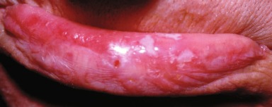
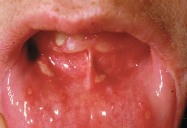
Aphthous stomatitis is a common, recurrent, idiopathic disorder of the mouth most often manifest by multiple small, “punched-out” ulcers ( Fig. 22.1 ).
Incidence
Recurrent aphthous stomatitis is a common disease, occurring in 20% to 60% of the general population. It is most common in young adults; the 60% prevalence was found in a survey of students attending professional schools.
History
A history of previous episodes is invariable. Recurrences are sometimes precipitated by trauma from biting or misguided toothbrushes. Some patients correlate outbreaks with emotional stress. Lesions are usually preceded by a 1-day prodrome of discomfort in the area of involvement. The ulcers are painful and sometimes interfere with eating.
Physical Examination
Lesions in aphthous stomatitis appear as 2- to 5-mm, round, punched-out ulcers with a yellowish necrotic surface and surrounding erythema. Lesions may be single but more often are multiple. The buccal and labial mucosae are the most common locations.
Aphthous stomatitis is the most common cause of oral ulceration.
Differential Diagnosis
Recurrent aphthous stomatitis is most often confused with herpes simplex infection . Recurrent herpes simplex rarely occurs inside the mouth. When it does, it appears as grouped small vesicles or erosions on an erythematous base. A Tzanck preparation or culture proves the diagnosis of herpes infection.
Herpes simplex rarely recurs inside the mouth.
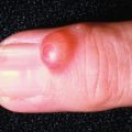
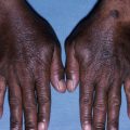
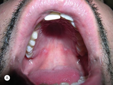
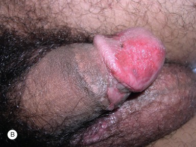
The oral ulcerations in Behçeťs syndrome are indistinguishable from those of aphthous stomatitis. However, Behçeťs syndrome is distinguished by its extraoral manifestations. The classic triad consists of oral ulcerations, genital ulcerations, and ocular inflammation (iridocyclitis) ( Fig. 22.2 ). Erythema nodosum, thrombophlebitis, arthritis, and neurologic and intestinal involvement may also occur. Patients with inflammatory bowel disease, particularly ulcerative colitis , occasionally have oral ulcerations that resemble aphthous stomatitis.
Oral ulcerations in Behçeťs syndrome look like aphthous stomatitis.
- ●
Herpes simplex infection
- ●
Behçeťs syndrome
- ●
Oral ulcers from inflammatory bowel disease
Laboratory and Biopsy
Usually, a biopsy is not required. If a biopsy is performed, the findings will not be diagnostic and will show only ulceration and nonspecific inflammation, composed primarily of lymphocytes. The only other laboratory test to consider is a complete blood count, to screen for the questionable association of iron or folate deficiency anemia in some patients with aphthous stomatitis.
Therapy
The variety of therapies that have been recommended for this disease indicates that a highly successful treatment is lacking. If an underlying iron or folate deficiency is detected, it should be corrected. The ulcerations are usually treated topically. Tetracycline suspension (250 mg/5 mL) “swished and swallowed” four times daily helps in some patients. Patients in whom tetracycline therapy fails are treated with topical steroids in a gel (e.g., fluocinonide, Lidex gel) or a special adherent base (e.g., triamcinolone, Kenalog in Orabase) applied three times daily or with a spray preparation (e.g., beclomethasone, Vanceril) applied three to four times daily. Intralesional steroids (triamcinolone, Kenalog-10) are useful in patients with large aphthous ulcerations. Oral prednisone is effective in aphthous stomatitis but should be used for only a short course in patients with severe, incapacitating disease. Colchicine and pentoxifylline (Trental) have also been reported to be helpful in preventing recurrent disease, but the clinical trials were not controlled.
Pain relief can be obtained with topical anesthetics such as dyclonine hydrochloride (Dyclone liquid) or topical lidocaine (viscous Xylocaine) used 20 minutes before meals. These preparations numb the entire mouth, including the taste buds, for 1 to 2 hours and allow for pain-free, albeit taste-free, dining.
Course and Complications
For minor aphthous stomatitis, spontaneous healing occurs within 4 to 14 days. Large aphthous ulcers (major aphthous ulcers) take as long as 6 weeks to heal. Individual ulcers lasting much longer than that should be examined by biopsy to rule out malignancy. Recurrences are common and range in frequency from occasional to almost continuous. In most patients, the disease eventually remits, but the time course is highly variable – from 5 to 15 years or longer.
Pathogenesis
Factors implicated in the pathogenesis include emotional and physical stress, hormones, infection, and autoimmunity. An immune mechanism is the most favored cause. Circulating T lymphocytes cytotoxic against oral mucosa have been identified and appear to play a role.
Leukoplakia
- 1.
White plaques can signify cancer
- 2.
Indurated white plaques require biopsy
- 3.
Smoking is most frequent cause
Definition
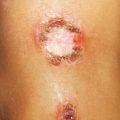
Leukoplakia literally means “white plaque” ( Fig. 22.3 ). Some clinicians simply use that as the definition of the disease, defining leukoplakia as “a white patch or plaque that cannot be characterized clinically or pathologically as any other disease.” Others use the term leukokeratosis to describe a white patch that is histologically benign, and reserve the term leukoplakia for a white patch or plaque in which epithelial dysplasia is present pathologically. The authors prefer the second definition. Either way, the important point to remember is that, for white plaques on mucous membranes, a dysplastic change should be considered a possible cause.