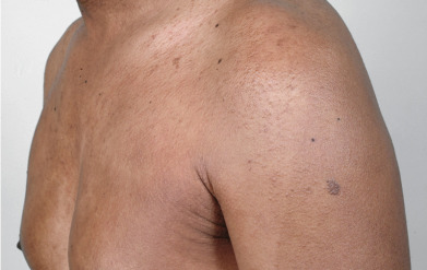
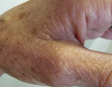
Localized cutaneous mucin deposition results in translucent pale to erythematous papules, as in digital myxoid cysts or focal cutaneous mucinosis. Papular mucinosis related to plasma cell dyscrasia with paraproteinemia causes papules with a distinct tendency to form linear arrays. Histologically, mucin is less apparent than an increase in fibroblasts and ropey collagen. This translates to induration of the skin over time, leading to a clinical appearance resembling systemic sclerosis.
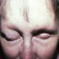
Myxedema results in subtle dermal mucin deposition often manifesting below the eyes, whereas pretibial myxedema can produce dramatic deposition within the skin of the dorsal foot as well as the pretibial region. Stasis-induced mucinosis can produce a similar appearance, but the mucin is confined to the upper dermis rather than affecting the full thickness of the dermis as is seen in thyroid disease.
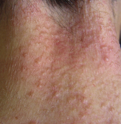
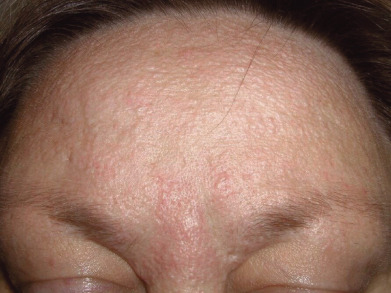
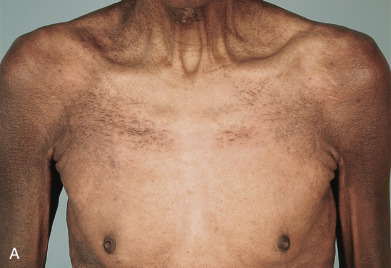
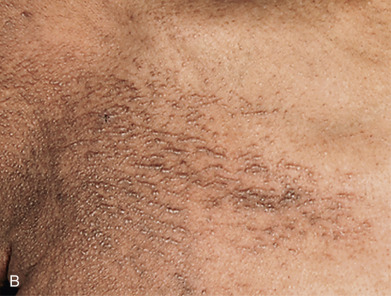
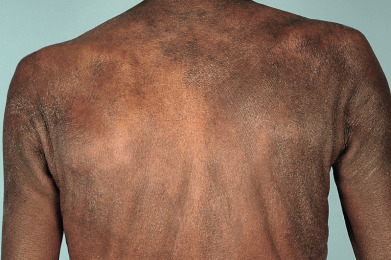
Follicular mucinosis can occur as erythematous, indurated, hairless plaques in benign alopecia mucinosa, as well as in the setting of mycosis fungoides. Dilated follicular orifices can give the appearance of comedones (blackheads), and sticky gelatinous material may be extruded from the lesions. This portion of the atlas will guide you through the various clinical presentations associated with mucinosis.
Related posts:

Stay updated, free articles. Join our Telegram channel

Full access? Get Clinical Tree


