This chapter includes a variety of conditions, all resulting from either inherited or acquired changes in the collagen or elastic tissue of the skin.
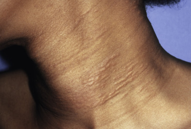
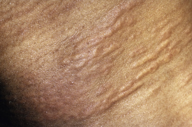
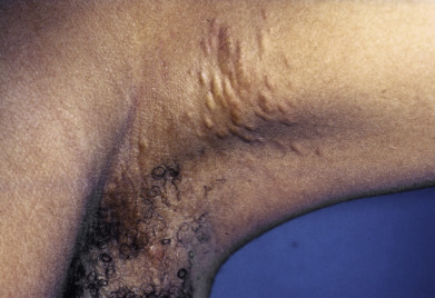
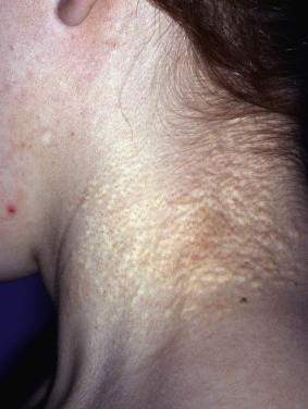
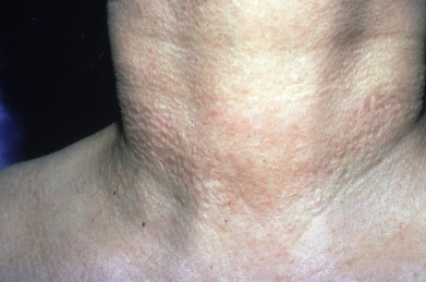
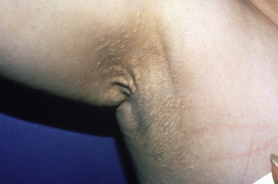
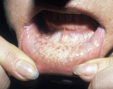
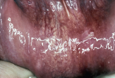
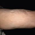
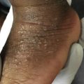
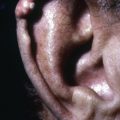
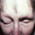
Some inherited syndromes with particularly unique skin findings include forms of Ehlers-Danlos syndrome (EDS). Affected individuals are noted to have doughy skin that is hyperextensible. Skin is prone to wide “fish-mouth” wounds, molluscoid pseudotumors, and atrophic scars. In contrast, those patients with inherited forms of cutis laxa produce widespread drooping, redundant skin with pendulous folds. Pseudoxanthoma elasticum (PXE) can present with yellowish papules predominantly on the lateral neck said to resemble “plucked chicken skin,” as well as lax skin akin to that seen in some forms of cutis laxa. Marfan syndrome is included here due to its underlying genetic mutation in fibrillin-1 that affects connective tissues. In Marfan syndrome characteristic physical findings include tall stature, high-arched palate, and arachnodactyly. Knowledge of the skin findings of these genetic conditions is important to aid in an early diagnosis, especially given that they all have the potential to involve internal organs such as the cardiovascular system and lungs.
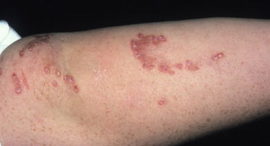
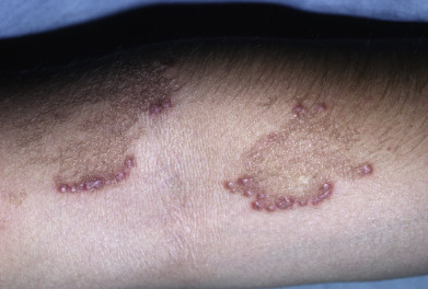
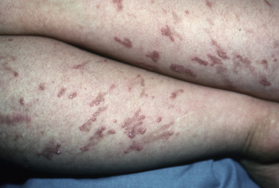
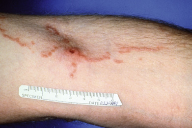
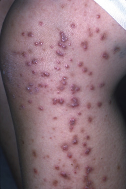
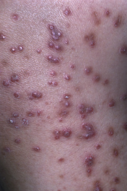
More localized skin conditions are also represented here, including the serpiginous keratotic papules seen in elastosis perforans serpiginosa. This rare condition is more common in those with trisomy 21 (Down syndrome), but can also be seen in patients with some of the aforementioned connective tissue conditions such as EDS and Marfan syndrome.
Skin biopsy with subsequent staining for connective tissue fibers can be necessary and diagnostic for some of these conditions such as PXE. However, those patients with a suspected inherited condition with known gene mutation should be offered confirmatory genetic testing from the blood.
This portion of the atlas features these connective tissue diseases, among others, that affect the dermal fibrous and elastic tissues.