Bacterial infections create a multitude of skin lesions, from pustules, to necrotic nodules, to ulcers. These infections range from the superficial crusted erosions of impetigo to the systemic purpuric plaques of meningococcemia. This chapter categorizes these bacterial conditions into gram positive, gram negative, and others, including rickettsial diseases.
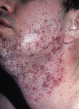
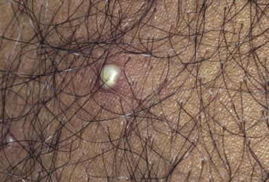
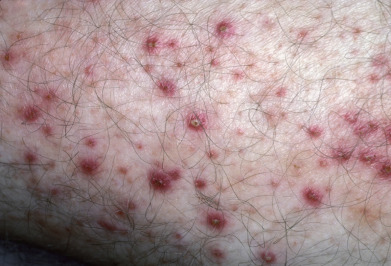
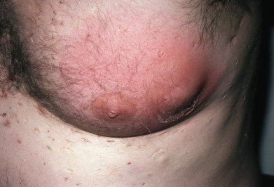
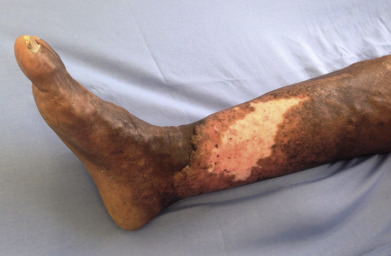
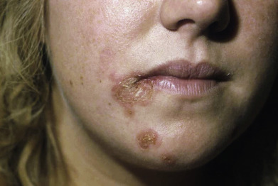
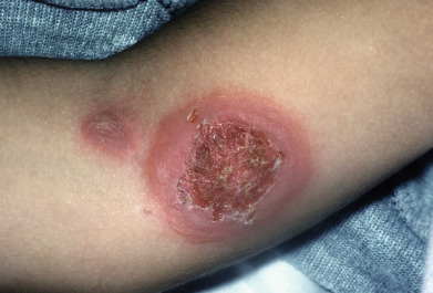
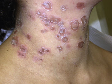
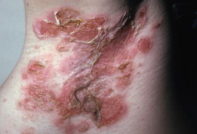
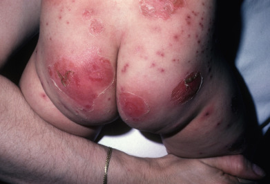
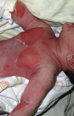
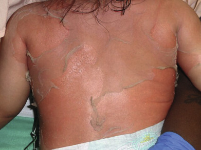
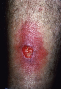
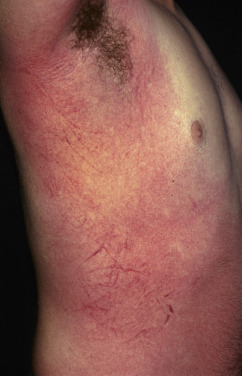
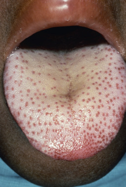
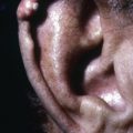
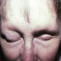
Recognizing the signs of bacterial infections is critical because most conditions will require treatment, whether topical or systemic. Staphylococcal skin infections can present with pustules, crusting, furuncles, or bullae in the case of bullous impetigo. Toxin-mediated conditions, such as staphylococcal scalded skin syndrome or toxic shock syndrome, will manifest as sunburn-like transient erythema accentuated in the folds. Streptococcal infections can present with pustules and crusting, as well as the firm painful erythematous plaques of cellulitis or erysipelas, or the ulcerative lesions in the case of ecthyma.
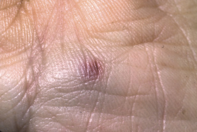
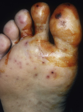
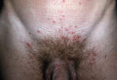
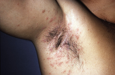
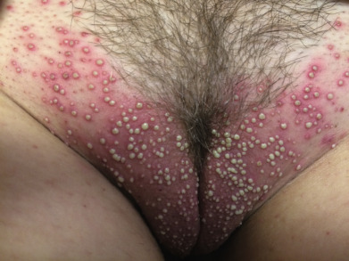
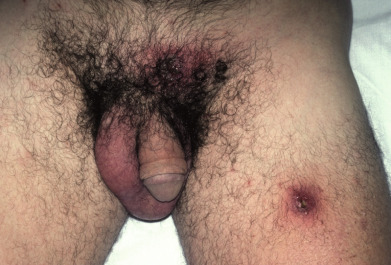
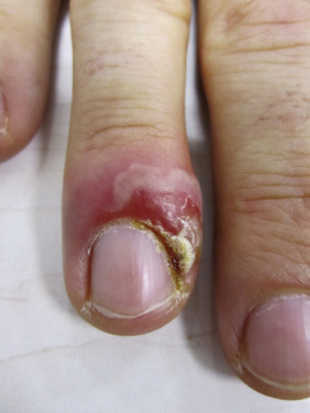
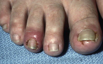
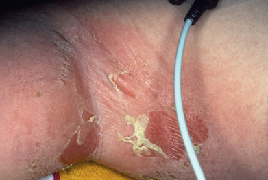
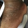
The anatomic locations involved can be a clue to which bacterial infections are most likely, such as in blistering distal dactylitis, perianal streptococcal infections, intertrigo, or gram-negative toe web infections.
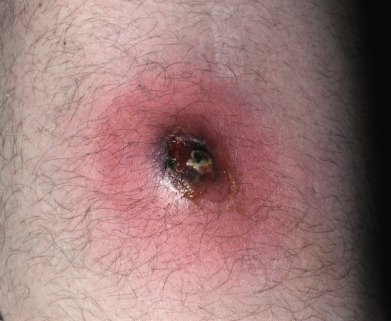
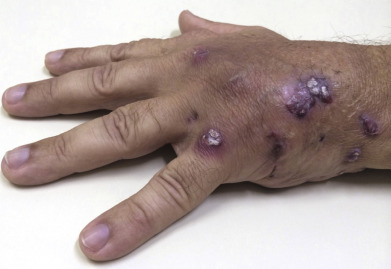
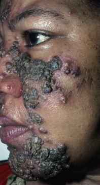
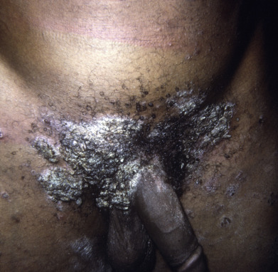
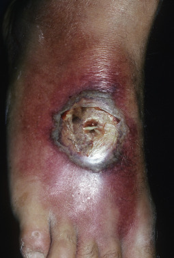
Certain bacterial infections, along with deep fungal infections and atypical mycobacteria, should be included on the list of causes when evaluating a patient with isolated or multifocal necrotic dusky papules, nodules, ulcers, and eschars. This differential diagnosis is especially important in the setting of an immunocompromised host. These bacterial infections can include disseminated diseases from more common opportunistic organisms such as pseudomonas and those rarer conditions such as tularemia or anthrax.
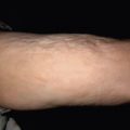
Lastly, more distinctive conditions are included, such as the petechial exanthem of Rocky Mountain spotted fever, angiomatous papules of bacillary angiomatosis, and the expanding annular plaques of erythema migrans seen in Lyme disease.
Depending on the infections suspected, the workup for these conditions can include surface cultures, tissue cultures, and skin biopsy samples with subsequent special stains. This portion of the atlas contains images of common, uncommon, superficial, and disseminated bacterial infections and their many manifestations in the skin.
Related posts:
 Atopic Dermatitis, Eczema, and Noninfectious Immunodeficiency Disorders
 Seborrheic Dermatitis, Psoriasis, Recalcitrant Palmoplantar Eruptions, Pustular Dermatitis, and Erythroderma
 Abnormalities of Dermal Fibrous and Elastic Tissue
 Errors in Metabolism
 Melanocytic Nevi and Neoplasms
 Genodermatoses and Congenital Anomalies
Stay updated, free articles. Join our Telegram channel

Full access? Get Clinical Tree


