Figure 14.1
Centrofacial melasma. Brown, reticular patches are noted on the forehead, cheeks, upper lip, nose, and chin
Differential Diagnosis
The patient’s clinical presentation was most consistent with melasma. Postinflammatory hyperpigmentation may present with brown macules and associated erythema, scale, and pruritus secondary to photosensitivity reactions or irritant contact dermatitis. Solar lentigines are secondary to ultraviolet radiation and present as round, flat, well-circumscribed macules in sun exposed sites. Drug-induced photosensitivity due to thiazides or tetracyclines may present as irregularly shaped hyperpigmentation. Actinic lichen planus often presents on the temples and may involve the neck and intertriginous sites. Exogenous ochronosis presents with hyperpigmentation followed by progressive darkening with superimposed pigmented papules. Acquired bilateral nevus of Ota-like macules (Hori’s nevi) presents with multiple brown to gray to blue macules, primarily on the malar region. It is usually more common in Asian females (Chung 2008).
Wood’s Lamp Examination
Wood’s lamp examination (365 nm) shows accentuation or darkening of the hyperpigmented patches.
Histopathology
Melasma is typically a clinical diagnosis; however, if a biopsy were performed, pathology would show epidermal melanin in basal and suprabasal keratinocytes. The number of melanocytes is not increased; however, the melanocytes that are present are larger, more dendritic, and more active. Inflammation is typically absent (Chung 2008).
Diagnosis
Epidermal melasma
Case Treatment
The likely etiology of melasma, including her oral contraceptive usage, pregnancy, and sun exposure was discussed. A triple combination compound consisting of hydroquinone 6 % + tretinoin 0.025 % + fluocinolone 0.01 % cream was started every other night to the dark areas only. The patient was recommended to increase the cream to nightly after 1 week if she had no burning or irritation. A broad spectrum sunscreen with an SPF of 30 was recommended daily. Daily use of Polypodium leucotomos was recommended as a supplement. A 3 month follow-up was recommended. After mild improvement was seen, she was started on a series of three 35 % glycolic acid peels every 4 weeks in addition to her topical regimen.
Discussion
Melasma is a common acquired disorder of hyperpigmentation that is found mostly in Latina, Southeast Asian, and African American women with Fitzpatrick skin types III-V. Women are more prone to developing the disease than men, especially after the woman has experienced childbirth—leading to the common term, “the mask of pregnancy.” Melasma is worsened by excess exposure to the sun; therefore, it is more common in those living in areas with intense ultraviolet radiation exposure (Sheth and Pandya 2011a). A recent multicenter survey from nine countries found that 41 % of women had onset of the disorder after pregnancy but before menopause. Only 8 % noted spontaneous remission, and 25 % had onset after starting their contraceptive (Ortonne et al. 2009).
The exact cause of melasma is unknown. The high incidence among family members suggest a genetic component, and various surveys from around the world report 10–70 % of family members being involved (Ortonne et al. 2009). Sun exposure is likely an exacerbating factor because of the ultraviolet induced upregulation of melanocyte proliferation, migration, and melanogenesis (Sheth and Pandya 2011a). While melasma is known to occur with hormonal changes, clinical evidence to date does not clearly associate serum hormone levels to melasma. Perez et al examined the link between circulating hormones and found that nulligravid women with melasma had significantly higher serum levels of luteinizing hormone and lower levels of estradiol than their counterpart controls, though further research is needed (Perez et al. 1983). Several studies have noted the onset of melasma with oral contraceptive use and pregnancy. Therefore, it is recommended that patients who develop melasma while taking an oral contraceptive should stop the medication and avoid future use of such drugs when possible (Sheth and Pandya 2011a). However, stopping the culprit oral contraceptive will not necessarily reverse melasma. Some studies have suggested that mild abnormalities in thyroid function are associated with oral contraceptive or pregnancy related melasma; therefore, it is reasonable to consider checking thyroid function tests in melasma patients (Lutfi et al. 1985).
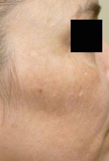
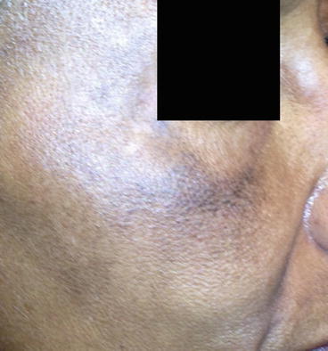
Melasma presents as symmetrically distributed hyperpigmented macules that coalesce into reticular patches. There are three distribution patterns. Centrofacial melasma, the most common pattern, involves the forehead, cheeks, upper lip, nose, and chin (Fig. 14.1). The malar pattern is limited to the cheeks and the nose (Fig. 14.2), and the mandibular pattern is specific to the jawline. Histologically, melasma may be divided into three subtypes. In epidermal melasma, melanin is increased in the epidermis, and patients present with tan to brown hyperpigmentation. In dermal melasma, melanin is found in superficial and mid dermal macrophages, which often congregate around small, dilated vessels, and patients present with a bluish discoloration (Fig. 14.3). Dermal melasma is typically harder to treat. In mixed melasma, melanin is found in both the epidermis and the dermis. In most cases, the number of melanocytes is not increased, yet the melanocytes that are present are larger, more dendritic, and more active. Inflammation is sparse or absent (Kang et al. 2002).


Figure 14.2
Malar melasma. Brown, reticular patches are noted on the medial cheeks and nose

Figure 14.3
Dermal melasma. Brown-bluish discoloration is noted on the bilateral malar cheeks
The excess melanin can be visually localized to the epidermis or the dermis by use of a Wood’s lamp. Epidermal pigment is enhanced during examination with a Wood’s light, whereas, dermal pigment is not. Lesions that have both enhancing and nonenhancing areas have a mixed pattern.
Treatment
Melasma is often difficult to treat and has a significant negative impact on patients’ quality of life. The ultimate goal of melasma management is to lighten the affected area enough so that it is even with the rest of the skin. This can be difficult when treating patients with skin of color.
Stay updated, free articles. Join our Telegram channel

Full access? Get Clinical Tree


