Case 1
Clinical Presentation
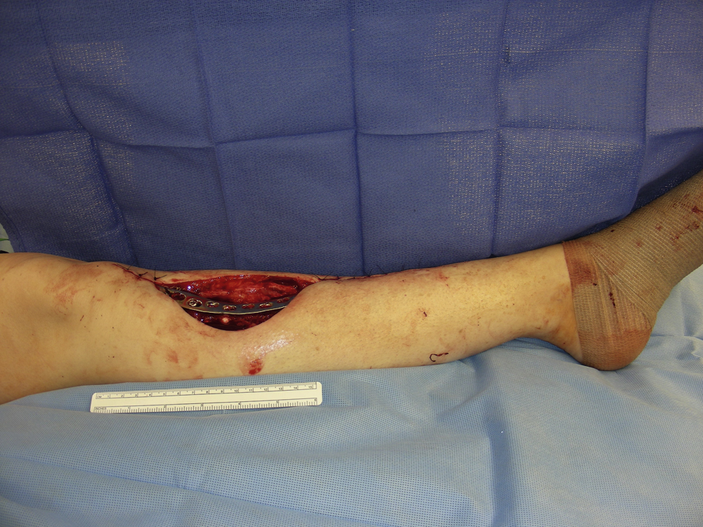
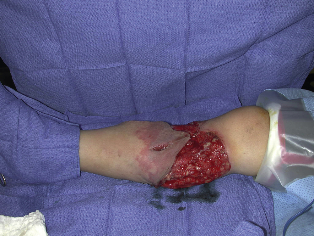
An 11-year-old White male sustained a significant crush and avulsion injury to his right upper leg as a result of a motor vehicle accident. He had extensive full-thickness skin loss over the proximal third of his leg with the exposed underlying tibia measuring 7 × 2 cm. The soft tissue wound was debrided by the trauma service and his surgical care was then transferred to the plastic surgery service for definitive soft tissue reconstruction ( Fig. 43.1 ). Soft tissue coverage was planned after definitive debridement.
Operative Plan and Special Considerations for Reconstruction
For this relatively large soft tissue wound with the exposed tibia in the proximal third of the leg, a classic local muscle flap, such as a medial gastrocnemius muscle flap, can be selected to cover the exposed tibia. The medial gastrocnemius muscle is a type I muscle flap and receives a blood supply primarily from the medial sural artery off the popliteal artery. The rest of the wound can be closed by an adjacent skin rearrangement and a split-thickness skin graft as a one-stage reconstruction. Because of significant crushing injury to the adjacent skin, any perforator-based skin flaps would not be an option. In addition, a distant flap, such as a reversed anterolateral thigh perforator flap, would not reach the proximal tibial location.
Operative Procedures
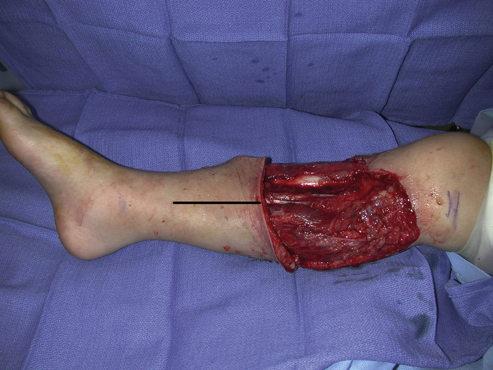
Under general anesthesia with the patient in the supine position, the right proximal tibial wound was debrided and unhealthy looking and traumatized skin was excised. All colonized tissues were sharply removed. The open wound appeared to be fresh and clean after a definitive debridement performed by the plastic surgery service ( Fig. 43.2 ).

The proposed incision for exposure of the distal medial gastrocnemius muscle was marked and the flap dissection was performed under tourniquet control ( Fig. 43.3 ). The skin incision was made through the skin, subcutaneous tissues, and fascia to expose the medial gastrocnemius muscle. In the proximal third of the leg, the medial surface of the medial gastrocnemius muscle was easily separated from the soleus muscle. The plantaris tendon was visualized between the gastrocnemius muscle and the underlying soleus muscle. The dissection went distally along the medial boarder of the medial gastrocnemius until its tendon joined the Achilles tendon. The tendon of the muscle was divided several centimeters distal to the muscle belly and the medial half of the gastrocnemius muscle was dissected from distal to proximal direction along the raphe between the medial and lateral gastrocnemius muscle bellies. During the flap dissection, the lessor saphenous vein and sural nerve were visualized and protected. Once the medial gastrocnemius muscle was elevated adequately, it was rotated medially to cover the exposed upper tibia. The flap was temporarily inset into the wound and the entire exposed upper tibial was completely covered. One drain was placed under the flap and another in the donor site.
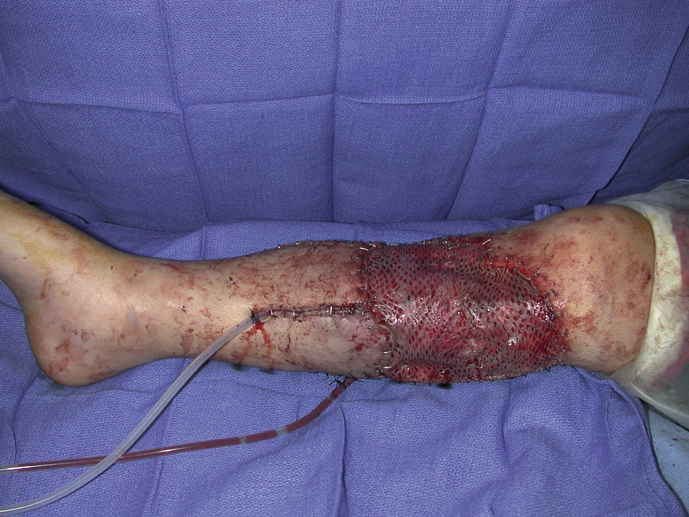
The flap was approximated with the adjacent subcutaneous tissue with several interrupted half-buried 3-0 Monocryl sutures. Additional local skin rearrangements were done for some portions of wound closure and the rest of the wound including the muscle flap was covered with split-thickness skin grafts. Split-thickness skin grafts were harvested with a dermatome from the right lateral thigh and meshed to 1:1.5 ratio. The incision for the flap exposure was closed in two layers and all skin grafts were secured with skin staples ( Fig. 43.4 ).
Follow-Up Results
The patient did well postoperatively without any issues related to the flap reconstruction and wound closure. He was discharged from hospital on postoperative day 5. The right upper leg wound healed uneventfully. He was followed by the plastic surgery service for routine postoperative care.
Final Outcome
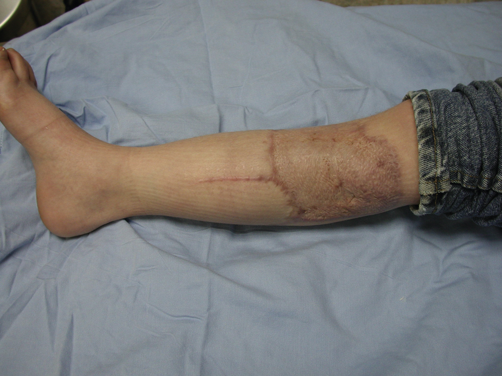
During further follow-up, the right upper tibial wound flap reconstruction site has healed well with good contour and minimal scarring. There were no wound breakdown, recurrent infection, or contour issues related to the soft tissue reconstruction ( Fig. 43.5 ). The patient has resumed his regular activities and has returned to school as a normal student.
Pearls for Success
This is a classic example where a medial gastrocnemius muscle flap is used to cover a complex proximal tibial wound. With a combination of the local muscle flap, local skin rearrangement, and skin graft, such a complex upper tibial wound can be reconstructed successfully. A medial gastrocnemius muscle flap is a work horse to provide good soft tissue coverage for an upper tibial wound. If not traumatized, the muscle flap can be reliable and expanded several centimeters after fascial scoring on the surface of the muscle belly. If needed, the origin of the muscle flap can be divided from the medial condyle of the femur under direct vision once the medial sural artery has been identified. This maneuver can also add several centimeters to the flap advancement. Once the muscle origin has been divided, the flap’s further advancement should be done very carefully to prevent avulsion injury to the pedicle because the pedicle length of the medial sural artery is quite short.
Case 2
Clinical Presentation
A 58-year-old White female underwent orthopedic debridement of a previous left proximal tibial fracture site for nonunion. New open reduction and internal fixation with a large reconstruction plate were performed. Unfortunately, this left an 11 × 5.5 cm soft tissue wound mostly in the proximal third of the left leg with exposed fracture site and reconstruction plate. The plastic surgery service was asked by the orthopedic trauma service to provide soft tissue coverage for the fracture site and exposed reconstruction plate to facilitate wound closure ( Fig. 43.6 ). The patient had several comorbidities including chronic occlusive pulmonary disease for which she received steroid therapy. Her leg was also relatively small with significant muscle atrophy and the fracture union and soft tissue healing therefore remained a challenge.