Groups
Reason for stiffness
Clinical/laboratory data
Treatment
I
(а) Aftermath of plaster immobilization of fractures of the femur; less frequently, fixing of the knee by bandaging the fractures of the lower leg
The stiffness is not associated with a cicatricial process in the knee joint; there are no rough unions or malunions between muscle and bone (myofasciodeses)
Passive–active development of movement using an external fixation device
(b) Complication of lengthening of the femur (extension stiffness) or lower leg (flexion stiffness)
Conservative treatment, including redressment attempts over 2–3 months, have failed
II
Aftermath of inflammatory process in the knee joint or intra-articular fracture
The femur muscles are intact or have no marked secondary fibrous changes or atrophy. The points of pathological fixation of the muscles to the femur are not available or not marked. A cicatricial process occurs in the knee joint cavity and para-articular tissues
Arthroscopic release of the knee joint with subsequent passive–active development of movement using an external fixation device
III
United fractures of the distal third and distal part of the femur
The stiffness is not associated with a cicatricial process in the knee joint or it is not marked and is secondary in character. Marked signs of myofasciodesis: cicatricial union of muscles and tendons with bone, union of tissues
“Semiclosed” with a local process or open myolysis with reconstruction of the muscles sliding. If required, release of the knee joint. Subsequent development of movement in the knee joint using an external fixation device
IV
United open fracture, after extensive injury of the soft tissues of the distal femoral muscle; osteomyelitis in remission
The stiffness is associated with a cicatricial process in the para-articular tissues; polylocal myofasciodeses
Open arthrolysis, myolysis with reconstruction of the muscles sliding with subsequent passive–active development of movement using an external fixation device
V
Malunions and nonunions of the distal third and distal part of the femur
The stiffness is associated with a cicatricial process in the para-articular tissues; polylocal myofasciodeses
One-stage operative treatment: Open arthrolysis, myolysis with reconstruction of the muscles sliding; rigid bone fragments fixation with subsequent passive–active development of movement using an external fixation device
Two-stage treatment (is more preferable): The first step is bone union with restoration of an anatomical axis of a segment (even at shortening kept). The second stage is open arthrolysis, myolysis with reconstruction of the muscles sliding with subsequent passive–active development of movement using an external fixation device
11.1.1 Soft Tissue Correction with External Fixators
Severe flexion contractures of the knee are a major obstacle to functional ambulation and weight bearing. Such contractures are particularly common in pediatric patients especially in arthrogryposis, congenital pterygium contractures, and multiple other congenital and acquired problems. Management of these deformities are quite challenging (Hosny and Fadel 2008; DelBello and Watts 1996; Heydarian et al. 1984). Gradual correction of the deformity has been reported previously using various external fixators (Herzenberg et al. 1994; Gillen et al. 1996; Ilizarov 1990; Damsin and Ghanem 1996; Saleh et al. 1989). Several authors have used an Ilizarov circular fixator (Damsin and Ghanem 1996), a monolateral fixator (Herzenberg et al. 1994), hinged distraction apparatus (Saleh et al. 1989), or computer software-based fixators.
“Reconstruction of the muscles sliding” involves various variants and modifications of operations to mobilize and reconstruct the quadriceps (Ebraheim et al. 1993; Paley 2002). Lengthening of the distal tendon of the quadriceps in adults is undesirable as it will result in restricted active extension of the lower leg. Besides, external fixation allows a gradual increase in flexion of the lower leg to the necessary angle in the postoperative period (Window 11.1).
Window 11.1
Apart from the indicators given in Table 11.1, it is necessary to take into account:
1.
The degree and duration of the stiffness and the patient’s age.
2.
The patient’s attitude to treatment and his/her desire to restore the amplitude of the knee joint movement. Some patients insist on the maximum possible restoration of joint movement, for others restoration of 30–45 % of movement amplitude is sufficient for their social rehabilitation. However, 90° of flexion is usually accepted functional level.
3.
The intensity of changes to the articulating surface of the femur. For example, in extension stiffness, a 40 % shortening of the belly of the rectus and median muscles due to their atrophy and fibrous changes prejudices the possibility of restoration of full active extension of the lower leg.
4.
The presence and characteristics of a chronic inflammatory process of the femur or knee joint.
5.
The presence of another orthopedic pathology: nonunions or deformities, including shortening of the femur or lower leg.
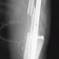
If extension stiffness occurs during femur lengthening, especially during formation of a regenerate in the distal third of the segment and the closed manipulation failed, external fixation can be used. For this purpose, a transosseous Ilizarov module is mounted on the lower leg (Fig. 11.1a) or the module is based on two half-pins and a wire (Fig. 11.1b). The module is connected by a hinge subsystem to the basic support. This approach should be used if extension stiffness forms during treatment of femoral fractures, specially open injuries to the distal third of the segment. This approach ensures that the emergence of rough cicatricial unions of tendons and muscles with the bone in the place of the fracture is avoided (Trick 11.1).
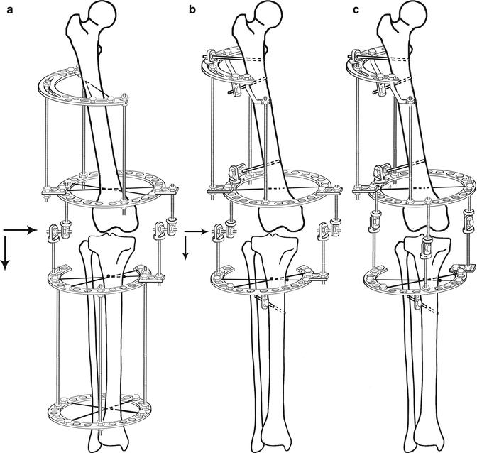
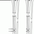
Fig. 11.1
(a–c) Schemes for an Ilizarov device (a) and a combined (hybrid) device (b) for reduction of the lower leg. The external supports of the module fixed to the femur are located parallel to the axis of the femoral condyles. Distraction starts on the 3rd to the 5th day at a rate of 0.25 mm six to eight times a day. The distraction rate is decreased if pain or signs of hyperextension of the great vessels and nerves occur. In lateral (external, internal) dislocations and subluxations, the distraction first must be uniform on all three hinges. After radiographic confirmation of the presence of the necessary diastasis for unhindered movement of the lower leg in the horizontal plane, the subsystem connecting the modules is remounted. The remounting depends on the type of dislocation: anterior, posterior, medial, or lateral. After reduction of the dislocation, the knee joint is fixed in the mid-physiological position for 2–3 weeks. After that the device can be used to develop movements in the knee joint (c)
Trick 11.1
In most cases, all supports of the device fixing the femur are located perpendicular to the anatomical (mid-diaphyseal) axis of the femoral bone. Accordingly, the distal basic ring (as well as all supports) is not parallel to the knee joint plane. Therefore, to install axial hinges, an additional support fixed at the necessary angle to the distal basic support of the femur must be introduced into the assembly. Another possibility is to use complex hinges, one part serving for fixation to the distal basic support of the femur device and the other the axial hinge itself.
11.1.1.1 Surgical Technique with Ilizarov Circular Fixator
For closed reduction of lower leg dislocations, the first stage involves mounting a transosseous module based on wires or a hybrid module (Fig. 11.1) on the femur. A second module that can also be based on wires or a hybrid module is mounted on the lower leg. A hinge-distraction system is installed between the modules. If the dislocation of the shin is a complication of femur lengthening, an additional transosseous module is mounted on the lower leg and connected by a hinge subsystem to the basic frame.
One of the conditions for successful external fixation for knee joint stiffness is to use the reference positions described in the atlas for insertion of transosseous elements.
The second mandatory condition of configuration of the device for increasing knee joint ROM is installation of axial hinges strictly according to an axis of rotation of a knee joint (Fig. 11.2).

Fig. 11.2
(a–d) Orientation of the axis of knee flexion–extension in frontal (a), sagittal (b), and transverse (c) planes. Axial hinges are installed between the transosseous modules fixing the femur and the lower leg 2 cm from the joint surface and at the junction of the middle and posterior thirds of the femoral condyle (d)
Along with it there is a widely accepted opinion that movements in a knee joint have more complex trajectory than it can be provided by the one-axial hinges. The trajectory of movements in a knee joint can be presented as superposition of points on certain segments of arches of a circle of femoral bone condyles and segments on condyles of a tibial bone. Due to a difference of radiuses, lengths of arches of condyles of femoral bone and segments of tibial condyle movement in a knee joint are carried out on several trajectories with change of the centers of rotation. Thus, the trajectory of movement in a knee joint represents a complex curve, which is different for external and internal condyles. This provides a rotational component of moving (Iwaki et al. 2000) (Fig. 11.3). Therefore, the use of the devices having the virtual hinge, for example, hexapods, is perspective for knee joint stiffness treatment.
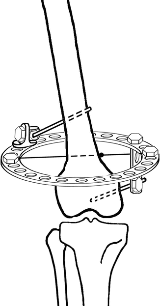
Fig. 11.3
Additional supports for the femur at lower leg lengthening
The formation of persistent flexion stiffness of the knee joint that does not respond to conservative treatment can be a complication of lengthening of the lower leg. The lack of loading on the extremity can negatively affect the distraction regenerate; thus, the stiffness needs to be eliminated as soon as possible. For this purpose, a transosseous module is mounted on the femur as shown in Fig. 11.1a, or a ring support based on a wire and two half-pins is used (Fig. 11.3).
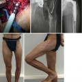
If patellofemoral synostosis or fibrous union of the patella with the femoral bone has occurred, open or arthroscopic mobilization is required. Extension stiffness of the knee joint is frequently present together with a nonunion, deformity, defect, or shortening of the femur in clinical practice. Simultaneous restoration of the anatomy and function of the injured extremity is the priority in planning the rehabilitation of the patient. At the same time, the operation for single-stage restoration of the knee joint function must be performed with the participation of a surgeon with experience of such interventions. Otherwise, the treatment should be divided into stages.
A single-stage operation to treat a traumatic injury simultaneously with mobilization of the knee joint involves the following:
1.
In angular and rotational deformity exceeding 15–20°, a shortening of 20–40 mm, osteotomy is recommended with subsequent gradual correction of the deformity.
2.
In hypertrophic defect, pseudoarthroses and anatomical shortening of the femur by 2–3 cm, microdistraction is performed to eliminate the inequality in the lengths of the extremities and to restore the anatomy of the bone simultaneously with restoration of the knee joint movement.
3.
If external fixation of a nonunion of the femur involves an intervention with an open stage (removal of a metal structure, osteoplasty), it is combined with an operation to mobilize the knee joint.
4.
In atrophic nonunions of the femur and shortening of the segment by 40 mm, the bone fragments are openly reduced using, according to the indications, osteoplasty, and corticotomy is performed with osteoclasia of the femoral bone to eliminate inequality in the lengths of the extremities (Kornilov et al. 1992).
5.
The operation to treat shortening of a lower extremity that is accompanied by severe stiffness of the knee joint after malunion of intra-articular fractures not more than 1.5 years after trauma starts with surgery to the knee joint. The congruity of the joint surfaces is restored and the joint is mobilized. The second stage involves lengthening of the segment (Reutov et al. 2000).
In replacement of a segmental defect of the femur by the Ilizarov method, operative approaches to improve the function of the knee joint must be postponed at least until adaptation of the transposed fragment to the basic fragment and their stabilization. Arthrolysis and myolysis can be performed in a single step with open adaptation of the transposed and basic bone fragments. Multiple solid myofasciodeses, filling the knee joint cavity with healing tissue, and osteomyelitis favor a two-stage treatment for restoration of the weight-bearing ability of the extremity and then improve knee joint function.
After closed operations the knee joint is stabilized in the position achieved by maximum elimination of the stiffness. However, to reduce pain, we have to reduce the position in the joint achieved by the end of the operation by 30–50 %. It often happens that after open arthrolysis and myolysis, to avoid excessive skin stretching, the skin is taken in with the knee joint flexed less than what was achieved during the operation. To reduce the risk of soft tissue necrosis after surgery, the knee joint is stabilized in a position that ensures good blood supply to the wound edges. If there is a marked cicatricial process occurring in the area of the knee joint, the knee is stabilized in a position close to full extension.
A diastasis of 5–6 mm is created between the joint surfaces in two or three stages. It is important to note that due to the flexure of the transosseous elements, the amount of separation on hinges will not correspond to the joint space. Therefore, the effectiveness of the distraction should be monitored radiographically. Radiographic monitoring of the installation of the axial hinges is also necessary.
Related posts:
 Distraction Arthroplasty for Ankle Osteoarthritis
 Hybrid Lengthening Techniques: Lengthening and Then Nailing (LATN) and Lengthening and Then Plating (LAP)
 Congenital Pseudarthrosis of the Tibia: Redefined (Congenital Crural Segmental Dysplasia)
 Lengthening over Nails (LON): Femur and Tibia
 Pelvic Support Osteotomy (PSO): Indications, Limits and Complications
 Reconstruction of Segmentary Defects in Chronic Osteomyelitis Using the Combined Technique
Stay updated, free articles. Join our Telegram channel

Full access? Get Clinical Tree


