Author
Year
N (patients)
Good results (%)
Conclusion
Van Valburg et al.
1995
11
100
50 % increased joint space
Van Valburg et al.
1999
17
76
2 years to see benefits
Marijnissen et al.
2002
57
75
1 year to see benefits
Randomized study favors DA
Ploegmakers et al.
2005
22
73
Good results at >7 year F/U
Paley et al.
2008
32
67
Recommend adjuvant procedures
Tellisi et al.
2009
25
74
91 % had some pain relief
Hinged distraction preserves ankle ROM
Patients >60 years old had good results
Lamm and Gourdine-Shaw
2009
3
MR showed increased joint space and decrease in subchondral cysts
Intema et al.
2011
26
Disappearance of subchondral cysts directly related to good clinical result
16.3 Surgical Technique
16.3.1 Overview
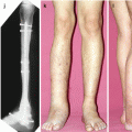
There are various techniques used to perform ankle distraction arthroplasty. Van Valburg et al. (1995) introduced the classic method using two tibial rings attached to the leg with two tensioned wires per ring. The foot ring was attached with four tensioned wires. This was a static external fixator (it has no hinges) with four rigid connections across the ankle joint. Distraction of 5 mm was performed gradually at 1 mm per day. Dutch authors also leave the external fixator in place for a minimum of 3 months but often more than 4 months (Van Valburg et al. 1995, 1999). The technique we use at our institute differs in many aspects. We use a streamlined external fixator (RAD frame, Small Bone Innovations, Morrisville, PA) with minimal points of fixation. The fixator is articulated allowing free range of motion at the ankle joint. Like other authors, we recommend adjuvant procedures at the time of frame application, if indicated (Paley et al. 2008; Lamm and Gourdine-Shaw 2009). These include arthrotomy for osteophyte resection and microfracture, Achilles tendon lengthening, and supramalleolar osteotomy for malalignment. Ankle distraction is applied acutely in the operating room. Ankle range of motion starts early in the postoperative period. The frame is worn for a minimum of 10 weeks.
16.3.2 Preoperative Evaluation
Preoperative workup includes an alignment and mobility exam. Weight-bearing x-rays are used to establish the degree of arthritis and quantify malalignment. Osteophytes are also identified. MR is often used to recognize the location and degree of cartilage thinning and to localize subchondral cysts. The subtalar joint condition should also be evaluated to distinguish any contribution to the patient’s overall pain.
A good candidate for ankle distraction includes any patient who has ankle arthritis and some meaningful joint mobility. Most patients are self-selected in that they have been offered ankle fusion and have forcefully refused arthrodesis. These patients want to maintain their ankle motion and decrease their pain, goals that ankle distraction can obtain. We have not identified, with significance, which patients are more likely to improve. An unpublished review was performed on our patients who achieved the most improvement in AOFAS scores after the distraction procedure. Pre- and postsurgical MR scans in these patients illuminated certain trends: these top-performing patients were between 30 and 40 years of age, had more intra-articular cartilage on pre-op MR scans, and had more mobility in the ankle joint presurgery. All of their MR scans demonstrated increases in joint space and disappearance of subchondral cysts consistent with previous studies (Lamm and Gourdine-Shaw 2009). Perhaps younger patients with more mobility and less arthritis are the ideal candidates, but our clinical series (Tellisi et al. 2009) indicated that patients over 60 years old fared best. Based on these findings, we do not use age, degree of arthritis, or absolute motion as inclusion criteria.
Contraindications are relative and include inflammatory arthritis, severe tibiotalar stiffness, and abnormal joint geometry. Our experience with rheumatoid arthritis (RA) has yielded a high failure rate with most patients requiring ankle fusion. RA is not a mechanical disease so it stands to reason that removing stress is not helpful. Patients who have severe stiffness of the ankle joint effectively have a fused ankle. They are wearing out the adjacent joints but continue to have tibiotalar joint pain. Ankle fusion will relieve their pain and not further compromise the adjacent joints. Advanced flattening of the talus, intra-articular pilon malunion with joint incongruence, and joint subluxation without realignment are probably best solved with ankle fusion.
16.3.3 Frame Application
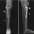
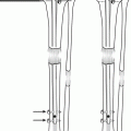
The surgical technique we use for ankle distraction arthroplasty has been reported (Inda et al. 2003; Tellisi et al. 2009). Patients receive spinal anesthesia with IV sedation. The ipsilateral iliac crest is prepped and draped and 60 cc of bone marrow is aspirated from it. A pneumatic thigh tourniquet is used for the open portion of the surgery and is removed for the external fixator application. An anterior arthrotomy is created in most cases and anterior osteophytes are resected. Osteophytes that are accessible in the medial and lateral gutters are removed as well. All areas of visible cartilage degeneration are drilled or picked to produce microfractures of the subchondral bone. After the wound is closed, the tourniquet is deflated. The bone marrow aspirate is concentrated using any number of commercially available systems and is then injected into the ankle joint. (The injection of bone marrow aspirate concentrate into the ankle joint is a technique we use at our institute. There is no previous investigation analyzing the efficacy of this intervention; however, we are engaged in a prospective study evaluating this technique. Other centers that perform ankle distraction also inject adjuvant treatment into the joint including viscosupplementation, recombinant human growth hormone (rhGH), and platelet-rich plasma (PRP). We all feel that adding these additional interventions could be beneficial.) A circular external fixator, the Ring Fix system (Small Bone Innovations, Morrisville, PA), is applied to the ankle. The tibial ring is mounted to the distal tibia using two 6 mm hydroxyapatite coated, tapered half pins (Biomet, Parsippany, NJ). The pin sites are predrilled with a 4.8 mm drill bit and the blunt tipped pins are inserted by hand. The hinges are then aligned with the axis of rotation of the ankle by placing a wire along a line from the tip of the medial malleolus to the tip of the lateral malleolus. We then ensure that the center of the hinges rests against the ends of the wire. Final hinge position is confirmed on lateral fluoroscopy (Fig. 16.1). The foot ring is attached to the foot using three tensioned K-wires (Fig. 16.2). The typical pattern used is two crossing wires through the calcaneus and one wire through the talar neck (Fig. 16.3
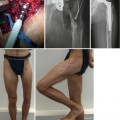
 Hybrid Lengthening Techniques: Lengthening and Then Nailing (LATN) and Lengthening and Then Plating (LAP)
 Hindfoot Reconstruction by the Combined Technique
 Lengthening over Nails (LON): Femur and Tibia
 Pelvic Inlet Reconstruction for Obstruction Associated with Lumbosacral Agenesis Utilizing Distraction Osteogenesis and Circular External Fixation
 Pelvic Support Osteotomy (PSO): Indications, Limits and Complications
 Reconstruction of Segmentary Defects in Chronic Osteomyelitis Using the Combined Technique


Related posts:
Hybrid Lengthening Techniques: Lengthening and Then Nailing (LATN) and Lengthening and Then Plating (LAP)
Hindfoot Reconstruction by the Combined Technique
Lengthening over Nails (LON): Femur and Tibia
Pelvic Inlet Reconstruction for Obstruction Associated with Lumbosacral Agenesis Utilizing Distraction Osteogenesis and Circular External Fixation
Pelvic Support Osteotomy (PSO): Indications, Limits and Complications
Reconstruction of Segmentary Defects in Chronic Osteomyelitis Using the Combined Technique
Stay updated, free articles. Join our Telegram channel
Full access? Get Clinical Tree
