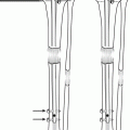
Fig. 4.1
Saw bone model demonstration of LATN. (a) Axial view showing that external fixation is peripherally placed in order to avoid contact with the IMN. The black lines depict the course of the external fixation pins > the blue dot depicts the future IMN location. (b) The TSF may be used to optimally align the bone fragments. (c) The IMN is inserted while the frame is in place. (d) Contact between internal and external fixation is avoided
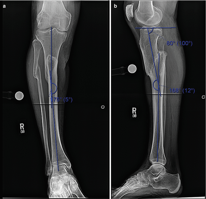
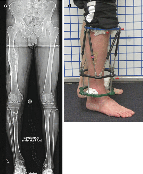
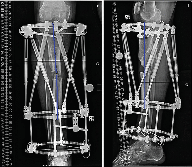
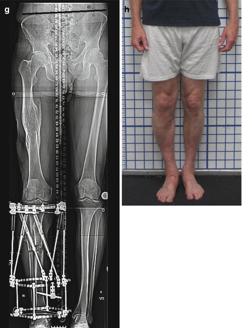
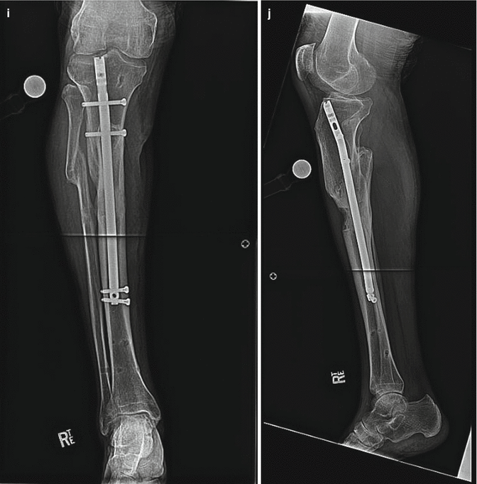
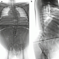
Fig. 4.2
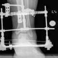
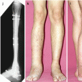
A 48-year-old male with malunion consisting of varus, recurvatum, and shortening that was treated with LATN. (a) AP x-ray showing 5° of varus deformity. (b) Lateral x-ray showing 12° of recurvatum (apex posterior) deformity. (c) Erect leg radiograph showing right leg shortening of 24 mm. (d) Clinical photo at end of distraction. (e) AP x-ray at end of distraction showing lengthening and correction of deformity. (f) Lateral x-ray showing the same. (g) Erect leg x-ray showing the same. (h) Clinical photo 4 months after IMN insertion. (i) AP x-ray showing the same. (j) Lateral x-ray showing the same
4.2.1 LATN: Clinical Experience
We published a retrospective case-matched comparison (Rozbruch et al. 2008a) of patients lengthened with LATN (39 limbs in 27 patients) vs. the classic technique (34 limbs in 27 patients). The LATN group wore the external fixator for less time (12 versus 29 weeks) and had a lower external fixation index (EFI) (0.5 versus 1.9) and a lower bone healing index (BHI) (0.8 versus 1.9) than the classic group (Fig. 4.3). One deep infection developed in the LATN group after the regenerate was fully united. This was successfully treated by removing the IMN and administering antibiotics. We concluded that LATN confers advantages over the classic method including shorter times needed in external fixation, quicker bone healing, and protection against refracture. There are also advantages over the lengthening over a nail (LON) and internal lengthening nail techniques.
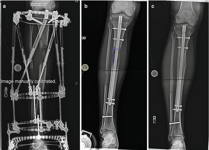
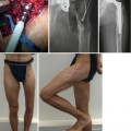
Fig. 4.3
Tibia lengthening of 7 cm in a 28-year-old man. (a) At the end of distraction. (b) Six weeks after IMN insertion. (c) Six months after IMN insertion
4.2.2 LATN of Tibia: Surgical Technique
4.2.2.1 Tibia and Fibula Osteotomy
The fibula osteotomy is performed using a multiple-drill-hole technique with a 1.8-mm wire and then completed with an osteotome. A three-ring Taylor Spatial Frame (TSF) (Smith and Nephew, Inc, Memphis, TN) is applied using a rings-first method (Rozbruch et al. 2006a; Taylor 2007). The proximal ring is stabilized with a 1.8-mm tensioned transverse wire, a 1.8-mm tibia-fibula wire, an anteromedial half pin, and an anterolateral half pin. The configuration of this proximal ring fixation is unique in that the bone fixation is placed peripherally within the proximal tibia to allow future insertion of an IMN avoiding any contact with the external fixation pins (Fig. 4.1a). The 1.8-mm wires are placed more posterior in the tibia than is typical. The half pins are inserted using a cannulated wire technique for precision. The anteromedial half pin is peripheral and runs in an anterior to posterior direction. The anterolateral half pin is peripheral and runs in an anterolateral to posterior central direction. The proximal ring is the reference ring and TSF mounting parameters (Taylor 2007) are measured in relation to this ring. The origin (Taylor 2007) is placed at the level of deformity within the diaphysis. When there is no deformity, we assign the origin to the center of the bone at the level of osteotomy 10 to 12 cm distal to the knee joint. Next, a ring block consisting of two rings connected with four rods is applied to the mid-distal tibia orthogonal to the tibial diaphysis. The distal ring is stabilized with a transverse 1.8-mm wire 1.5 cm proximal to the ankle joint, a 1.8-mm tibia-fibula wire 2 cm proximal to the ankle joint, and an anteromedial to posterolateral 6-mm half pin. The middle ring is typically left with no fixation. In cases where there is deformity correction, we may insert a wire or half pin off the middle ring to achieve optimal leverage during the correction. The proximal and middle rings are then connected with six TSF struts whose lengths are recorded (Fig. 4.1b). The struts are then removed for the tibial osteotomy.
The tibial osteotomy is performed in a percutaneous fashion using a multiple-drill-hole technique. Distraction is started on postoperative days 7–10. The distraction schedule is made using the TSF Internet-based software using the total residual method (Smith and Nephew, Inc, Memphis, TN). Care is taken to correct all deformity at the osteotomy site prior to nail insertion.
4.2.2.2 Insertion of Intramedullary Nail and Removal of Frame
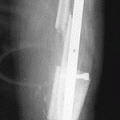
Once length and deformity correction are achieved, the second-stage surgery is performed without the use of a tourniquet. The external fixator is prepped into the surgical field, and betadine-soaked sponges are placed around all pin sites. The frame is covered with sterile towels. To prevent proximal migration of the fibula, we insert a 4.5-mm solid syndesmosis screw 1 cm proximal to the distal tibia pin fixation. An IMN with an extra hole 8 cm from the proximal end is used in order to be able to insert two proximal interlocking screws. A minimal incision technique for IM nail insertion is used. A guidewire was passed across the regenerate and into the distal fragment ending at the syndesmosis screw. Serial reaming is performed until cortical chatter is achieved, and a nail 1 mm smaller than the last reamer used is inserted. We do not open the regenerate site and we do not remove any reaming particles. The IMN is locked with two screws in both the proximal and distal fragments. The external fixator is then removed without risk of tibial displacement or shortening (Fig. 4.1c). The pin sites were irrigated but not curettaged and are not closed. While in some cases, we use a full length IMN, there are situations where we use a short custom-made IMN. Such situations include an abnormal intramedullary canal in the distal bone or a multilevel reconstruction. In all circumstances, there is no contact between internal and external fixation in order to minimize the chance of developing infection (Figs. 4.2 and 4.3).
4.3 Lengthening and Then Plating (LAP)
The lengthening and then plating (LAP) technique Harbechuski et al. (2012) (Figs. 4.4 and 4.5
 Distraction Arthroplasty for Ankle Osteoarthritis
 Hindfoot Reconstruction by the Combined Technique
 Lengthening over Nails (LON): Femur and Tibia
 Pelvic Inlet Reconstruction for Obstruction Associated with Lumbosacral Agenesis Utilizing Distraction Osteogenesis and Circular External Fixation
 Pelvic Support Osteotomy (PSO): Indications, Limits and Complications
 Reconstruction of Segmentary Defects in Chronic Osteomyelitis Using the Combined Technique


Related posts:
Distraction Arthroplasty for Ankle Osteoarthritis
Hindfoot Reconstruction by the Combined Technique
Lengthening over Nails (LON): Femur and Tibia
Pelvic Inlet Reconstruction for Obstruction Associated with Lumbosacral Agenesis Utilizing Distraction Osteogenesis and Circular External Fixation
Pelvic Support Osteotomy (PSO): Indications, Limits and Complications
Reconstruction of Segmentary Defects in Chronic Osteomyelitis Using the Combined Technique
Stay updated, free articles. Join our Telegram channel
Full access? Get Clinical Tree
