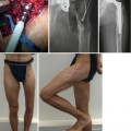
Fig. 14.1
Illustration of the pathological anatomy demonstrated for CCSD type Vb. (a) X-ray of tibial and fibular pseudarthrosis with sclerosis of more than 30 %. (b) Resected tibial pseudarthrosis. The rectangular field shows the area were specimen was taken out for histological examination. (c) Light microscopically hyperplastic fibrotic periosteum is evident. (d) Immunohistochemical staining (S-100) shows neurogenic cells surrounding the capillaries in the periosteum. (e) Electron microscopically the capillaries are obliterated by surrounding neurogenic cells (Hermanns et al. 1999 and 2005)
This phenomenon leads to a constriction of the vessels and reduction of the perfusion in periosteum and bone (Fig. 14.1). It results in a degenerative fibrosis of the periosteum giving rise to a vicious cycle and decreases the vascular perfusion of the bone. The consequence is fracture of the weakened bone and a subsequent nonunion (Boyd 1982; Camurati 1930; Hermanns et al. 1999; Hermanns-Sachweh et al. 2005; Morrissy 1982; Weber 2006). My theory is supported by a remarkable animal experiment of Wright and co-authors (Wright et al. 1991).
14.3 New Definition: Congenital Crural Segmental Dysplasia (CCSD)
The old term congenital pseudarthrosis of the tibia is wrong because:
The pseudarthrosis of the tibia occurs in 90 % of all cases not until the first years of life (Andersen 1973; Hefti et al. 2000; Murray and Lovell 1982).
The pseudarthrosis is not restricted to the tibia but affects in 60 % of all cases also the fibula.
The fibula is affected alone with a pseudarthrosis in 19 % without affection of the tibia (Keret et al. 2000).
14.4 Clinical Evaluation
1.
Postpartum without pseudarthrosis (90 %).
2.
Postpartum with pseudarthrosis (10 %).
3.
Forms 1 and 2 which have had unsuccessful prior surgery.
4.
The diagnosis of CCSD should be considered in children who in the absence of trauma present with a fracture or nonunion or procurvatum and varus deformity of the lower leg, especially if the deformity is increasing.
5.
The diagnosis is also supported by the presence of neurofibromatosis (incidence of 50 %).
14.4.1 Diagnostic Tools
The new own technique of radiological measurement of the extent of sclerosis, independent of different tibial lengths and magnification factor, and a method to determine the extent of resection ensures a successful healing process. Furthermore, it allows to collect data pre- and postoperatively for comparison with own results and the results of other authors (Fig. 14.2) (Andersen 1972; Boyd 1982; Edvardsen 1973; Mahnken et al. 2001). Length extent of sclerosis (%) = length of sclerosis: length of bone × 100. Width extent of sclerosis (%) = width of sclerosis: width of diaphysis × 100. In our clientele the resection of >90 % of the sclerotic bone ensures a healing rate of 93 %.
Fig. 14.2
(a) Illustration to the method for evaluation of the preoperative/postoperative extension of sclerosis. (A) Length of the diaphyseal sclerosis proximal of the pseudarthrosis. (A′) Length of the diaphyseal sclerosis distal from the pseudarthrosis. (B) Length of the tibia proximal of the pseudarthrosis. (B′) Length of the tibia distal of the pseudarthrosis. (C) Width of the cortical sclerosis at the level of the pseudarthrosis, proximal. (C′) Width of the cortical sclerosis at the level of the pseudarthrosis, distal. (D) Width of the tibial diaphysis at the level of the pseudarthrosis, proximal. (D′) Width of the tibial diaphysis at the level of the pseudarthrosis, distal. (E) Width of the medullary canal at the level of the pseudarthrosis, proximal. (E′) Width of the medullary canal at the level of the pseudarthrosis, distal. (F) Width of the diaphysis near the metaphysis, proximal. (G) Width of the cortical sclerosis near the metaphysis, proximal*. (H) Width of the medullary canal near the metaphysis, proximal. * The width of the cortex was measured when at this level sclerosis was no longer present. (b) Illustration to the method for evaluation of the postoperative extension of the sclerosis. (I) Length of the diaphyseal sclerosis proximal of the resection. (I′) Length of the diaphyseal sclerosis distal of the resection. (J) Length of the tibia proximal to the resection. (J′) Length of the tibia distal to the resection. (K) Width of the diaphysis at the level of resection, proximal. (K′) Width of the diaphysis at the level of resection, distal. (L) Width of the cortical sclerosis at the level of resection, proximal. (L′) Width of the cortical sclerosis at the level of the resection, distal. (M) Width of the medullary canal at the level of resection, proximal. (M′) Width of the medullary canal at the level of resection, distal
MRI: to detect the thickened periosteum and confirm the diagnosis. It is also useful in planning the extent of surgical resection of the lower leg segment (Mahnken et al. 2001).
14.4.2 Classification According to Weber (2006)
This new classification (Fig. 14.3) (Weber 2006) shows several advantages in comparison to older classifications (Andersen 1972, 1973; Boyd 1982; Boyd and Sage 1958; Crawford and Bagamery 1986):

Severity of disease increases with the types.
Relation to treatment and prognosis is given.
Independence from time of diagnosis (any stage).
Only X-rays are needed for classification.
The important role of fibula is respected.
Type I is a stage of indifference.
It is characterised by the typical procurvatum-varus deformity of the lower leg without fracture or pseudarthrosis and without progression of the deformity.
Types II and III are the stages of prefracture.
It is characterised by impending fracture of increasing deformity in type II and cystic lesion in type III.
Types IV to VI describe the stages of pseudoarthrosis of the tibia.
The differences between types IV, V and VI are in the extent of the longitudinal sclerosis.
Subtypes of types II–VI
(a)
Fibula without fracture.
(b)
Fibula with fracture (different stages: persisting fibular fracture/pseudarthrosis with hypoplasia/dislocation of the lateral malleolus including a subluxation of the distal talofibular joint/increasing valgus deformity of the ankle).
14.4.3 Scoring
The score system (Weber 2006) (Tables 14.1 and 14.2) which leads to six different classes gives the advantage to compare different cases of different authors according to their results of treatment due to evaluation of the main problems of the disease:
Table 14.1
Classification and score system of CCSD
CCSD type | Score | ||
|---|---|---|---|
I | Indifference stage, procurvatum-varus deformity without progress, no fractures | 32 | |
IIa | Prefracture stage, progressive deformity, no fibular fracture | 29 | |
IIb | Prefracture stage, progressive deformity, + fibular fracture | 26 | |
IIIa | Prefracture stage, cystic lesion, no fibular fracture | 23 | |
IIIb | Prefracture stage, cystic lesion, + fibular fracture | 20 | |
IVa | Tibial pseudarthrosis, sclerosis <30 %, no fibular fracture | 17 | |
IVb | Tibial pseudarthrosis, sclerosis <30 %, + fibular fracture | 14 | |
Va | Tibial pseudarthrosis, sclerosis between 30 –50 %, no fibular fracture | 11 | |
Vb | Tibial pseudarthrosis, sclerosis between 30 –50 %, + fibular fracture | 8 | |
VIa | Tibial pseudarthrosis, sclerosis >50 %, no fibular fracture | 5 | |
VIb | Tibial pseudarthrosis, sclerosis >50 %, + fibular fracture | 2 | |
Leg length discrepancy | 0–20 % | In comparison to the healthy leg | 2 |
21–40 % | 1 | ||
>41 % | 0 | ||
Contracture of the upper ankle joint | No | 2 | |
Mild | 1 | ||
Severe | 0 | ||
Contracture of the knee joint | No | 2 | |
Mild | 1 | ||
Severe | 0 | ||
Localisation of pseudarthrosis | Middle | Third of lower leg | 4 |
Middle-distal | 2 | ||
Distal | 0 | ||
Deformity of the upper ankle joint | 0–5° | In comparison to standard | 2 |
6–10° | 1 | ||
>11° | 0 | ||
Osteoporosis | No | 2 | |
Mild | 1 | ||
Severe | 0 | ||
Number of preoperations | 0 | 2 | |
1–4 | 1 | ||
>4 | 0 | ||
Compliance of the child | Good | 2 | |
Moderate | 1 | ||
Bad | 0 | ||
Compliance of parents | Good | 2 | |
Moderate | 1 | ||
Bad | 0 | ||
Muscle function | Good | 2 | |
Moderate | 1 | ||
Bad | 0 | ||
Table 14.2
Six different classes of CCSD according to the scores
Class | Score |
|---|---|
6 | 0–9 |
5 | 10–18 |
4 | 19–27 |
3 | 28–36 |
2 | 37–45 |
1 | 46–54 |
1.
Different types of the Weber Classification (types I to VI = high to less points)
2.
Extent of length deficiency
3.
Amount of contractures at knee and ankle
4.
Location of pseudarthrosis
5.
Deformity of ankle joint
6.
Extent of osteoporosis
7.
Number of previous operations
8.
Compliance of patient and parents
9.
Muscle function
14.5 Treatment Options
14.5.1 Conservative Treatment
Conservative treatment is indicated only when there is no fracture and no progression of the deformity (Type I):
1.
Ankle-Foot Orthesis = lower leg/foot orthotic device with isometrical hinges at upper ankle joint (indications: if the apex of the deformity is typically located in the middle to the distal third of the lower leg and no increase of deformity can be detected or fracture risk is low).
2.
Knee-Ankle-Foot Orthesis = a knee-ankle-foot orthotic device with isometrical hinges at the knee and upper ankle joint (indications: if the deformity is located more proximally, if increasing deformity of lower leg can be detected and if increasing fracture risk is given).
14.5.2 Time of the Surgical Treatment
I disagree with a frequently expressed opinion that a surgery should not be performed before the end of puberty. The delay of surgical treatment until puberty leads to the development of significant problems as follows:
Hypoplasia (foot, entire leg)
Muscle atrophy
Increasing leg length discrepancy
Osteoporosis
Contractures
Dislocation of the lateral malleolus including
Subluxation of the distal talofibular joint
Increasing valgus deformity of the ankle
14.5.3 Surgical Techniques
Instruments
1.
Image intensifier
2.
Ring fixators
3.
Weber-Cable Device
4.
Intramedullary rods
Related posts:
 Distraction Arthroplasty for Ankle Osteoarthritis
 Hybrid Lengthening Techniques: Lengthening and Then Nailing (LATN) and Lengthening and Then Plating (LAP)
 Lengthening over Nails (LON): Femur and Tibia
 Pelvic Inlet Reconstruction for Obstruction Associated with Lumbosacral Agenesis Utilizing Distraction Osteogenesis and Circular External Fixation
 Pelvic Support Osteotomy (PSO): Indications, Limits and Complications
 Reconstruction of Segmentary Defects in Chronic Osteomyelitis Using the Combined Technique
Stay updated, free articles. Join our Telegram channel

Full access? Get Clinical Tree


