Inflammatory Eruptions of Unknown Cause
Overview
Pityriasis rosea, granuloma annulare, and lichen planus are unique inflammatory conditions of unknown etiology. Each has various clinical presentations, and distinctive patterns of anatomic distribution. Most often the diagnosis can be readily made based upon the typical signs and symptoms of these curious dermatioses.
There are conflicting reports about the association of these entities with certain infectious and metabolic conditions. For example, pityriasis roses is thought to represent a viral exanthem possibly caused by a herpesvirus. An association has been noted between lichen planus and hepatitis C, chronic active hepatitis, and primary biliary cirrhosis and widespread granuloma annulare is considered by some investigators to be associated diabetes mellitus.
 pityriasis rosea
Classic
Atypical or inverse
Pityriasis rosea-like eruption in association with various drugs
 Granuloma Annulare
Childhood
Adult
Subcutaneous
Disseminated
 Lichen Planus
Hypertrophic
Atrophic
Erosive (mucosal)
Follicular (lichen planopilaris)
Other eruptions resembling lichen planus are seen in the following conditions:
Drug-induced
Lichenoid reactions associated with graft-versus-host disease
LP associated with hepatitis C
Pityriasis Rosea
Basics
Pityriasis rosea (PR), which means “fine pink scale,” is an acute, benign, self-limiting eruption with a characteristic clinical course. PR tends to occur in the spring and fall, and although it may occur at any age, it is seen mostly in young adults and older children. The cause is unknown; however, the occasional clustering of cases and seasonal appearances suggest an infectious, transmissible origin. A single outbreak tends to elicit lifelong immunity, although repeat cases have reportedly been observed.
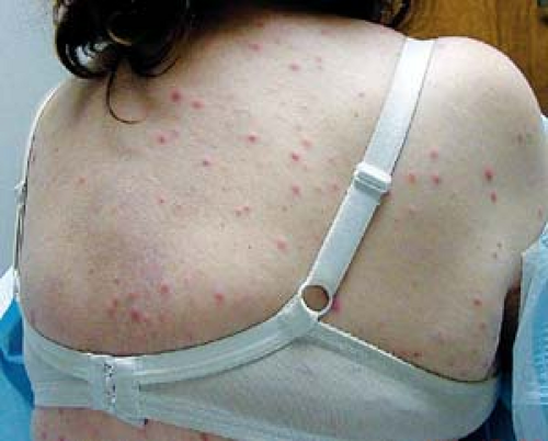
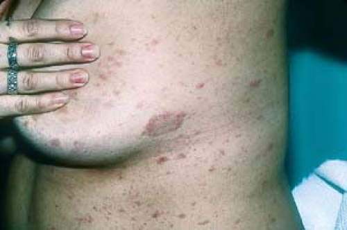 4.1 Pityriasis rosea. This patient has a herald patch on her chest. Other, smaller, elliptical lesions can be seen. |
Pathogenesis
A viral etiology has been suggested, but this has not been confirmed. Despite the prevailing opinion that PR is caused by an infectious agent, it does not appear to be contagious; household contacts and schoolmates usually do not develop the eruption.
Description of Lesions
The typical course of PR often begins with the larger herald patch (Fig. 4.1), a 2- to 5-cm, scaly lesion that may exhibit central clearing and therefore may mimic, and often be confused with, tinea corporis.
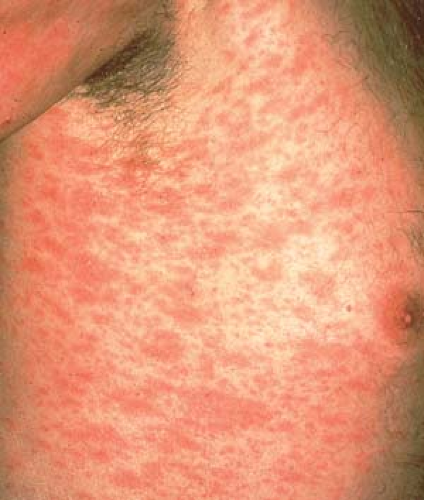
The herald patch is followed in several days to 2 weeks by multiple characteristic oval or elliptic erythematous patches with fine, thin scale on their surfaces (Fig. 4.2).
Individual lesions often develop a thin, circular, collarette of scale (see “Introduction: Reaction Patterns, Shapes, and Configurations”). Lesions in very dark-skinned patients may lack the typical pink color of PR and may appear darker than the surrounding skin.
 4.2 Pityriasis rosea. Note the characteristic elliptic (“football”) shape of lesions. |
Distribution of Lesions
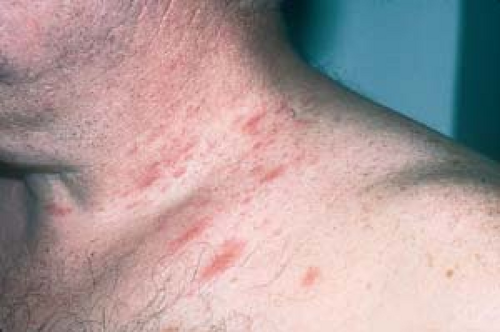
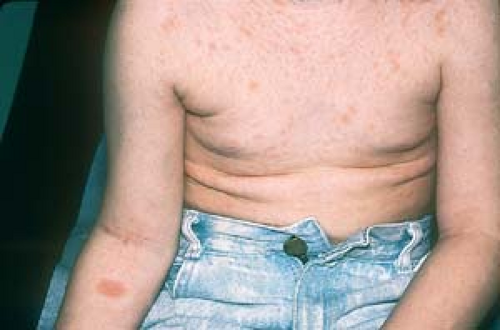
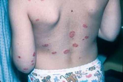
The initial lesion, the herald patch, is most commonly observed on the trunk, neck, or extremities (Fig. 4.3). The absence of the herald patch does not necessarily rule out the diagnosis; it may be absent or hidden in an obscure location such as the scalp or groin.
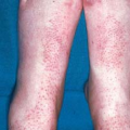
Subsequent lesions appear on the trunk, neck, arms, and legs in an “old-fashioned bathing suit” distribution.
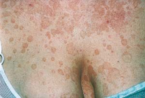
The long axis of the lesions runs parallel to skin tension lines. This gives a so-called “Christmas tree” pattern on the trunk (Fig. 4.4).
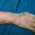
 4.3 Pityriasis rosea. The herald patch is on this child’s flexor forearm. |
Clinical Manifestations
Itching is usually absent or mild but may be severe in a minority of patients.
PR is a self-limiting, benign disorder; it usually lasts for 6 to 8 weeks.
On resolution, postinflammatory pigment changes (hyperpigmentation) can occur, particularly in dark-skinned people (Fig. 4.5).
Recurrences are rare.
 4.4 Pityriasis rosea. Here the lesions are more exuberant and the “Christmas tree” pattern is evident. |
Clinical Variants
PR can occur with less typical presentations such as those in which the herald patch is not noted by the patient or clinician. Moreover, in dark-skinned patients, the lesions may be vesicular and uncharacteristically pruritic.
The eruption may be limited in its distribution, or it may present in an inverse fashion involving the groin (Fig. 4.6), axillae, or distal extremities (inverse PR).
Infrequently, eruptions resembling those of PR also can occur in association with many drugs. These include barbiturates, bismuth, captopril, clonidine, diphtheria toxoid, gold, isotretinoin, ketotifen, levamisole, metronidazole, and d-penicillamine.
Diagnosis
The diagnosis is made clinically in most cases. In general, laboratory tests are not necessary or helpful, with the following exceptions:
A potassium hydroxide (KOH) examination may be helpful when only the herald patch is present, to help rule in or rule out tinea corporis; similarly, a KOH examination may be used to distinguish tinea versicolor (see Chapter 7, “Superficial Fungal Infections”) from PR.
A rapid plasma reagin (RPR) or venereal disease research laboratory (VDRL) test should be performed in all patients with PR.
A skin biopsy may be performed when the eruption is atypical, the diagnosis is uncertain, or the disease has not resolved after 3 to 4 months.
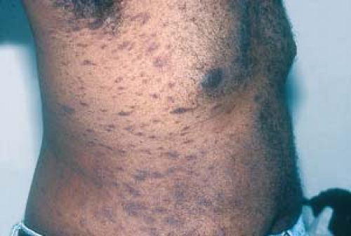 4.5 Pityriasis rosea. Note the hyperpigmented lesions in this African-American patient. |
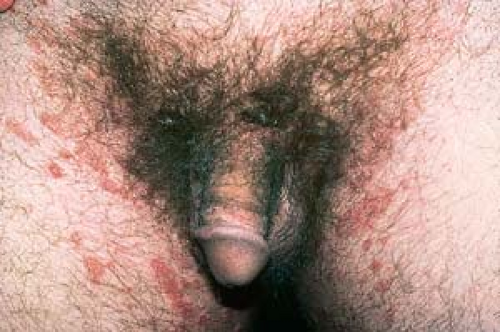 4.6 Pityriasis rosea. Atypical (inverse). The larger herald patch is present in the left inguinal area. |
Guttate Psoriasis
Guttate psoriasis often follows streptococcal pharyngitis, and its abrupt onset and appearance can easily be confused with PR.
Scale is generally thicker than that of PR.
 4.7 Guttate psoriasis. Here the plaques are thicker and more round or oval in shape than those seen in PR. |
Tinea Versicolor
Round or oval macules or patches are present.
Pigmentation may be varied (“versicolor”).
The KOH examination is positive.
Drug Eruption or Viral Exanthem
Lesions are redder, less scaly, and tend to be itchier than in PR.
There may be other symptoms (e.g., fever).
Nummular Eczema
“Coin-shaped” patches and plaques are present.
Lesions tend to be on the legs; they are less common on the arms.
This condition generally itches.
Parapsoriasis
Lesions may be similar to those of PR.
Despite its similar name, parapsoriasis is unrelated to psoriasis.
Secondary Syphilis
Lesions are often seen on the palms, soles, and face, as well as the trunk, and may be indistinguishable from PR.
The RPR or VDRL test is positive.
Parapsoriasis should be considered in PR that persists for longer than 8 weeks.
 4.8 Secondary syphilis. Papulosquamous lesions appear on the trunk, similar to PR. (Image courtesy of Ed Zabawski, D.O.). |
 4.9 Tinea versicolor. The oval, tan patches are positive on KOH examination. |
Treatment is often unnecessary, because PR is usually a self-limiting, asymptomatic condition with no sequelae.
All that is usually necessary is to advise the patient about the usual course of the rash and its noncontagious nature.
Exposure to sunlight or administration of ultraviolet B by a dermatologist may speed resolution of the eruption.
Follow-up or referral to a dermatologist should be made if the rash persists more than 8 weeks.
In cases of severe pruritus, oral antihistamines or topical steroids may help alleviate the itching. The sedative effect of the antihistamines may help the patient sleep better at night. On occasion, systemic steroids (prednisone, 0.5 to 1 mg/kg/day for 7 days) can be used in patients with severe pruritus.Related posts:

Stay updated, free articles. Join our Telegram channel

Full access? Get Clinical Tree