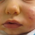
Fig. 14.1
Impetigo. Superficial erosions on the cheeks of an adolescent male
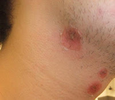
Fig. 14.2
Impetigo. Erosions with superficial desquamation on the neck
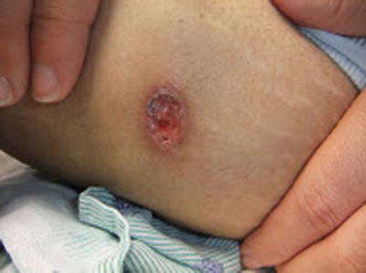
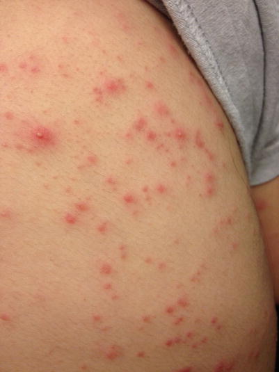
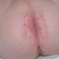
Fig. 14.3
Ecthyma. Punched-out ulcer with purulent exudate on the thigh of a teenage girl
S. aureus is the most common cause of bacterial folliculitis, which presents with superficial non-scarring pustules on an erythematous base, pierced by a central hair. In contrast, deep staphylococcal folliculitis presents with painful plaques and nodules that heal with scarring; impetigo and furuncles are often associated. A circle of surrounding desquamation is a helpful identifying feature [3]. Both methicillin-sensitive S. aureus (MSSA) and MRSA are causative. Folliculitis barbae (sycosis barbae) involves the deep portions of the hair follicles of the beard area (Fig. 14.4) [4]. Staphylococcal folliculitis often involves the scalp, face, and intertriginous areas in children [5].
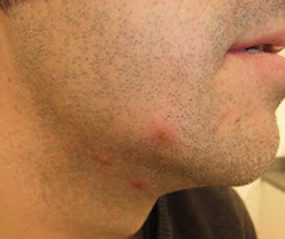
Fig. 14.4
Folliculitis barbae (sycosis barbae). Folliculocentric pustules in the beard area. Culture grew S. aureus
Abscesses are collections of pus in the skin and soft tissues that present as fluctuant or non-fluctuant nodules with surrounding erythema and edema (Fig. 14.5). Purulent drainage may be seen, and pain and tenderness are common. A furuncle is an abscess of the hair follicle that extends into the dermis and subcutaneous tissue (Fig. 14.6), while a carbuncle is a coalescence of multiple furuncles [6]. Given their close relationship to folliculitis, carbuncles and furuncles are seen on non-glabrous skin, most commonly of the axillae, groin, buttocks, and head and neck. Although abscesses can be polymicrobial, S. aureus is the most common cause, accounting for up to 75 % of cases, and both MRSA and MSSA are causative [7].
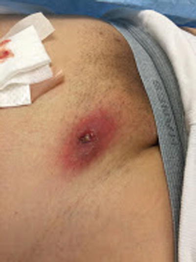
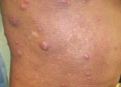
Fig. 14.5
Furuncle. Tender, fluctuant nodule with purulent drainage (Photo courtesy of Sylvia Hsu, MD)
Fig. 14.6
Furunculosis. Multiple abscesses are present in a young adult with severe atopic dermatitis
Similar to bullous impetigo, staphylococcal scalded skin syndrome (SSSS) is caused by cleavage of desmoglein 1 by exfoliative toxins. In contrast, SSSS is due to dissemination of these toxins from a distant nidus of infection, and results in sterile bullae. Flaccid blisters occur most prominently in the intertriginous areas, buttocks, hands, and feet, over a background of diffuse erythema (Fig. 14.7). Mucous membranes are spared. Systemic symptoms, including fever, are common. SSSS occurs almost exclusively in children under 6 years of age, and neonates are particularly susceptible (Fig. 14.8) [8].
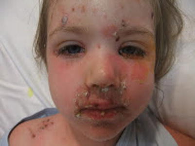
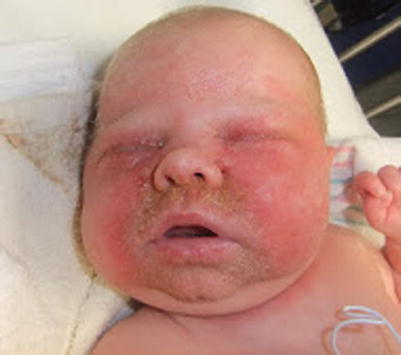
Fig. 14.7
Staphylococcal scalded skin syndrome. Superficial erosions and crusting on a background of diffuse erythema in a 4-year-old girl
Fig. 14.8
Staphylococcal scalded skin syndrome. A neonate with diffuse erythema, crusting, purulence, and characteristic “sad man” facies
In children, the most common sites for cellulitis are the extremities and the head and neck. Periorbital, orbital, and preseptal cellulitis are particularly more common in children than in adults. Erythema, edema, and warmth involving the skin and deep soft tissues is characteristic. Purulence, lymphangitis, lymphadenopathy, purpura, bullae, and necrosis are variable features. Among cases of cellulitis with positive cultures, S. aureus is the most common isolate, and MRSA and MSSA are equally causative [9].
Infections Mainly Caused by Streptococcus pyogenes
Scarlet fever is an exanthem that occurs in association with pharyngitis due to Streptococcus pyogenes, a group A beta-hemolytic Streptococcus (GABHS) which elaborates erythrogenic toxins. The median age of affected patients is 4 years old. Erythema begins in the flexural areas and spreads to the rest of the body but spares the circumoral area, palms, and soles. The erythema is composed of pinpoint papules that result in a rough “sandpaper” like texture to the skin which later desquamates. Pastia’s lines (linear petechiae of the flexures), strawberry tongue, fever, and lymphadenopathy are present in the majority of patients. Signs and symptoms consistent with streptococcal pharyngitis, including tonsillar edema and the absence of cough and coryza, are typical. Untreated, scarlet fever with pharyngitis may precede the development of acute rheumatic fever [10].
Perineal streptococcal dermatitis (PSD) presents as a well-demarcated erythema that starts at the anus and spreads centrifugally toward the genitals. Eighty percent of patients are between 2 and 7 years of age. Rectal itching, pain, and bleeding are common, but systemic symptoms are absent [11]. GABHS is the causative pathogen in the vast majority of cases, although other streptococci and S. aureus have been reported to cause a similar clinical picture.
Blistering distal dactylitis (BDD) is an infection localized to the volar fat pad of the distal phalanx that presents with a non-tender, fluid-filled, bulla. Dorsal extension may occur, with involvement of the nail folds. GABHS is the most common isolate from cultures, although staphylococci have also been reported in the literature. Children between 2 and 16 years of age are most commonly affected [12].
In contrast to cellulitis, erysipelas involves only the upper dermis and superficial lymphatics. As a result, erysipelas presents with sharply demarcated and elevated erythematous plaques. Systemic symptoms and a very acute presentation are frequent. The most common sites of involvement are the lower extremities. Although both streptococci and staphylococci can cause erysipelas, streptococci are more frequently pathogenic [13].
Toxic shock syndrome (TSS) may be caused by S. aureus or S. pyogenes, and is characterized by an acute, rapid illness with fever, hypotension, and multisystem organ involvement. Superantigens underlie the pathogenesis of TSS by binding directly to molecules of MHC class II. Since they are not processed by antigen-presenting cells, superantigens trigger massive T-cell activation and release of cytokines. The major staphylococcal superantigens are TSST-1 and enterotoxins, while the major Streptococcal superantigens are the pyrogenic exotoxins [14].
In streptococcal TSS, fever and localized pain are the most common presenting signs. Children with streptococcal TSS present with bacteremia, osteomyelitis, and cellulitis; unlike in adults, necrotizing fasciitis and myositis are uncommon. Varicella is an important risk factor for the development of invasive GAHBS infections and TSS. Staphylococcal TSS presents with abrupt fever and generalized myalgia, among other constitutional symptoms. In menstrual cases of staphylococcal TSS, tampons serve as a nidus of infection and may result in recurrent disease in patients who fail to develop neutralizing antibodies against bacterial toxins. Non-menstrual cases may begin due to localized skin and soft tissue pyodermas, including postoperative wound infections.
Mucocutaneous findings include diffuse erythroderma that develops within 24–48 h, conjunctival injection and hemorrhages, and beefy red edematous mucous membranes. Desquamation occurs within 7–14 days. Mucocutaneous findings are much more common in staphylococcal TSS than in streptococcal TSS [14].
Necrotizing fasciitis is much less common in children than adults. The average age of the pediatric patient who develops necrotizing fasciitis is 8 years old. Mortality in children overall is less than 10 %, but is much higher in neonates. Risk factors include varicella, immunosuppression, chronic illness, and recent trauma or surgery. Polymicrobial infections are less common in children than in adults, and the most frequently isolated pathogen is GABHS. Erythema, warmth, and pain out of proportion to physical findings are the early signs of necrotizing fasciitis; shock and fever develop within 1–2 days. The subtlety of skin findings may lead to misdiagnosis: superficial findings may be minimal despite rapid underlying necrosis of soft tissue, due to spread of bacteria along fascial planes. In late disease, bullae, necrosis, and crepitus are present [15, 16].
Specific Investigations
For diagnosis |
Gram stain |
Culture |
Skin biopsy |
Rapid antigen testing (S. pyogenes) |
Serology (S. pyogenes) |
Laboratory evaluation: BUN and creatinine, LFTs, CBC, metabolic panel, creatinine kinase (necrotizing fasciitis and TSS) |
Imaging: CT and MRI (necrotizing fasciitis) |
For treatment |
CBC (for linezolid) |
The diagnosis of impetigo can be confirmed by Gram stain and culture of exudate from lesions [6]. Skin biopsy is generally not necessary for diagnosis, but histopathology demonstrates subcorneal pustules with neutrophilic exocytosis overlying dermal edema in non-bullous impetigo. In contrast, bullous impetigo demonstrates fluid-filled blisters and subtle superficial acantholysis. Ecthyma is characterized by ulceration with diffuse infiltrates of neutrophils. Cocci are not always present, but can be highlighted by Gram stain if they are [17].
Differentiation of bacterial folliculitis from sterile folliculitis can be made by culture or Gram stain of pustule contents. Histopathology of acute lesions demonstrates intrafollicular abscess or suppuration, while chronic or longstanding lesions may demonstrate follicle rupture with surrounding foreign body granulomas or scar. Staphylococcal folliculitis often features subcorneal pustules with abscess of the follicular infundibulum in superficial lesions and deeper portion of the follicle and dermis in deep lesions. Gram-positive cocci may sometimes be identified in the follicular lumen [5, 6].
Purulent exudate or drainage from furuncles or carbuncles may be submitted for culture or gram stain if diagnostic confirmation is necessary [6]. Histopathology demonstrates collections of neutrophils extending through the dermis and into the subcutaneous tissue. Ruptured follicles, granulomatous foreign body reaction, and collections of gram-positive cocci may also be seen [17].
When SSSS is suspected, cultures should be obtained from all potential foci of infection, including nasopharynx, blood, and urine [6]. Skin biopsy is sometimes used to distinguish SSSS from Stevens-Johnson syndrome (SJS) or toxic epidermal necrolysis (TEN), although SJS and TEN almost always feature prominent mucosal involvement. Histopathology of frozen or paraffin-embedded specimens demonstrates a pauci-inflammatory blister within the stratum granulosum with acantholysis [18].
In cellulitis and erysipelas, blood cultures are not useful, as they are negative in 95 % of cases [19]. Cultures of needle aspirates and tissue obtained from skin biopsy are also low-yield, positive in less than 40 % of cases [20, 21]. Histopathology of cellulitis is not specific, but demonstrates prominent edema with abundant interstitial neutrophils; necrosis, abscess, and vasculitis may also be seen. Erysipelas demonstrates similar changes, but with more edema and lymphatic dilation, and these changes are more superficial [17].
The diagnosis of scarlet fever is based on the clinical appearance of the exanthem in tandem with microbiologically confirmed streptococcal pharyngitis. Throat culture is the reference standard for the diagnosis of streptococcal pharyngitis, has a sensitivity of over 90 %, and will also identify less common causes such as group C and G streptococci. Rapid antigen testing permits point-of-contact testing and early institution of therapy; sensitivity is above 70 % and specificity is over 95 % [22]. If rapid antigen testing is obtained first and is negative, throat culture should be submitted. Serologies for antistreptococcal antibodies such as antistreptolysin O, antideoxyribonuclease, and streptokinase are only useful for confirmation of infection in the convalescent period, and are not affected by antibiotic therapy [23].
Children with PSD almost always have concomitant presence of pharyngeal GABHS, although symptomatic pharyngitis is usually absent. Thus, either site may be tested, but testing of both sites is not necessary. Standard bacterial culture and rapid antigen testing are both highly sensitive methods for diagnosis [24].
Culture or gram stain of blister fluid can be used for diagnostic confirmation of BDD and discrimination from herpetic whitlow [12]. Skin biopsy reveals abscess, edema, and overlying epidermal necrosis [17].
In staphylococcal TSS, blood cultures are positive in less than 5 % of children, but cultures from the nidus of infection are usually positive. In streptococcal TSS, cultures from blood and the site of infection are positive in the majority of cases. Laboratory evaluation reveals elevated blood urea nitrogen and creatinine, abnormal liver function tests, hypocalcemia, hypoalbuminemia, anemia, thrombocytopenia, elevated creatinine kinase, and increased immature neutrophils [14]. If skin biopsy is performed, nonspecific but supportive histological features may be demonstrated, including foci of spongiosis with dyskeratotic keratinocytes and exocytosis of neutrophils [17].
Once clinically suspected, definitive diagnosis of necrotizing fasciitis is only made during surgical exploration, based on the findings of “dishwater” discharge, necrosis without bleeding, and a lack of resistance to fascial dissection. Laboratory findings are nonspecific and similar to those seen in TSS. Plain radiographs are not helpful, as they are insensitive for detecting gas in soft tissues, but computed tomography (CT) and magnetic resonance imaging (MRI) are useful in discrimination from nonnecrotizing soft tissue infections. Imaging should not delay surgical or medical therapy, which should be initiated based on clinical impression [16]. Histologic specimens demonstrate diffuse infiltrates of neutrophils with hemorrhage, thrombosis, necrosis, and vasculitis [17].
Impetigo and Ecthyma
For bullous and non-bullous impetigo with limited involvement, topical therapy is sufficient. For impetigo with extensive involvement, numerous lesions, or for ecthyma, oral antibiotics should be given [6].
In a Cochrane systematic review of 57 trials, topical antibiotics were superior to placebo for the treatment of impetigo, but no topical antibiotic was clearly superior. Topical mupirocin was more effective than oral erythromycin, but otherwise topical antibiotics and oral antibiotics did not demonstrate significantly different cure rates. Among oral antibiotics, penicillin was inferior to erythromycin and cloxacillin. A small trial showed oral cephalexin was the most effective therapy, with no associated treatment failures; treatment with oral erythromycin only resulted in one treatment failure out of 25 patients, whereas penicillin V was inferior, with a 24 % treatment failure rate. In an open trial comparing erythromycin and dicloxacillin for impetigo, over 90 % were improved or cured.
In a randomized trial comparing topical mupirocin, topical bacitracin and oral cephalexin, there was no treatment failure with mupirocin or cephalexin, and cephalexin produced the highest cure rate; bacitracin was inferior, with treatment failure occurring in six of nine patients. For impetigo due to MSSA, topical retapamulin was over 85 % effective (compared to 52 % efficacy for placebo.) For impetigo due to MRSA, topical retapamulin was only 64 % effective while linezolid demonstrated a cure rate of over 90 %.
Oral erythromycin estolate | 30–40 mg/kg/day in 3 divided doses for 10 days | A |
TMP-SMX | 8 mg/kg plus 40 mg/kg/day in 1–2 doses for 3–5 days | B |
Oral clindamycin | 20 mg/kg/day in 3 divided doses for 7 days | E |
Doxycycline | 2–4 mg/kg/day in 2 divided doses for 7 days | E |
Linezolid | Treatment for 10 days: | A |
<5 years old: 10 mg/kg q8 h | ||
5–11 years old: 10 mg/kg q12 h | ||
≥12 years old: 600 mg q12 h |
Oral co-trimoxazole (trimethoprim/sulfamethoxazole), clindamycin, doxycycline, and linezolid can be used for the treatment of impetigo due to MRSA. Doxycycline should not be used in children under the age of 8 years due to risk of tooth discoloration. For non-bullous impetigo, treatment was successful in 85 % of children who received intramuscular benzathine penicillin and in 85 % of children who received either 3- or 5-day courses of co-trimoxazole; however, 90 % of cultures were positive for S. pyogenes with our without S.aureus.
As mentioned in the discussion of first-line therapies, a large Cochrane review showed that, among oral antibiotics, penicillin was inferior to erythromycin and cloxacillin. A randomized trial showed topical bacitracin was inferior to topical mupirocin and oral cephalexin, with treatment failure occurring in six of nine patients. Thus both should be reserved as third-line therapies and used only if the first- and second-line therapies are not options.
Staphylococcal Folliculitis Including Folliculitis Barbae (Sycosis)
Topical antibiotic therapy is sufficient first-line treatment for most cases of bacterial folliculitis. In a large prospective study of patients with pyoderma including bacterial folliculitis, mupirocin ointment was effective in almost all patients, and resulted in cure in almost 75 % of patients. Topical clindamycin has a long history of successful use for bacterial folliculitis, and most isolates from bacterial folliculitis due to S. aureus are sensitive to clindamycin. Topical retapamulin has demonstrated efficacy in high-quality studies for the treatment of pyoderma, including impetigo and secondarily-infected dermatitis due to MSSA and MRSA. Thus, the use of retapamulin in bacterial folliculitis is reasonable as well.
Medication | Dosing (duration of treatment: 7–10 days) | Evidence level |
|---|---|---|
Cephalexin | 25–50 mg/kg/day po in 3–4 divided doses | A |
Dicloxacillin | 25–50 mg/kg/day po in 4 divided doses | A |
TMP-SMX | 8–12 mg/kg/day po in two divided doses based on TMP | A |
Clindamycin | 40 mg/kg/day po divided in 3–4 doses | A |
Doxycycline | 25–50 mg/kg/day po in 4 divided doses | B |
For patients with extensive skin involvement, folliculitis refractory to topicals, or folliculitis barbae (sycosis), oral antibiotics should be administered for 7–10 days. For most patients, cephalosporins or antistaphylococcal penicillins are sufficient, but for patients with risk factors or cultures positive for MRSA, clindamycin, TMP-SMX, or a tetracycline class antibiotic (in patients older than 8 years) should be used. These recommendations are based upon evidence from randomized controlled trials and prospective open studies of children and adults with skin and soft tissue infections, largely due to S. aureus.
Adjunctive treatments to eliminate carriage of S. aureus, to reduce the risk of recurrence, are reasonable for children with a history of pyoderma including bacterial folliculitis, impetigo, and furunculosis. However, a systematic review of multiple topical and oral antibiotics did not find sufficient evidence for the use of these agents to eradicate nasal or extranasal carriage of MRSA. Nonetheless, in an open study of children and adults with a history of community-acquired pyoderma and nasal or extranasal MRSA or MSSA colonization, mupirocin with or without bleach baths or chlorhexidine was effective in eradicating colonization over a 4-month period. The best regimen, mupirocin with bleach baths, was over 70 % effective. However, recurrent infections still occurred in 36 % of these patients, despite eradication of S. aureus colonization at 4 months.
Furuncles and Carbuncles
In pediatric patients presenting to the emergency department with furuncles, the use of TMP-SMX following standard incision and drainage did not improve resolution of the acute infection or reduce the risk of long-term recurrence. However, the risk of short-term recurrence (within 30 days) was lower in the group that received post-drainage antibiotics. Additionally, clinical non-response in patients with furuncles due to MSSA or MRSA was associated with lack of receipt of incision and drainage, rather than directed antimicrobial therapy. Therefore, for children with a single furuncle or carbuncle less than 2 cm in size, incision and drainage alone, without antibiotic treatment, is sufficient.
Medication | Dosing (duration of treatment: 5–7 days) | Evidence level |
|---|---|---|
TMP-SMX | 8–12 mg/kg/day po in two divided doses based on TMP | A |
Clindamycin | 40 mg/kg/day po divided in 3–4 doses | A |
Tetracycline class antibiotic: | Doxycycline: | B |
Doxycycline, minocycline | 4 mg/kg/day po divided in two doses | |
Maximum single dose: 100 mg | ||
Linezolid | 30 mg/kg/day po divided in three doses | B |
Cephalexin | 25–50 mg/kg/day po in 3–4 divided doses | A |
Dicloxacillin | 25–50 mg/kg/day po in 4 divided doses | A |
For children with multiple lesions, or a single abscess greater than 2 cm in size, cellulitis, immunosuppression, systemic signs or symptoms, indwelling device, or without clinical response to incision and drainage alone, oral antibiotics should be administered. In children and adults with abscesses greater than 2 cm in diameter, due to MRSA and MSSA, the cure rates were higher in patients who received TMP-SMX along with incision and drainage compared to surgery alone (80 % versus 74 %, respectively). In contrast, in a study of patients with abscesses due to MRSA, in 88 % of cases cephalexin offered no additional benefit to incision and drainage alone. Therefore, when empiric antibiotic therapy is given for furuncles and carbuncles, coverage for MRSA should be selected in patients with risk factors for MRSA or from areas with high prevalence of MRSA: clindamycin, tmp-smx, tetracycline class antibiotics, or linezolid are acceptable. Otherwise, coverage of MSSA is acceptable: an antistaphylococcal penicillin or cephalosporin. Duration of therapy is 5–7 days. For patients who fail to respond to incision and drainage and oral antibiotics, or with extensive involvement or systemic toxicity, parenteral therapy with one of the first-line agents, or vancomycin, should be used.
The evidence for the use of these antibiotics is supported by randomized controlled trials and prospective open studies with children and adults.
Medication | Dosing | Evidence level |
|---|---|---|
Azithromycin | 10 mg/kg po weekly for 12 weeks with maximum dose 500 mg | B* |
Oral clindamycin, topical mupirocin and chlorhexidine | Clindamycin: | B* |
20–40 mg/kg/day po in 3–4 divided doses for 21 days | ||
Mupirocin: | ||
Nasal application for 5 days | ||
Chlorhexidine: | ||
Skin disinfection daily for 21 days | ||
Rifampin | 10 mg/kg/day po for 10 days with maximum daily dose 600 mg | B* |
In an open study of patients with three or more episodes of furunculosis due to MSSA, suppressive azithromycin was 80 % effective in preventing recurrence.
Another prospective study demonstrated 87 % efficacy in preventing recurrence in patients with a history of four or more prior episodes, following combination therapy with clindamycin, mupirocin, and chlorhexidine.
In patients with recurrent furunculosis with nasal swab cultures positive for S. aureus, a course of rifampin, added to standard antibiotic therapy with TMP-SMX at the time of an episode, resulted in elimination of the carrier state and prevention of recurrence in over 70 % of cases. Clindamycin and rifampin provide coverage of MRSA.
Staphylococcal Scalded Skin Syndrome (SSSS)
In a recent American study of antibiotic susceptibilities of S. aureus cultured from children with SSSS, 86 % of isolates were sensitive to oxacillin (14 % of isolates were MRSA), and 52 % of isolates were susceptible to clindamycin. Based on this information, empiric therapy of SSSS should include a penicillinase-resistant penicillin in combination with clindamycin until culture results are available to tailor therapy. Although clindamycin inhibits toxin production by S. aureus, it should not be used as monotherapy [52].
Medication | Dosing | Evidence level |
|---|---|---|
Penicillinase-resistant penicillin: | Nafcillin: | D |
Nafcillin, oxacillin, dicloxacillin | Neonates: 25 mg/kg/dose IV every 6–12 h | |
Infants and children: 100 to −200 mg/kg/day IV in divided doses every 6 h with | ||
Maximum daily dose 12 g | ||
Clindamycin | Neonates: | D |
5 mg/kg/dose IV every 6–12 h | ||
Infants and children: | ||
20–40 mg/kg/day IV divided every 6–8 h | ||
Maximum single dose 600 mg | ||
Cephalosporins: | Ceftriaxone: | D |
Ceftriaxone, cefpodoxime, cefdinir | 50 mg/kg IV once daily | |
Maximum daily dose 1 g |
Based on a retrospective review of 39 neonates with SSSS, there were no significant differences in response to therapy between the beta-lactamase-resistant semisynthetic penicillins, cephalosporins, or combination therapy with both. Thus, these are first-line agents.
Medication | Dosing | Evidence level |
|---|---|---|
Vancomycin | 40–60 mg/kg/day IV divided every 6–8 h | E |
Maximum daily dose: 2–4 g | ||
Macrolides: | Erythromycin: | E |
Erythromycin, clarithromycin | Neonates: | |
10 mg/kg/dose IV every 8–12 h | ||
Infants and children: | ||
15–20 mg/kg/day IV divided every 6 h | ||
Maximum daily dose: 4 g |
Vancomycin should be reserved for patients who fail to respond to standard, first-line therapy, or when susceptibility testing identifies MRSA. Macrolides including erythromycin can also be used effectively for the treatment of SSSS.
Retrospective data suggest that systemically unwell children may benefit from the use of FFP or IVIG to neutralize exotoxin antibodies. However, patients treated with IVIG for SSSS have longer hospital stays than patients receiving standard therapy.
Scarlet Fever
Scarlet fever, like acute rheumatic fever, toxic shock syndrome, acute glomerulonephritis, and pediatric autoimmune neuropsychiatric disorder associated with GAS (PANDAS), is a nonsuppurative complication of GAS tonsillopharyngitis. The treatment of scarlet fever is the same as that for streptococcal pharyngitis. Antibiotic treatment is most helpful in altering the disease course if started within the first 48 h of illness. Antibiotic treatment also reduces the risk of acute rheumatic fever [58].
Medication | Dosing | Evidence level |
|---|---|---|
Penicillin | Penicillin V: | A |
250–500 mg po 2–3 times daily for 10 days | A | |
Penicillin G benzathine 900,000 U mixed with penicillin G procaine 300,000 U IM single dose (patients <27 kg) | A | |
Penicillin G benzathine | ||
1.2 million U (patients>27 kg) | ||
Amoxicillin | 50 mg/kg/day po in 2–3 divided doses for 10 days | A |
Maximum daily dose: 1 g | ||
Cephalexin | 25–50 mg/kg/day po in 2 divided doses for 10 days | A |
Maximum daily dose: 1 g |
Penicillin is the preferred first-line treatment for streptococcal pharyngitis. Intramuscular penicillin is the only drug with data demonstrating prevention of rheumatic fever, but a 10-day course of oral penicillin is considered equivalent for this purpose. Meta-analysis has demonstrated that the risk of bacteriologic and clinical failure is reduced by treatment with cephalosporins such as cephalexin compared to penicillin. Thus, cephalexin and other first-generation cephalosporins are first-line treatments. However, second- and third-line cephalosporins are associated with emerging resistance.
Medication | Dosing | Evidence level |
|---|---|---|
Azithromycin | 12/mg/kg/dose po on day 1 followed by 6 mg/kg/dose po on days 2–5 | A |
Maximum single dose: 250/500 mg | ||
Clarithromycin | 7.5 mg/kg/dose po twice daily for 10 days | A |
Maximum dose 250 mg | ||
Clindamycin | 7 mg/kg/dose po 3 times daily for 10 days | A |
Maximum dose 300 mg |
Based on a multiple randomized controlled trials, there are no significant differences in the efficacy of macrolides compared to penicillin in children with GAS pharyngitis. However, resistance rates can be as high as 20 %, and the incidence of adverse events in children is higher with macrolides than with penicillin. In patients allergic to penicillin and cephalexin, another acceptable and effective alternative agent is clindamycin.
Perineal Streptococcal Dermatitis
In a large retrospective review of children up to 12.5 years of age, treatment with amoxicillin was associated with a recurrence rate of 12.4 %. The recurrence rate of PSD following treatment with penicillin or amoxicillin was 38 %, while the recurrence rate following treatment with a beta-lactamase resistant antibiotic was lower at 28 %. In 13 of 14 patients treated with cefuroxime, clinical improvement was rapid and GABHS was not isolated from the perianal skin. In comparison, GABHS was found in 8 of 15 patients following treatment with penicillin.
Penicillin V | 50,000–100,000 U/kg/day, divided in 3 doses, for 10 days | B |
Blistering Distal Dactylitis
Cephalexin | 25–50 mg/kg/day po in divided doses for 14 days | E |
Oral penicillin V | 25–75 mg/kg/day po in 3–4 divided doses for 10 days with maximum daily dose 2,000 mg | E |
Penicillin G benzathine | <27 kg: 600,000 units IM × 1 | E |
≥27 kg: 1.2 million units IM × 1 |
While the typical age in children is between 2 and 16 years old, cephalexin was successful in patients down to infancy.
Dicloxacillin | 50–75 mg/kg/day po in 4 divided doses for 10 days | E |
Erythromycin | 30–50 mg/kg/day po divided q 6–12 h (max 4 g/day) | E |
In reports, dicloxacillin was used successfully for staphylococcal disease.
Erysipelas
Medication | Dosing (duration of treatment: 5–10 days) | Evidence level |
|---|---|---|
Penicillin | 25–50 mg/kg/day po in 3–4 divided doses | A |
Amoxicillin | 25–50 mg/kg/day po in 3 divided doses | A |
For the treatment of streptococcal and staphylococcal skin and soft tissue infections, ampicillin and ceftriaxone were both over 90 % effective in terms of clinical and bacteriologic response rates.
In children with infections due mainly to S. aureus and S. pyogenes, outpatient treatment with once-daily ceftriaxone is highly effective and associated with equivalent outcomes of intravenous administration during hospitalization. If the diagnosis of nonpurulent cellulitis, rather than erysipelas, is still under consideration, then cefazolin is preferred over ceftriaxone for coverage of MSSA.
Early studies demonstrated roughly equivalent efficacy for macrolides compared to cephalosporins and antistaphylococcal penicillins in the treatment of uncomplicated skin and soft tissue infections. However, given the emerging resistance of erythromycin-resistant group A Streptococcus in children, this agent is not a preferred therapy for erysipelas.
Cellulitis
For children with non-purulent cellulitis, empiric treatment should include coverage for beta-hemolytic streptococci and MSSA. Children with purulent cellulitis, including primary or secondary non-drainable abscess, should receive empiric coverage for MRSA. These recommendations apply to non-facial cellulitis in infants, children, and adolescents [6, 37].
Medication | Dosing (duration of treatment: 5 days) | Evidence level |
|---|---|---|
Cephalosporins: | Cephalexin: | A |
Cephalexin, cefadroxil, cefuroxime, cefaclor | 25–50 mg/kg/day po in 3–4 divided doses | |
Clindamycin | 20–30 mg/kg/day po in 4 divided doses | A |
Dicloxacillin | 25–50 mg/kg/day po in 4 divided doses | A |
TMP-SMX | 8–12 mg/kg/day po in two divided doses based on TMP component | A |
Tetracycline class: | Doxycycline: | B |
Doxycycline, minocycline | 4 mg/kg/day po divided in two doses | |
Maximum single dose 100 mg | ||
Amoxicillin-clavulanate | 25 mg/kg/day of the amoxicillin component in 2 divided doses po | A |
For nonpurulent cellulitis, empiric therapy can be selected from one of the following: a cephalosporin, clindamycin, or an antistaphylococcal penicillin such as dicloxacillin. Symptomatic improvement should occur within 48 h and cutaneous signs should improve within 72 h. If not, MRSA should be suspected and clindamycin, or amoxicillin with TMP-SMX, or amoxicillin with doxycycline or minocycline should be given. These regimens can also be initiated in patients with risk factors for MRSA infections.
In a randomized controlled trial, TMP-SMX in combination with cephalexin was not superior to cephalexin alone for nonpurulent cellulitis in children and adults, with cure rates above 80 % for both regimens. A variety of cephalosporins other than cephalexin have been tested in high-quality studies and demonstrated similar efficacy for uncomplicated cellulitis in children and adults. Amoxicillin-clavulanate was as effective as cefaclor in an early study, but its current use is preferred for coverage of S. pyogenes rather than S. aureus. Thus when used for cellulitis, an antistaphylococcal agent should be added.
For patients with purulent cellulitis, empiric therapy with clindamycin, TMP-SMX, or—in children older than 8 years of age—minocycline or doxycycline. Once the culture results of the abscess, culture, or exudate are available, therapy should be tailored. In adults and children with purulent and non-purulent cellulitis, cure rates were similar for clindamycin in comparison to TMP-SMX. Cases of purulent cellulitis included MRSA infections. In a separate open study of adults with purulent MRSA skin and soft tissue infections, the majority of isolates were sensitive to tetracycline class antibiotics, clindamycin, and TMP-SMX.
For patients who respond to empiric therapy, a total of 5 days of antibiotic treatment is sufficient. However, for patients with slow response, therapy may be extended to 14 days. For patients with systemic toxicity, progression of signs or symptoms, or immunosuppression, parenteral therapy with the first-line antibiotics, their equivalents, or vancomycin, should be used in place of oral treatment.
In retrospective and open prospective studies, linezolid was as effective as vancomycin for the treatment of complicated cellulitis, or cellulitis in patients requiring hospitalization. This drug provides coverage of MSSA, MRSA, and beta-hemolytic streptococci, but is reserved as an alternative agent for patients intolerant of or refractory to first-line therapies.
Streptococcal Toxic Shock Syndrome (TSS)
Medication | Dosing (duration of treatment: at least 14 days) | Evidence level |
|---|---|---|
Treatment should consist of a beta-lactam agent AND clindamycin | ||
Clindamycin | 30–40 mg/kg/day IV divided every 6–8 h | D |
Carbapenems: | Meropenem: | E |
Meropenem, imipenem | 10–20 mg/kg/dose IV every 8 h | |
Beta-lactam with beta-lactamase inhibitor: | Piperacillin-tazobactam: | D |
100 mg piperacillin/kg/dose IV every 8 h with maximum daily dose 16 g | ||
Piperacillin-tazobactam | ||
Ticarcillin-clavulanate | ||
Penicillin G | 200,000–300,000 units/kg/day IV divided every 4 h with maximum daily dose 24 M units | D |
First-line therapy for streptococcal TSS is a beta-lactam agent and clindamycin. Clindamycin can be combined with either a carbapenem or a penicillin with beta-lactamase inhibitor. Although S.pyogenes is highly susceptible to penicillin, monotherapy is associated with high mortality rates. In a retrospective studies, use of clindamycin, including as part of combination therapy with a beta-lactam, was associated with improved outcomes and decreased mortality in children with severe invasive GAS infections.
Once the diagnosis of streptococcal TSS has been established, treatment should be switched to a combination of clindamycin and penicillin G to complete at least 14 days of therapy. GAS isolates with inducible resistance to macrolides or lincosamides are uncommon in the United States, but penicillin is used as part of dual therapy for coverage of potential clindamycin resistance.
Medication (in combination with Clindamycin) | Dosing | Evidence level |
|---|---|---|
Vancomycin | 45–60 mg/kg/day IV divided every 6–8 h with maximum daily dose 4,000 mg | E* |
Daptomycin | 4–10 mg/kg/day IV | E* |
Clindamycin may be combined with one of the alternative agents for therapy, vancomycin and daptomycin. These drugs are useful in patients with allergy to penicillins and/or carbapenems.
IVIG has been used based on the idea that passively transferred antibodies can neutralize streptococcal toxins. However, retrospective data show that children with streptococcal TSS do not have improved outcomes following adjunctive treatment with IVIG. Hyperbaric oxygen has also been used in small numbers of patients with streptococcal TSS.
Staphylococcal Toxic Shock Syndrome (TSS)
Based on retrospective data, all patients with suspected staphylococcal TSS should receive empiric therapy with clindamycin and vancomycin. Once culture and sensitivity results are available, treatment can be narrowed and tailored. For patients with staphylococcal TSS due to MSSA, clindamycin should be combined with an antistaphylococcal penicillin. For patients with staphylococcal TSS due to MRSA, clindamycin should be combined with vancomycin or linezolid. Treatment should be continued for a total of 7–14 days of antimicrobial therapy.
The use of clindamycin is important given that vancomycin and antistaphylococcal penicillins do not suppress toxin synthesis by S. aureus.
As stated above, the use of clindamycin is important, given that vancomycin and antistaphylococcal penicillins do not suppress toxin synthesis by S. aureus. Anecdotal evidence supports the use of linezolid, based on successful clinical response as well as suppression of toxin synthesis.
Unlike in streptococcal TSS, little evidence currently supports the use of IVIG in staphylococcal TSS. Superantigens produced by S. aureus may be less susceptible to inhibition by IVIG than those produced by S. pyogenes.
Necrotizing Fasciitis
As in adults, early (within the first 24 h of diagnosis) surgical debridement is critical in the treatment of necrotizing fasciitis and improving survival. In addition, empiric antibiotic therapy should consist of three antimicrobials: clindamycin, a carbapenem or beta-lactam with beta-lactamase inhibitor, and an antibiotic with activity against MRSA [6]. Treatment should be tailored once culture and susceptibility results are available, if possible. In the setting of GAS infection, treatment can be narrowed to the combination of penicillin and clindamycin [97].
For treatment of necrotizing fasciitis, earlier studies reported the need for aggressive surgical debridement, such as fascial excision. However, more recent studies have advocated conservative surgical management, which is described as separation of gangrenous skin from surrounding healthy tissue, wound washing, frequent application of dressings with antibiotic ointment, and healing by granulation. Compared to aggressive management, conservative debridement is associated with shorter hospital stays, improved cosmesis and function with minimal scarring or restricted movement, and shorter duration to healing.
Medication | Dosing | Evidence level |
|---|---|---|
Carbapenem: | Meropenem: | D |
Meropenem, imipenem, or ertapenem | 20 mg/kg/dose IV every 8 h | |
Beta-lactam with beta-lactamase inhibitor: | Piperacillin-tazobactam: | D |
100 mg piperacillin/kg/dose IV every 8 h with maximum daily dose 16 g | ||
Piperacillin-tazobactam, | ||
Ampicillin-sulbactam | ||
Clindamycin | 40 mg/kg/day IV divided every 8 h | D |
Active agent against MRSA: | Vancomycin: | D |
Vancomycin, daptomycin, or linezolid | 15 mg/kg/dose IV every 6–8 h | |
Penicillin G | 300,000 units/kg/day IV divided every 6 h | D |
Prompt antibiotic treatment, along with aggressive supportive treatment (fluid resuscitation and electrolyte replacement) should also be initiated at the time of presumptive diagnosis. Empiric therapy should be broad and provide coverage of gram-positive (especially GAS), gram-negative, and anaerobic organisms.
Based on retrospective data, children with invasive soft tissue infection due to S. pyogenes were more likely to have a favorable outcome if clindamycin was used in combination with a beta-lactam antibiotic; the use of a beta-lactam alone was associated with a 68 % failure rate. Clindamycin should be included in all treatment regimens.
Medication | Dosing | Evidence level |
|---|---|---|
Intravenous immune globulin (IVIG) | 1 g/kg IV on day 1; 0.5 g/kg IV on days 2 and 3 | A* |
Hyperbaric oxygen | 2–3 daily 90-min sessions at 3 atm | D* |
Postexposure prophylaxis: | 25–75 mg/kg/day po in divided doses every 6–8 h | E |
Penicillin VK |
In a randomized, controlled trial of adults with streptococcal toxic shock syndrome with or without necrotizing fasciitis, a greater than threefold reduction in mortality was associated with IVIG treatment. Hyperbaric oxygen may be useful as an adjunctive treatment for necrotizing fasciitis. Several retrospective studies have demonstrated a survival benefit with decreased need for aggressive debridements.
The incidence of invasive GAS infections is dramatically increased in household contacts of patients with severe infections such as streptococcal toxic shock or necrotizing fasciitis. Routine screening and chemoprevention for contacts of index patients are not recommended. However, for immunocompromised patients who are household contacts of patients with necrotizing fasciitis due to GAS, prophylaxis should be considered.
Pseudomonas Infections
Clinical Features
Pseudomonas aeruginosa is a gram-negative aerobic bacillus that can cause mild infections in immunocompetent hosts or severe hospital-acquired infections, especially in immunocompromised hosts.
Folliculitis and hot foot syndrome are benign, self-limited infections that occur following exposure to contaminated water. Pseudomonas folliculitis is characterized by tender or pruritic follicular papules, nodules, or pustules, often associated with low-grade fever and malaise (Figs. 14.9 and 14.10). Hot foot syndrome is common in children, and results in tender nodules on the soles. Both folliculitis and hot foot syndrome resolve without antibiotic therapy [109, 110].
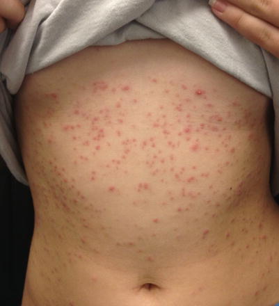
Fig. 14.9
Hot tub folliculitis. Papulopustules on the trunk in a child following exposure to pool water. Culture grew P. aeruginosa (Photo courtesy of Adam Rees, MD)
Ecthyma gangrenosum occurs in the setting of P. aeruginosa bacteremia and sepsis in immunocompromised patients, including those with neutropenia. It results from bacterial invasion of the media and adventitia of vessels with secondary ischemic necrosis. Single or multiple lesions begin as painless red macules, which then evolve into pustules, vesicles, and eventually ulcers with gangrene. Rapid progression is typical [111].
Noma neonatorum is a very rare gangrenous form of noma involving the mucocutaneous junctions of the oral, nasal, and anal area. It presents during the first few weeks of life in premature and low-birthweight neonates, and causes mutilating and disfiguring destruction of soft tissues, skin, and bone. In low-birthweight neonates, it is usually quickly fatal. P. aeruginosa is causative of the skin lesions and also results in septicemia [112].
Specific Investigations
For diagnosis |
Culture |
Histopathology with gram stain |
For treatment |
Renal function should be monitored during therapy with aminoglycosides and colistimethate |
Diagnosis of pseudomonas folliculitis or hot foot syndrome is clinical, but histopathology with gram stain or culture can be used to confirm cases, if necessary [110].
If ecthyma gangrenosum is suspected clinically, blood cultures should be obtained and prompt antibiotic therapy initiated. As bacteremia is not present in all patients, negative blood cultures do not exclude this diagnosis. Exudates from skin lesions should also be obtained for culture. Importantly, given the nonspecific clinical findings and the variety of organisms that may cause gangrenous ulcers in immunocompromised patients, skin biopsy for histopathology and bacterial, fungal, and mycobacterial tissue culture should be obtained. Biopsy of skin lesions shows hemorrhagic necrosis, gram-negative bacilli, and vasculitis [113].
In noma neonatorum, blood cultures, and cultures with susceptibility, testing from skin lesions should be obtained. Histopathology and tissue culture should also be considered [114].
Ecthyma Gangrenosum
Medication | Dosing | Evidence level |
|---|---|---|
Penicillins: | Piperacillin-tazobactam: | D |
Piperacillin-tazobactam | 100 mg piperacillin/kg/dose IV every 8 h with maximum daily dose 16 g | |
Ticarcillin | ||
Cephalosporins | Ceftazidime: | D |
Ceftazidime | 30–50 mg/kg/dose IV every 8 h with maximum daily dose 6 g | |
Cefipime | ||
Monobactams | Aztreonam: | D |
30 mg/kg/dose IV every 8 h with maximum daily dose 8 g | ||
Carbapenems: | Meropenem: | D |
Imipenem | 30–60 mg/kg/day IV in 3 divided doses | |
Maximum daily dose: 3 g | ||
Meropenem | ||
Aminoglycosides: | Amikacin: | D |
Gentamycin | 15–22.5 mg/kg/day IV divided every 8 h | |
Amikacin | ||
Fluoroquinolones: | Ciprofloxacin: | D |
Ciprofloxacin | 20–30 mg/kg/day IV divided every 12 h with maximum daily dose 800 mg | |
Levofloxacin |
When the clinical diagnosis of ecthyma gangrenosum is made, empiric combination antimicrobial treatment should be initiated and should include antipseudomonal agents. Retrospective and prospective data have suggested that multiagent empiric therapy, compared to empiric monotherapy, is associated with lower rate of mortality in patients with Pseudomonas bacteremia.
However, once results of culture identify Pseudomonas, antipseudomonal monotherapy should be selected based on susceptibility testing. Most studies examining directed combination therapy compared to monotherapy have not demonstrated survival benefit. Nonetheless, monotherapy with an aminoglycoside agent is not recommended, as retrospective data has revealed higher rates of treatment failure and mortality in P. aeruginosa bacteremia. Of note, a retrospective study found that combination therapy was linked to superior outcomes in children with Pseudomonas septicemia.
Only a few retrospective studies are available to support specific therapies for Pseudomonas in children, but the same antibiotics effective in adults are recommended in children. However, the use of fluoroquinolones should be carefully considered before electing this therapy in patients under 18 years of age, given the risk of adverse events involving joints and surrounding tissues. There is no current evidence that limited courses of treatment with fluoroquinolones cause sustained injury of this type in children, and these agents may be highly useful in children with serious infections due to susceptible isolates.
Retrospective studies support the use of colistin in severe, refractory cases due to multidrug-resistant P. aeruginosa, but treatment with this agent is limited by nephrotoxicity and ototoxicity. Alternatively, combination regimens, such as cefepime with amikacin, may be used.
Medication | Dosing | Evidence level |
|---|---|---|
Surgery | D |
In pediatric oncology patients, most with neutropenia, radical debridement was important in the adjunctive treatment of progressive lesions to prevent mortality. However, this is not warranted for infections with limited involvement.
Noma Neonatorum
Medication | Dosing | Evidence level |
|---|---|---|
Colistimethate | 2.5–5 mg/kg/day IV in 1–2 doses | E |
Given the rarity of noma neonatorum, treatment is only based on anecdotal evidence. In a patient with multidrug-resistant P. aeruginosa, colistin (colistimethate) was used successfully with complete healing. Therapy should be tailored based on in vitro susceptibility results, once available.
Medication | Dosing | Evidence level |
|---|---|---|
Amikacin | 15–22.5 mg/kg/day IV divided every 8 h | E |
Clindamycin | 20–40 mg/kg/day IV divided every 6–8 h | E |
Maximum single dose 600 mg | ||
Fluoroquinolones: | Ciprofloxacin: | E |
Ofloxacin, ciprofloxacin | 20–30 mg/kg/day IV divided every 12 h with maximum daily dose 800 mg |
Noma
Clinical Features
Noma is a polymicrobial infection associated mainly with anaerobic bacteria. Causative organisms include Fusobacterium species, Prevotella intermedia, alpha-hemolytic streptococci, Pseudomonas, Actinomyces, Peptostreptococci, and spirochetes. The incidence of noma is highest in sub-Saharan Africa and among infants and young children. Risk factors include poverty, malnutrition, poor sanitation and oral hygiene, proximity to livestock, nomadic lifestyle, and high prevalence of other infectious diseases such as malaria and tuberculosis [128]. Noma begins as an ulcerative gingivitis and then spreads rapidly causing destructive gangrene of the skin, muscle, and bone of the mouth and face. In the acute phase, fetid breath, facial swelling, and mouth soreness are characteristic. Bluish-black discolored patches follow gingivitis, with rapid progression to perforation and then gangrene. Mortality rates are high without prompt treatment [129].
Specific Investigations
For diagnosis |
Culture |
Histopathology |
For treatment |
No specific investigations |
Histopathology demonstrates ulceration with necrosis and surrounding epidermal hyperplasia. Numerous neutrophils may be present. Gram-positive cocci of various morphologies and gram-negative cocci bacilli may be seen. Cultures are polymicrobial, but Fusobacterium necrophorum has been observed most frequently [129].
Medication | Dosing | Evidence level |
|---|---|---|
Penicillin | Penicillin VK: | D |
25–50 mg/kg/day in 3 divided doses | ||
Penicillin G procaine: | ||
25,000–50,000 units/kg/day in divided doses 1–2 times/day | ||
Metronidazole | 30 mg/kg/day in 3–4 divided doses | D |
Amoxicillin | 80–100 mg/kg/day po divided every 8 h with maximum single dose 500 mg | D |
Streptomycin | 20–30 mg/kg/day IV divided every 12 h with maximum daily dose 2 g | D |
High-quality evidence regarding the optimal treatment of noma is lacking, and treatment recommendations are based on typical in vitro susceptibilities of causative organisms, as well as retrospective studies. Penicillin, either oral or parenteral, has been used successfully in combination with metronidazole or streptomycin. Amoxicillin may be substituted for penicillin in combination therapy with metronidazole. Metronidazole can also be used as monotherapy.
Surgery is often required as an adjunctive treatment to antibiotics in noma. During the acute infection, surgical debridement is employed to limit spread of the infection and to promote healing. Following resolution of the acute infection and necrosis, reconstructive surgery improves function and cosmesis.
Wound care is also important during the acute phase of infection, to prevent further spread and secondary infection. Antiseptic dressings and chlorhexidine oral rinses are recommended. Nutritional supplementation has improved outcomes during acute infection. Protein-enriched formulas, via oral or parenteral feeding, in addition to fluid and electrolyte replacement, are administered.
Neisseria Infections
Clinical Features
Gonococcemia, or disseminated gonococcal infection, is caused by bacteremic spread of the of the sexually-transmitted, gram-negative facultatively intracellular diplococcus Neisseria gonorrhoeae, resulting in a triad of tenosynovitis, cutaneous lesions, and polyarthralgias. Painless lesions which are few in number present as hemorrhagic macules, papules, vesicles, or pustules, and are transient (Figs. 14.11 and 14.12) . Fever, malaise, and multiple inflamed tendons of the wrist, fingers, ankle, and toes are common features. Purulent asymmetric oligoarthritis of the knees, wrists, and ankles without skin lesions may also occur [138].
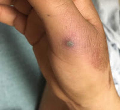
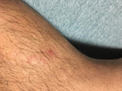
Fig. 14.11
Gonococcemia (disseminated gonococcal infection). Hemorrhagic vesicopustule on an acral surface in an adolescent patient (Photo courtesy of Yousuf Qureshi, MD)
Fig. 14.12
Gonococcemia (disseminated gonococcal infection). Cutaneous lesions are often sparse and subtle (Photo courtesy of Yousuf Qureshi, MD)
Neisseria meningitidis is the leading cause of bacterial meningitis in children and young adults in the United States, with a mortality rate of 13 %. Meningitis may occur with or without meningococcemia, and vice versa [139]. The typical initial presentation of meningitis due to N. meningitidis consists of the sudden onset of fever, nausea, vomiting, headache, and myalgias in an otherwise healthy patient. Rash and meningismus are also often present. However, clinical expression is highly variable and often incomplete. The first clinical symptoms are worrisome signs of early sepsis: leg pain, cold hands and feet, and pallor or mottling; 72 % of children demonstrate one of these features in the first 8 h after onset. Rash is present in up to 70 % of cases, is usually petechial, and often involves the trunk, lower extremities, and mucous membranes. Ecchymoses, painful purpura, and geographic areas of hemorrhagic necrosis may develop: these findings are indicative of purpura fulminans and disseminated intravascular coagulation (DIC) [140].
Chronic meningococcemia and chronic gonococcemia produce indistinguishable clinical findings, and cannot be differentiated except by the isolation of the causative organism [141].
Specific Investigations
For diagnosis |
Gram stain |
Culture (Thayer-Martin media) |
Nucleic acid amplification testing (NAAT) |
Histopathology |
For treatment |
If chloramphenicol is used, CBC should be monitored, given the risk of bone marrow suppression |
N. gonorrhoeae are recovered from less than 50 % of purulent synovial effusions due to disseminated gonococcal infections (DGI), positive blood cultures occur in less than one-third of patients, and skin lesions are always sterile. At least two sets of blood cultures should be obtained in suspected cases. In addition, synovial, skin, urethral or cervical, rectal, and pharyngeal specimens should be submitted for nucleic acid amplification testing (NAAT), the preferred method of diagnosis. If NAAT is not available, then cultures from these sites should be submitted. Cultures should be performed on Thayer-Martin media. If urethritis is present, gram stain of the urethral exudate should be examined for the gram negative intracellular diplococci, and diagnosis can be made based on this finding despite negative cultures. If skin biopsy is performed, vasculitis with thrombosis and pustules may be seen, but bacteria are rarely identified. All patients should undergo testing for other sexually transmitted infections, such as HIV, Chlamydia trachomatis, and syphilis serology, since coexistent infection with other sexually transmitted pathogens is common [142].
In meningococcemia, the frequency of positive blood cultures is 50–60 %, while the frequency of positive cerebrospinal fluid (CSF) cultures is 80–90 %, even in patients without meningeal signs [143]. Gram stain of CSF is valuable and may provide rapid confirmation of diagnosis. Latex agglutination, loop-mediated isothermal amplification (LAMP), rapid polysaccharide dipstick test, and PCR can also be performed on CSF [144–147]. Skin biopsy, when combined with culture and gram stain, demonstrates a sensitivity of 56 % in isolating N. gonorrhoeae. Routine histopathology demonstrates vasculitis, thrombotic vasculopathy, and necrosis [148]. Although the gold standard for diagnosis remains culture, antibiotic therapy should not be delayed by any diagnostic measure when meningococcemia is suspected.
Disseminated Gonococcal Infection
Medication | Dosing | Evidence level |
|---|---|---|
Ceftriaxone | 50 mg/kg IM or IV daily | B |
Maximum daily dose 1 g | ||
Duration of therapy: 7–14 days | ||
Cefotaxime | 50–180 mg/kg IV every 8 h | B |
Maximum single dose 1 g | ||
Duration of therapy: 7–14 days | ||
Azithromycin | 20 mg/kg po as a single dose | D |
Maximum dose: 1 g | ||
Doxycycline | 2–4 mg/kg/day divided every 12 h | D |
Duration of therapy: 7 days | ||
Maximum single dose: 100 mg |
Ceftriaxone or cefotaxime, intravenous or intramuscular, are the preferred first-line initial treatments for disseminated gonococcal infection. Oral azithromycin or doxycycline (in patients older than 8 years) should also be administered alongside the parenteral cephalosporin for dual coverage and to treat potential C. trachomatis coinfection. Dual coverage is recommended because of the increasing resistance of N. gonorrhoeae to cephalosporins.
Antibiotic therapy should be administered for 7 days, provided that clinical signs and symptoms of infection have resolved. In patients with purulent arthritis, parenteral therapy for up to 14 days in combination with joint drainage is recommended.
Medication | Dosing | Evidence level |
|---|---|---|
Cefixime | 8 mg/kg/day divided every 12 h | D |
Maximum single dose 400 mg | ||
Amoxicillin | 25–50 mg/kg/day in divided doses every 8 h | D |
Maximum single dose 500 mg | ||
Doxycycline | 2–4 mg/kg/day divided every 12 h | D |
Duration of therapy: 7 days | ||
Maximum single dose: 100 mg |
In patients who lack septic arthritis, respond promptly to first-line parenteral therapy, and for whom susceptibility testing demonstrates sensitivity to a second-line agent, the 7-day course of antibiotic therapy can be completed with cefixime, amoxicillin, or doxycycline (in patients over 8 years of age). Ciprofloxacin is also considered an appropriate agent for step-down therapy, but the use of fluoroquinolones is generally not recommended in patients under 18 years of age. If susceptibility testing is not available, step-down therapy with oral antibiotics is not recommended, and the course of therapy should be completed with parenteral ceftriaxone or cefotaxime.
Infected partners of patients should be treated with single-dose intramuscular ceftriaxone, single-dose oral azithromycin, or a 7-day course of oral doxycycline.
Meningococcemia
Treatment should be initiated within 30 min of presumptive diagnosis, and should be started immediately after blood cultures are obtained; treatment should not await lumbar puncture. Data from a prospective open trial studying children and adults (more than 80 % of patients were under 18 years of age) with epidemic disease, and retrospective data from pediatric studies, support the use of monotherapy with ceftriaxone. In the prospective study, the treatment failure rate was 9 % and the mortality rate was 6 %. Cefotaxime is equivalent to ceftriaxone, and these are the treatments of choice prior to available susceptibility testing. Seven days of parenteral therapy is effective and adequate.
Penicillin may be used for effective therapy if susceptibility testing indicates that the causative isolate of N. meningitidis is sensitive. However, penicillin resistance has been present for decades, and since therapy must be initiated before susceptibility testing, this is not a first-line treatment. For patients who are allergic to cephalosporins or penicillin, chloramphenicol has been shown to be as effective as ceftriaxone, with the same treatment failure and mortality rate for epidemic disease. However, given the more favorable side effect profile of beta-lactam antibiotics, as well as the possibility for high-level resistance in causative isolates, chloramphenicol is a second-line therapy.
Protein C activation is impaired in meningococcal infection and patients with purpura fulminans in particular have reduced levels of activated protein C (APC). A randomized controlled trial demonstrated that treatment with protein C concentrate led to dose-related increases in plasma APC levels and resolution of indicators of coagulation imbalances such as d-dimer levels in children with purpura fulminans due to meningococcemia. Mortality and amputation rates were not affected by treatment. However, an open study in a similar group of patients demonstrated improved survival and a decreased number of amputations.
Corynebacterial Infections
Clinical Features
Erythrasma is a chronic superficial infection caused by Corynebacterium minutissimum, a gram-positive, non-spore forming bacillus. It is rare in children. In adolescents and young adults, it is more common and involves the interdigital toe webs, axillae, and groin. Thin brown patches or plaques with or without maceration and scale are typical (Figs. 14.13 and 14.14). Occasional genital or perianal involvement may occur. Infection is usually asymptomatic but may be pruritic [162].
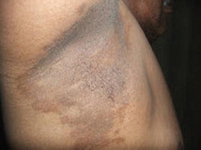
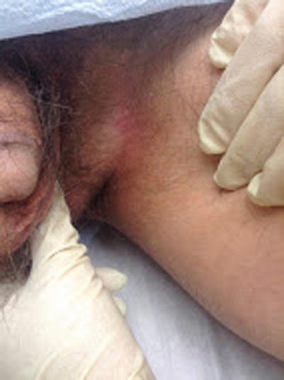
Fig. 14.13
Erythrasma. Thin brown plaques in the axillae
Fig. 14.14
Erythrasma. Erythematous patches in the groin with a nonspecific clinical appearance (Photo courtesy of Sylvia Hsu, MD)
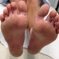
Pitted keratolysis presents as crateriform pits on the pressure-bearing aspects of the soles (Fig. 14.15) and is caused by Cornyebacterium species, Kytococcus sedentarius, or Dermatophilus congolensis. It is associated with malador, hyperhidrosis, pruritus, and sometimes burning sensation or tenderness [163].
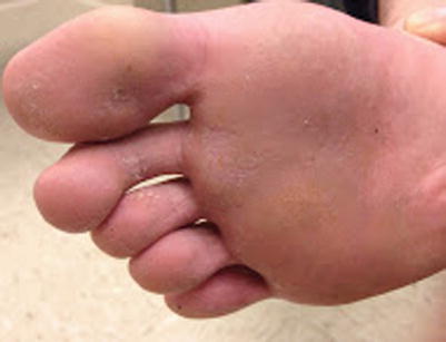
Fig. 14.15
Pitted keratolysis. Crateriform pits on the plantar surface (Photo courtesy of Sylvia Hsu, MD)
Trichomycosis presents as asymptomatic soft tan concretions of the axillary or pubic hair, and is associated with malodor and hyperhidrosis [164]. Thus, it may be seen in postpubertal adolescents. Cornyebacterium species including C. tenuis are causative. It may be associated with concomitant pitted keratolysis and erythrasma [165].
Specific Investigations
For diagnosis |
Gram stain |
Wood’s lamp |
Histopathology |
Culture |
For treatment |
No specific investigations required |
A gram stain of skin scraping will identify gram-positive rods and filaments in erythrasma. Examination with wood’s lamp in a dark room demonstrates coral-red fluorescence due to porphyrin production by C. minutissimum (Fig. 14.16). The causative bacteria can also be seen as small light blue colonies in the stratum corneum on histopathology; PAS, gram, or Giemsa stains can be used to highlight the organisms, but skin biopsy is usually performed to exclude inflammatory dermatoses [166]. Culture is rarely indicated, but can be performed.
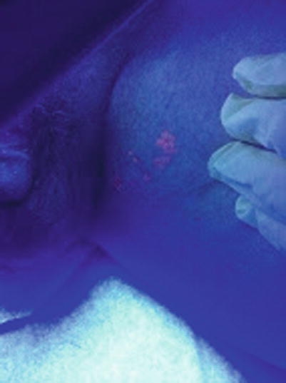
Fig. 14.16
Erythrasma. Characteristic coral red fluorescence during illumination with Wood’s lamp (Photo courtesy of Sylvia Hsu, MD)
Pitted keratolysis is generally a clinical diagnosis. Wood’s lamp is less sensitive for detection than in cases of erythrasma, but may be helpful if positive. If skin biopsy is performed, bacterial colonies can be identified in the stratum corneum [163]. Culture is possible, but rarely performed or necessary.
In trichomycosis, KOH or gram stain of the concretions demonstrates that they are composed of bacilli. Wood’s lamp is very useful and demonstrates fluorescence in most cases [165]. Bacterial culture is not necessary, but can be performed.
Erythrasma
Medication | Dosing | Evidence level |
|---|---|---|
Topical clindamycin 1 % | Twice daily for 2–3 weeks | D |
Topical erythromycin 2 % | Twice daily for 2–3 weeks | D |
Topical imadazole antifungals: | Twice daily for 6 weeks (on average) | A* |
Oxiconazole, miconazole, econazole |
Although there are no randomized trials studying the topical clindamycin and topical erythromycin for the treatment of erythrasma, they are considered first-line therapies given their long history of clinical efficacy, safety, availability, and low cost. Several randomized studies have examined the clinical utility of topical imidazole antifungals, finding that over 90 % of patients were cured. However, these studies also examined superficial fungal infections as well as co-infections. Additionally, the duration of successful treatment occurred within a wide range: between 7 and 60 days (approximately 40 on average in a study by Grigoriu et al.).
Oral antimicrobial therapy should be reserved for patients with extensive or refractory disease, given the potential for antibiotic resistance following failed oral therapy. In an early randomized trial, cure or improvement was obtained in 77 % of patients treated with oral erythromycin, compared to 87 % of patients treated with topical fusidic acid. A more recent double-blind study also found topical fusidic acid to have the highest cure rate (97 %) for erythrasma; single-dose clarithromycin and a 2-week course of erythromycin demonstrated cure rates of 67 % and 53 %, respectively. Based on this data, fusidic acid is the most effective evidenced-based treatment for erythrasma, but unfortunately is not available in the United States.
C. minutissimum produces natural porphyrins, accounting for the observed fluorescence under Wood’s light, a quality which can be exploited by photodynamic therapy. In a small trial, one or two red light irradiations resulted in complete response in only 3 of 13 patients. Tetracycline has also been used for erythrasma, but should be considered a third-line therapy, given its side effect profile and reduced efficacy compared to oral erythromycin. Tetracycline should be avoided in children younger than 8 years of age.
Pitted Keratolysis
Medication | Dosing | Evidence level |
|---|---|---|
Topical erythromycin 2 % | Twice daily for 10 days | C |
In 97 adults and children (youngest age 8 years) with pitted keratolysis, hyperhidrosis was an associated finding in nearly all patients. Topical erythromycin resulted in complete clearance of cutaneous findings and hyperhidrosis in all patients, with only a 6 % recurrence rate. Although hyperhidrosis is generally considered to be a predisposing factor for this infection, the authors postulated that hyperhidrosis may be secondary to eccrine gland dysfunction resulting from Kytococcus infection.
Mupirocin 2 % ointment has been effective for pitted keratolysis in case reports. In a small series, resolution of clinical signs and symptoms of pitted keratolysis resolved following treatment with clindamycin 1 %-benzoyl peroxide 5 % gel for 3–4 weeks.
Medication | Dosing | Evidence level |
|---|---|---|
Botulinum toxin injection | 50 U total for each plantar surface, with 2 U per each of 25 sites marked (each site 2 cm apart) | E* |
In two patients with disease refractory to antibiotics, botulinum toxin injection resulted in elimination of hyperhidrosis and pitted keratolysis within 14–30 days following treatment.
Trichomycosis
In general the most effective and rapid treatment of trichomycocis is to shave or removed the affected hair. Of note, however, recurrence is common if shaving is only performed once and without an adjunct treatment such as benozyl peroxide or sulfur-containing soap. Topical antibiotics such as clindamycin and erythromycin are also curative. Overall, these treatments have demonstrated over 90 % cure rates in a retrospective review.
Erysipeloid
Clinical Features
Erysipelothrix rhusiopathiae is a non–spore forming, gram-positive bacillus capable of causing self-limited soft tissue infection or serious systemic infection. Infection in humans is usually due to occupational exposure to domestic or marine animals. Erysipeloid is a localized cutaneous infection that follows minor trauma, and results in a subacute cellulitis appearing as a violaceous lesion with central clearing and a raised border (Sect. 14.17). Stiffness, pain, and local lymphangitis may be seen. If diffuse cutaneous disease, progression occurs from erysipeloid to widespread sites, sometimes with urticarial or bullous lesions. Systemic infection is rare, and associated with skin findings of erysipeloid or diffuse cutaneous disease, in addition to bacteremia and visceral involvement such as endocarditis [182].
Specific Investigations
For diagnosis |
Gram stain |
Culture |
For treatment |
No specific investigations required |
The best way to diagnose erysipeloid is based on clinical findings. Gram stain or culture of tissue or aspirates obtained from cutaneous lesions are often negative. Blood cultures should be obtained in suspected cases of diffuse cutaneous or systemic disease. E. rhusiopathiae grows within 2–3 days. Of note however, misidentification as Lactobacillus or Enterococcus species may occur [183].
Erysipeloid is typically an occupational infection seen in individuals exposed to livestock or involved in fishing. It is rare in children; [184] thus there are no controlled or comparative studies in pediatric patients, and treatment is based on in vitro data and clinical experience.
Penicillin and imipenem are the most active agents in vitro against E. rhusiopathiae. Other agents with activity include cephalosporins, fluoroquinolones, erythromycin, clindamycin, and linezolid. Although in vitro data supports the use of fluoroquinolones in localized cutaneous as well as diffuse cutaneous and systemic infections, this class of antibiotics should not be used in children under 18 years of age.
Localized cutaneous disease typically resolves within 3 weeks without treatment, but antimicrobial therapy is recommended to hasten resolution, alleviate symptoms, and reduce the risk of relapse.
Table 14.55


Related posts:

Stay updated, free articles. Join our Telegram channel
Full access? Get Clinical Tree
